Imaging - Radiologic Evaluation of Knee, Ankle, and Foot
1/100
There's no tags or description
Looks like no tags are added yet.
Name | Mastery | Learn | Test | Matching | Spaced | Call with Kai | Chat |
|---|
No analytics yet
Send a link to your students to track their progress
101 Terms
AP
lateral
PA "tunnel"
Tangential "sunrise" or "merchant"
What are routine radiographic views of the knee?
AP
What view is this?
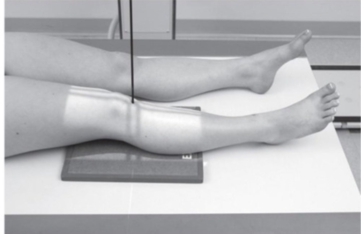
AP
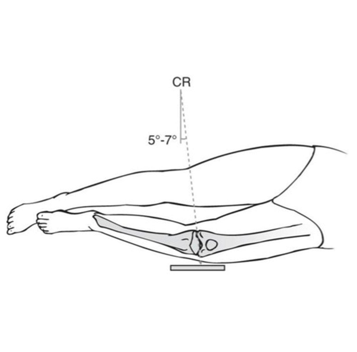
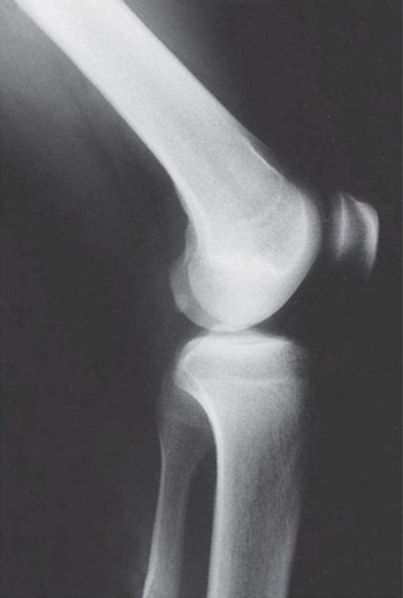
What view is this?
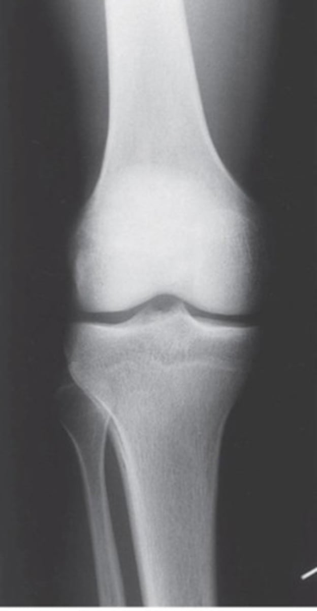
Lateral
What view is this?
Lateral
What view is this?
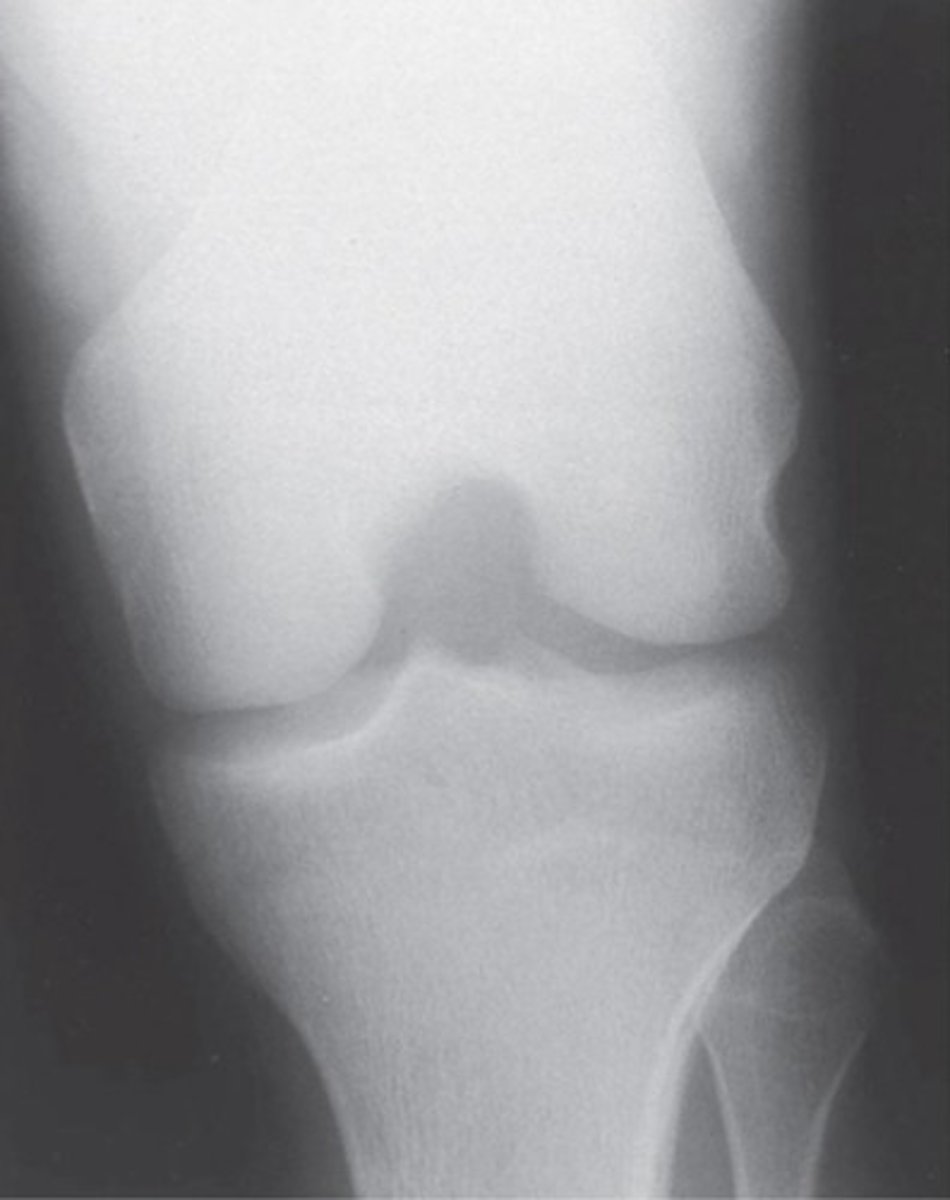
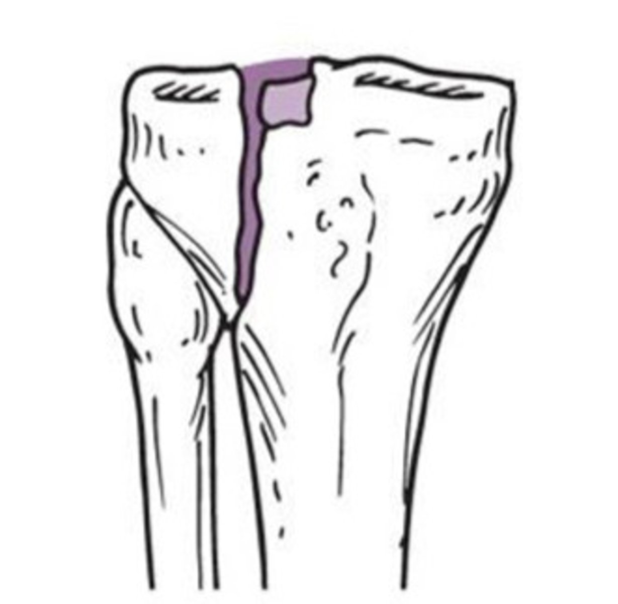
PA intercondylar
What view is this?
PA intercondylar
What view is this?
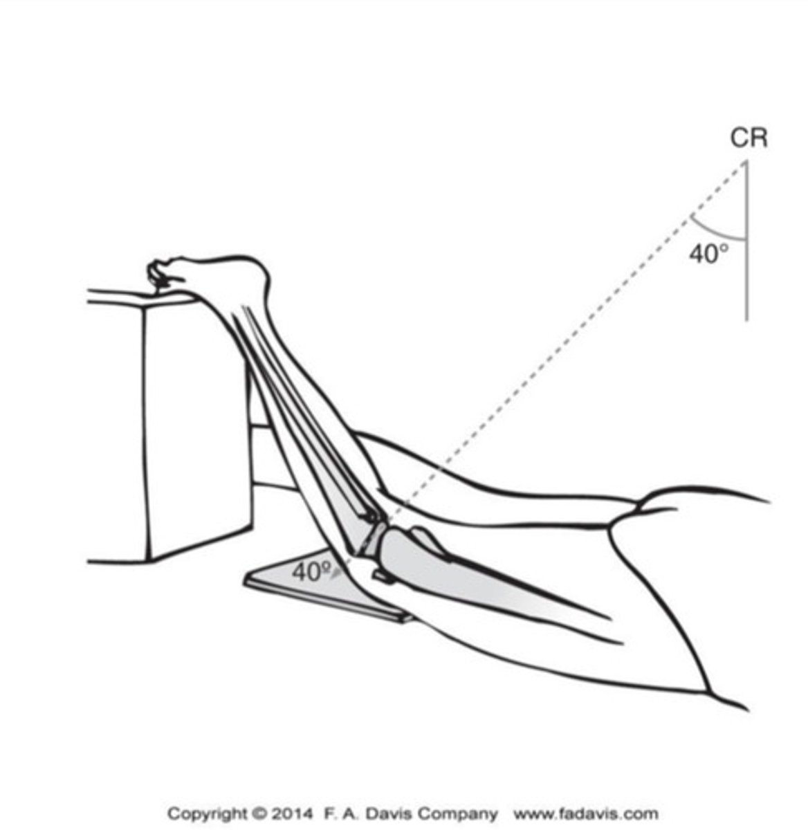
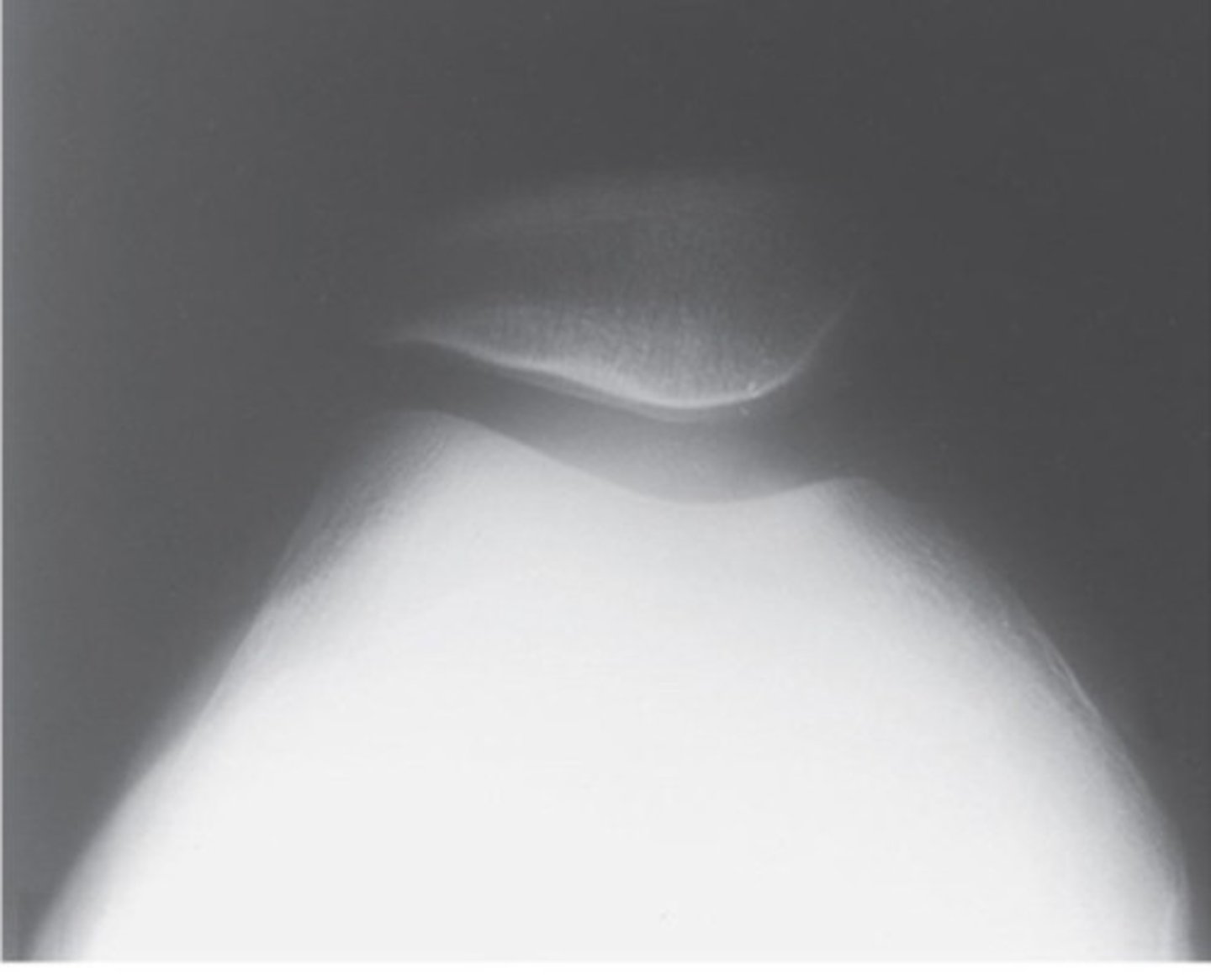
Tangential
What view is this?

Tangential
What view is this?
Distal femur - medial and lateral condyles
Proximal tibia - medial and lateral condyles, intercondylar eminence
Femorotibial joint space
Patellar position
Proximal fibula - head, neck, shaft
What can you see on an AP knee radiograph?
Distal femur - medial and lateral condyles
Proximal tibia - tibial tuberosity, articular surface, intercondylar
eminence
Femorotibial joint space
Patellar position (alta or baja) - length of patella
Proximal fibula - head, neck, shaft
What can you see on an lateral knee radiograph?
Distal femur - medial and lateral condyles and intercondylar fossa
Proximal tibia - medial and lateral condyles, intercondylar eminence
What can you see on an AP axial "tunnel" intercondylar fossa radiograph?
Patella - sulcus angle, congruence angle, position within
intercondylar sulcus, subtle subluxations
Femoral condyles - medial and lateral surface
What can you see on an tangential (sunrise or merchant) radiograph?
axial
sagittal
coronal
What are the planes of knee CTs and MRIs?
Meniscus lesions
Ligament injuries
Soft tissue injuries
Osteochondral abnormalities
What are common reasons to use an MRI to image a knee?
femoral condyles
patellofemoral articulation
Tibial plateau
Tibial tuberosity
What do you see on an axial view of a CT or MRI?
Patellar positioning
Patella tendon
femoral condyles
Tibia
What do you see on an sagittal view of a CT or MRI?
femur
tibia/tibial plateau
Intercondylar notch
Intercondylar eminence
What do you see on an coronal view of a CT or MRI?
Distal femur fracture
Proximal tibial fractures
Patellar dislocations
Patellar fractures
What are types of fractures and dislocations of the knee?
Joint effusion after direct blow or fall
Inability to walk without limping
Palpable tenderness over the patella or fibular head
Inability to flex the knee to 90 degrees
> 55 years old
When do you refer for a radiograph of the knee after trauma?
nondisplaced
What type of fracture does A show?
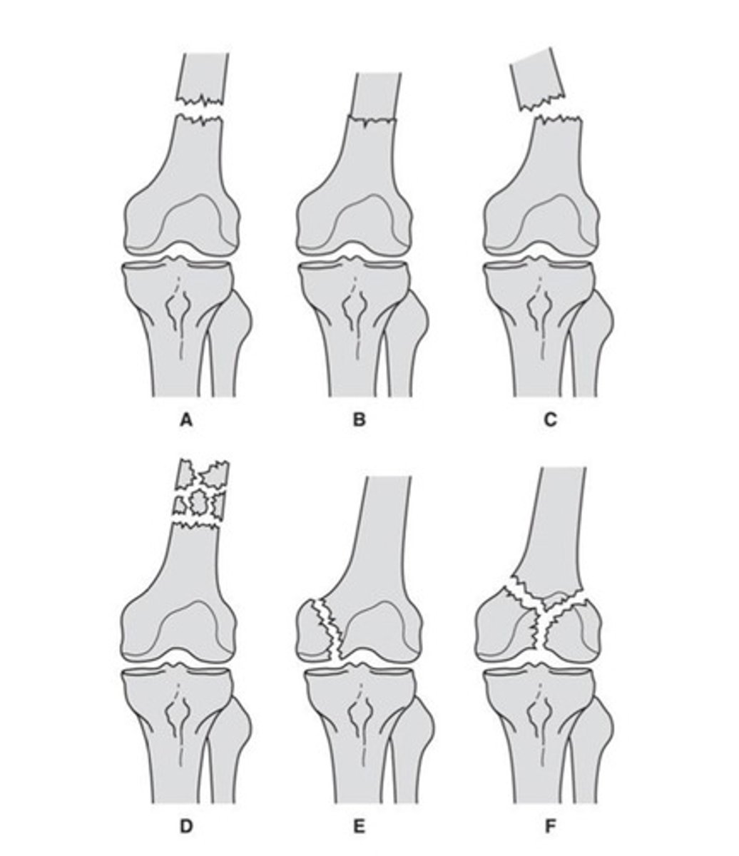
impacted
What type of fracture does B show?

displaced
What type of fracture does C show?

comminuted
What type of fracture does D show?

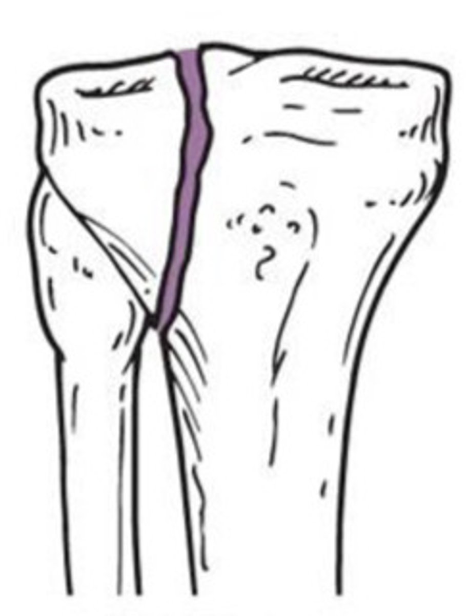
condylar
What type of fracture does E show?

intercondylar
What type of fracture does F show?

falling from height
car accident y
weaker bones
What causes a distal femur fracture?
ORIF
How do you treat a distal femur fracture?
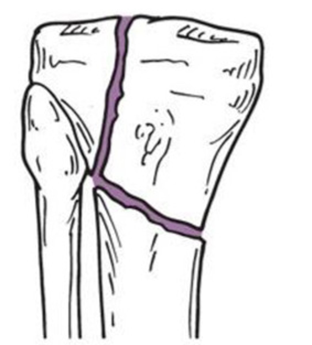
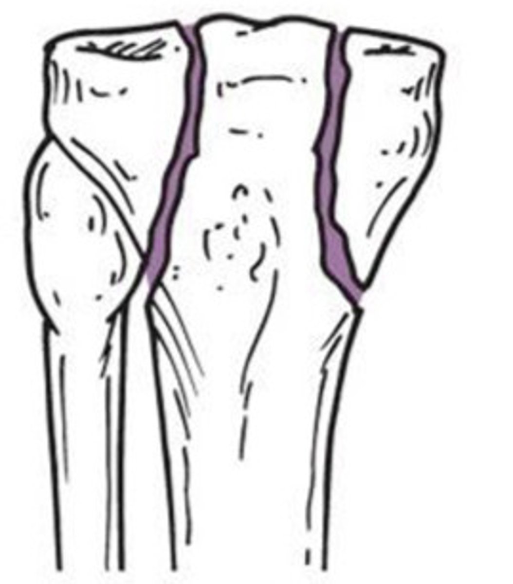
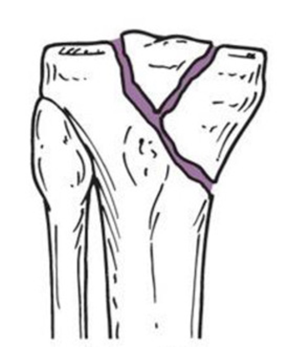
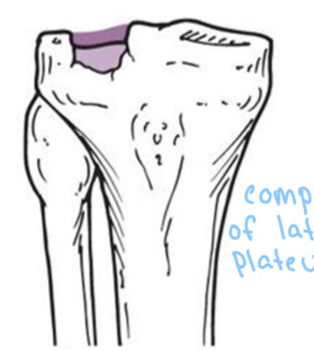
wedge or split fracture of lateral aspect of plateau
Type I proximal tibial fracture
lateral wedge or split fracture
Type II proximal tibial fracture
pure compression fracture of lateral plateau
Type III proximal tibial fracture
fracture involving medial plateau
Type IV proximal tibial fracture
fracture include split elements of both the medial and lateral condyles
Type V proximal tibial fracture
complex, bicondylar fracture
Type VI proximal tibial fracture
Type VI - dissociation of metaphysis and diaphysis
What type of fracture does this show?
Type V - Bicondylar fracture
What type of fracture does this show?
Type IV - split fracture, medial plateau
What type of fracture does this show?
Type III - central depression
What type of fracture does this show?
Type II - Split depression
What type of fracture does this show?
Type I - Split
What type of fracture does this show?
depends on amount of compression of plateau and possible joint instability
How do you treat a proximal tibial fracture?
Vertical
What type of patellar fracture is 1?

transverse, nondisplaced
What type of patellar fracture is 2?

transverse, displaced
What type of patellar fracture is 3?
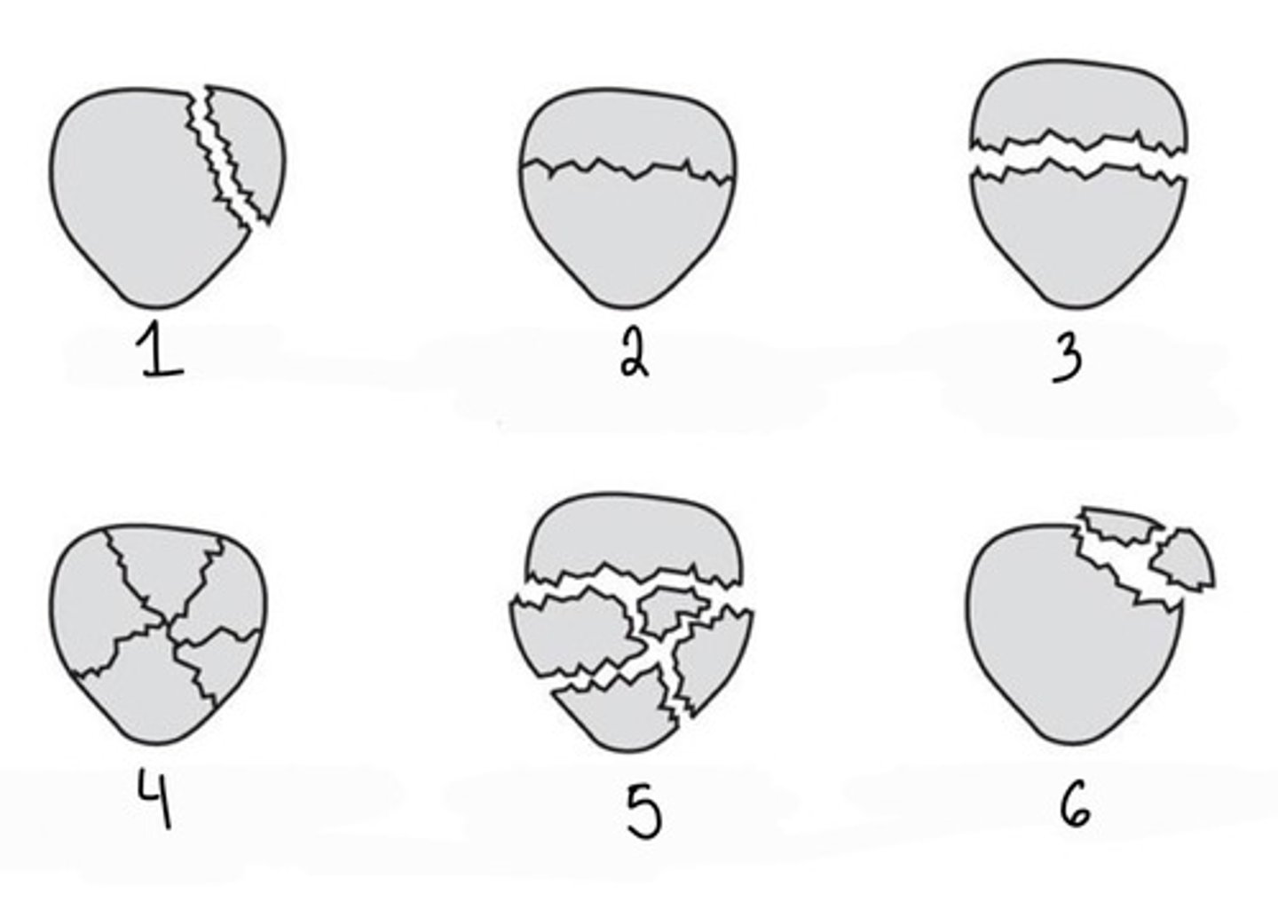
Comminuted nondisplaced
What type of patellar fracture is 4?

Comminuted displaced
What type of patellar fracture is 5?

Avulsed fragments
What type of patellar fracture is 6?

fall
car accident
forceful contraction of quads (avulsion)
What caused a patellar fracture?
injuries to arteries, nerves, and veins
What can dislocation or subluxation of the patella cause?
Osteochondral fracture
fracture to knee cartilage often seen in young athletes
Osteochondritis dissecans
osteochondral fracture that is now chronic, often seen in adolescent and early adulthood
Spontaneous osteonecrosis
sudden decreased blood flow to knee, often in area of WB of medial femoral condyle in older adults
shearing/turing force
What causes a meniscus tear?
MRI
What imaging is used to diagnose a meniscus tear?
normal meniscus
What type of meniscus is 1?

vertical tear
What type of meniscal tear is 2?
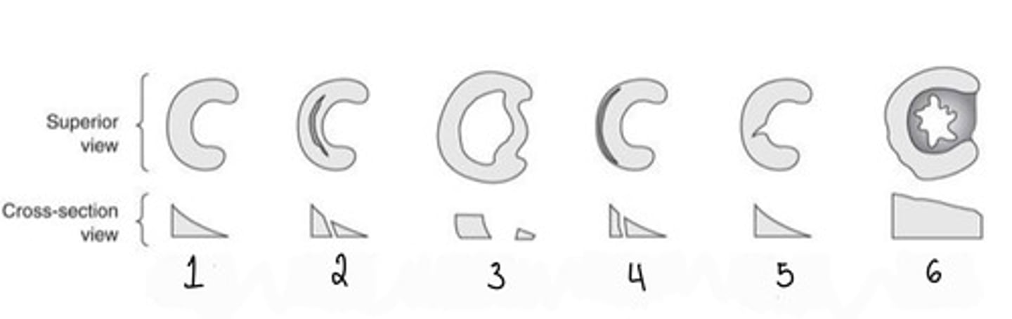
bucket handle tear
What type of meniscal tear is 3?

peripheral tear
What type of meniscal tear is 4?

horizontal tear
What type of meniscal tear is 5?

discoid meniscus
What type of meniscus is 6?

enlarged or thickened meniscus
What is a discoid meniscus?
stress x-ray
What type of imaging do you do see collateral injuries on?
radiograph for avulsion fracture
MRI to visualize ligaments
What type of imaging do you use to see cruciate ligaments?
Osgood-schlatters disease
enlarged and deformed tibial tubercle caused by repetitive trauma at distal tendon attachment
adolescent boys
Who is Osgood-schlatters disease most common in?
lateral radiograph
What imaging is Osgood-schlatter disease seen on?
reduction of joint space
sclerosis of subchondral bone
osteophytes at joint majins
subchondral cysts
noted varus or valgus joint deformity
What is the radiographic detection of DJD/OA?
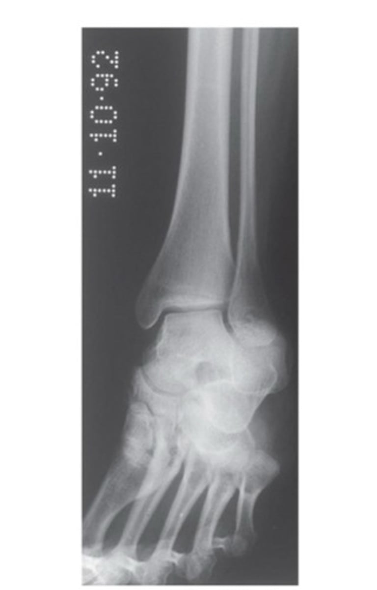
AP ankle
What view is this?
AP ankle
What view is this?
AP oblique ankle
What view is this?
AP oblique ankle
What view is this?
lateral ankle
What view is this?
lateral ankle
What view is this?
eversion
inversion
anterior drawer
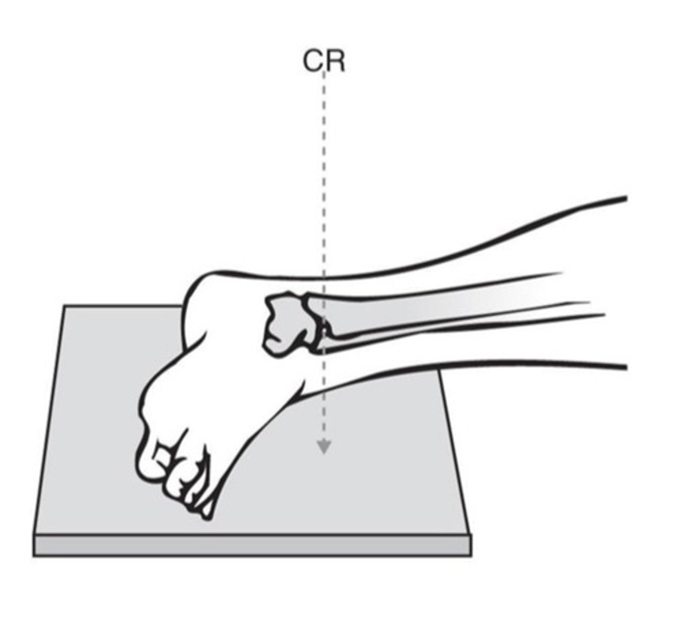
What are the three stress views of the ankle?
What view is this?
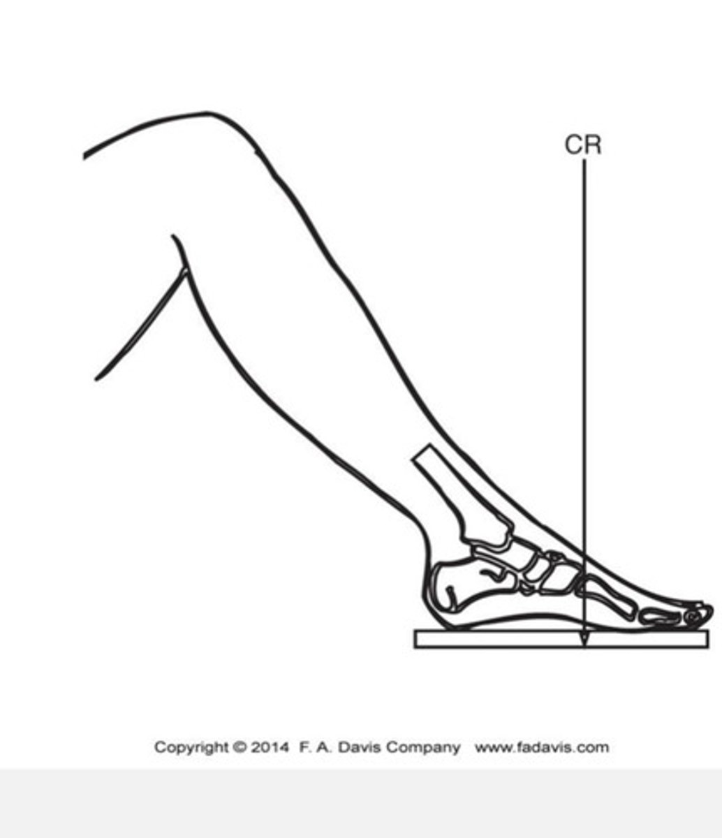
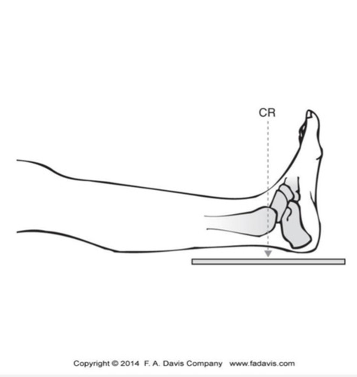
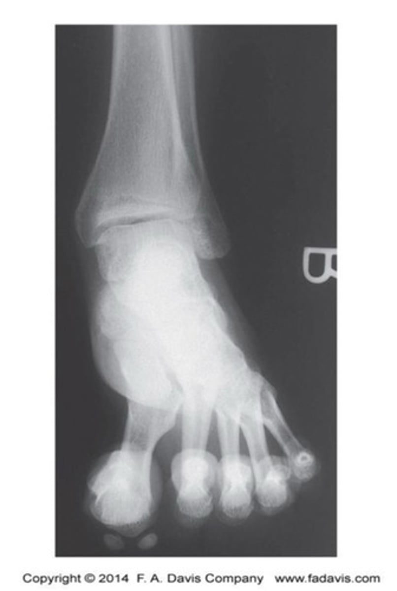
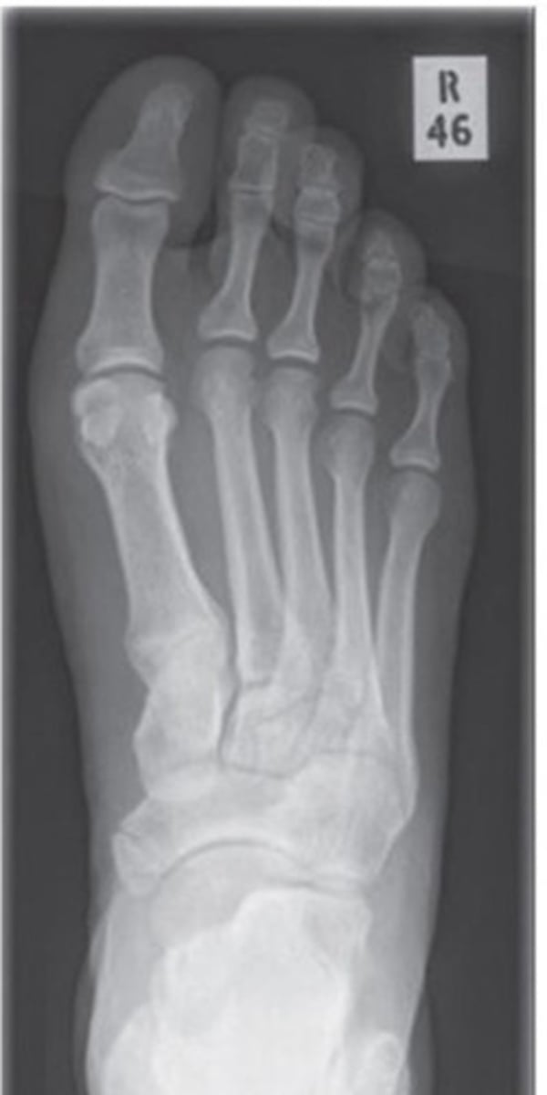
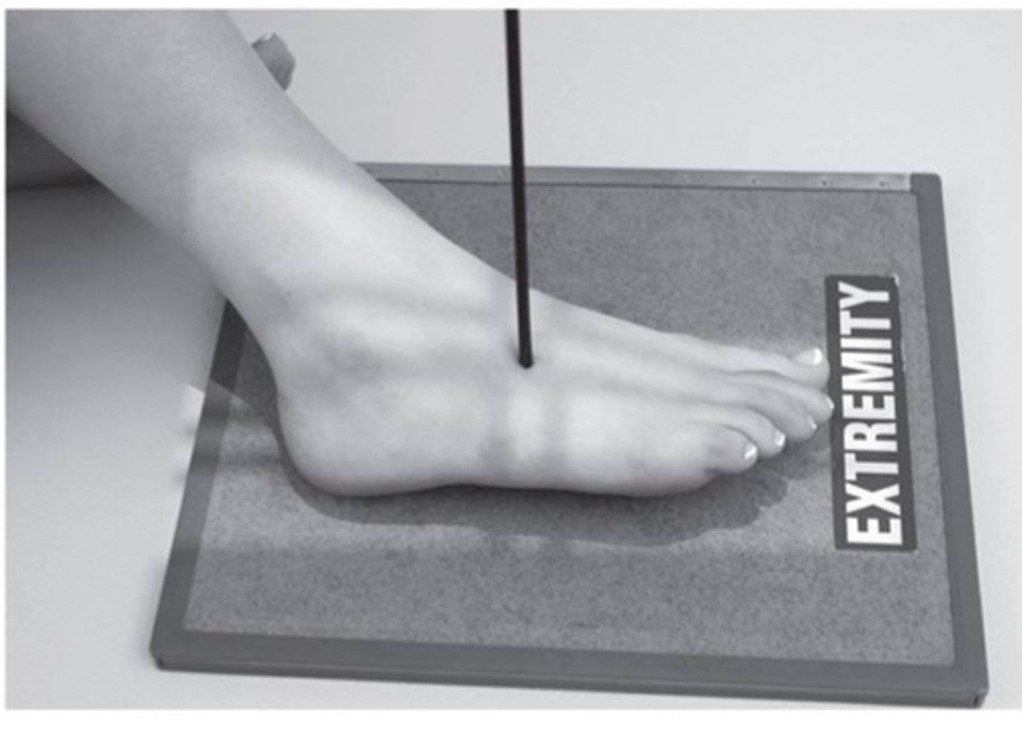
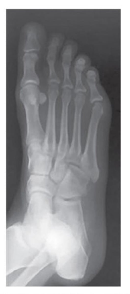
AP foot
What view is this?
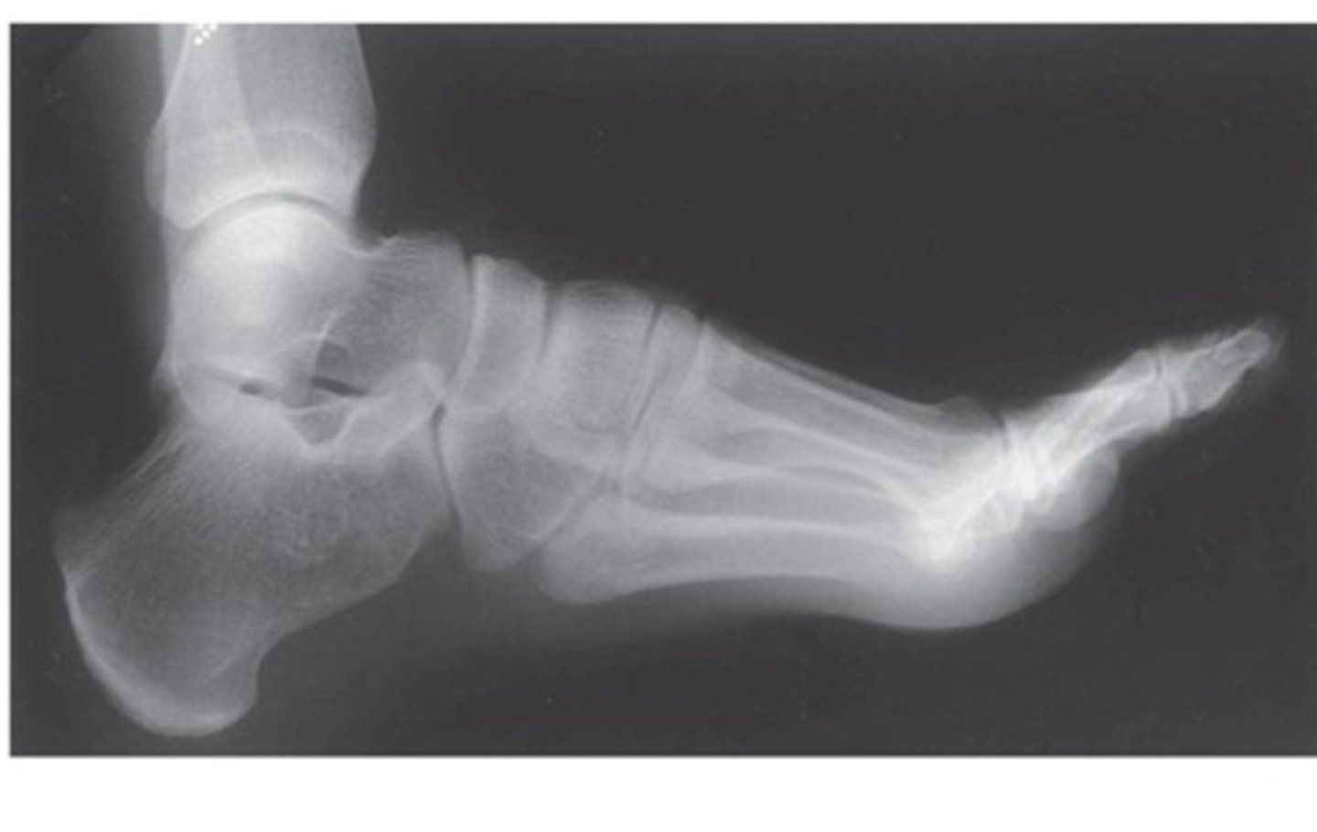
Lateral foot
What view is this?
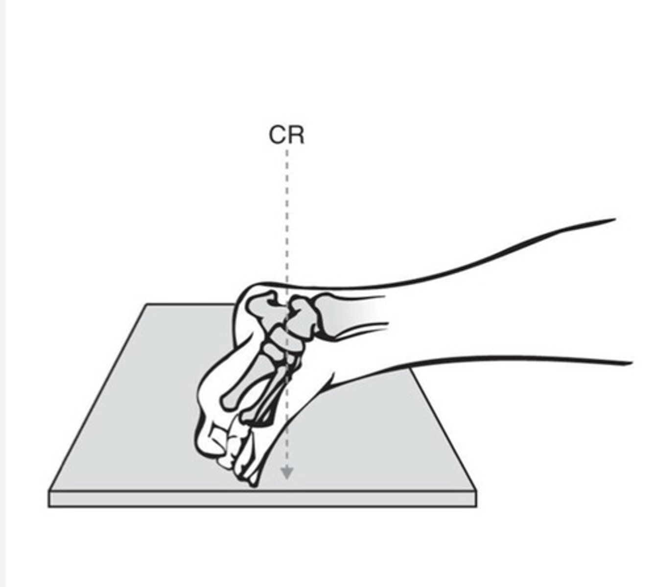
Lateral foot
What view is this?
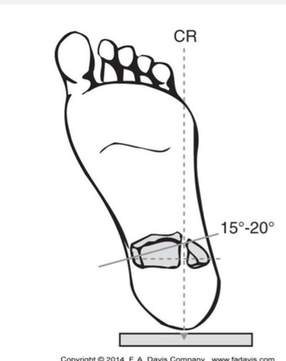
Oblique foot
What view is this?
Oblique foot
What view is this?
ankle tendons
sinus tarsi
tibiofibular ligament
Osseous structures
What structures are seen in the axial plane of the ankle?
achilles tendon
tibiotalar joint
subtalar joint
transverse tarsal joint
plantar fascia
sinus tarsi
What structures are seen in the sagittal plane of the ankle?
deltoid ligaments
calcaneofibular ligament
tarsal tunnel
talar dome
subtalar joint
plantar fascia in cross section
What structures are seen in the coronal plane of the ankle?
bone tenderness at the Posterior edge or tip of lateral malleolus
bone tenderness at the Posterior edge or tip of medial malleolus
inability to bear weight
What are the rules of the ottawa ankle rules?
bone tenderness at the base of the 5th metatarsal
bone tenderness at the navicular
inability to bear weight
What are the rules of the ottawa foot rules?
Trimalleolar fracture
fracture of both malleoli and posterior rim of tibia
inversion
What is the most common type of ankle sprains?
avulsion fracture suspected
When would you use an image with an ankle sprian?
MRI
What is the best imaging for tendon injury?
achilles tendon
fibularis longus/brevis post inversion injury
What are examples of tendon injuries?
Hallux valgus
first metatarsal deviates medially, great toe is deviated laterally
Pes Cavus
HIgh medial longitudinal arch
Pes planus "flat foot"
Rigid or flexible classification
Clubfoot
Congenital talipes equinovarus
intersection of a line drawn along the midshaft of the 1st metatarsal and a line bisecting the talus on the lateral view of the foot
How is the talometatarsal angle formed?
Distal tibia & fibula and joint space between
Proximal talus or talar dome
Ankle mortise
What can you see in a AP ankle view?
Distal tibia & fibula
Proximal talus or talar dome
Ankle mortise
What can you see in a AP oblique ankle view?
Distal tibia anterior tubercle and posterior malleolus
Distal fibula superimposed behind the tibia and talus
Talus, calcaneus, navicular and cuboid
Joint spaces
What can you see in a lateral ankle view?
Phalanges and IP joints
Metatarsals
Tarsals
Transverse tarsal joint
Tarsometatarsal joint
What can you see in the AP foot view?
Tibia and fibula
Transverse tarsal joint
Tarsometatarsal joint
Talus and calcaneus
Tarsal sinus & subtalar joint
Ability to measure Boehler's angle and the calcaneal inclination
What can you see in a lateral foot view?