The Musculoskeletal System (chp 8)
1/90
Earn XP
Description and Tags
muscles and skeleton
Name | Mastery | Learn | Test | Matching | Spaced |
|---|
No study sessions yet.
91 Terms
What are the types of Muscles?
Skeletal muscles
Smooth muscles
Cardiac muscles
What are skeletal muscles?
Muscles attached to the skeleton
Under conscious control and allows us to carry out a wide range of physical activities
Contraction Brings about movement in joints and gives bodies its form and contours
What are Smooth muscles?
muscles associated with the internal organs of the body and provide their movement (stomachs and intestines)
Under involuntary control
What are Cardiac muscles?
heart muscle
Contracts to reduce the space in chambers of the heart and push the blood from the heart into the blood vessels
What do muscles do?
they have special properties that together bring about movement at a joint
Contractibility
Extensibility
Elasticity
What is contractibility of the muscles?
all muscle has the ability to contract (shorten)
Muscles re designed so that when they contract, they reduce the distance between the parts they are connected to, or decrease the space they surround
What is extensibility?
ability to be stretched
What is elasticity?
ability to return to original length after stretching
How do the muscles work together? *3 points*
muscles are attached to the bone by fibrous elastic connective tissue called tendons. Attach bones in such a way it allow the bone to move when the muscles contract
Muscles can pull bones, but cannot move them apart. (1 directional of movement). There fore, muscles which move parts of the body work in pairs
Pairs of muscles where one muscle provides movement in one direction, and the other muscle in the other directed are called antagonists
What is the origin?
End of muscle fixed to stationary bone (bone doesn’t move in action)
What is insertion?
End of muscle fixed to moveable bone (moves in the action)
What is the belly?
the fleshy portion of the muscle between the tendons of origin and insertion
What is the Agonist muscle?
prime mover
Muscle that causes desired action
What is an antagonist muscle?
Muscle that has the opposite effect to the agonist
What is a synergist muscle?
muscles that help, indirectly, to steady the joint during movement (prevent unwanted movement)
What is a fixator?
When a synergist immobilises the joint
What is the structure of skeletal muscles?
Muscle cells are held together in bundles
Sheath of connective tissue surrounds each bundle so it can function as an INDIVIDUAL UNIT, and slide over one another as they contract
Towards the end f the muscle, the sheathes of connective tissue taper and form the tendon
What are muscle cells?
they are called, muscle fibres:
muscle cells which are cylinder in shape
Each muscle cell lies parallel to the next, about 10-100 micrometres in diameter and few mm to several cm long
Contain MANY nuclei
What is the structure of the muscle fibres?
In muscle cells, the thin plasma membrane surrounding the cells is called the sacrolemma
Contains cytoplasm which in muscle cells is called sarcoplasm
Inside the sarcoplasm of each fibre are thread-like myofibrils lying parallel to each other. Can be hundreds to many thousands of myofibrils in each fibre
What is the structure of the myofibrils?
composed of many smaller myofilaments made of protein
These are very important as they allow contraction of the muscle
What are the two types of myofilaments?
Myosin: thick myofilaments
Actin: thin myofilaments
What is the structure of thin Actin myofilaments?
composed mostly of action
Contain tropomyosin and troponin
Double-stranded coil
What is the structure of thick Myosin myofilaments?
composed mostly of myosin
Rod-shaped (tail) with head
How do the myofilaments work?
when stimulated by NERVE IMPULSES, there is enough energy, the thick and thin filaments slide pass each other which SHORTENS the myofibril
What is the structure of myofibrils and what do they give?
Arrangement of thick and thin filaments in the myofibril gives balanced effect to the muscle
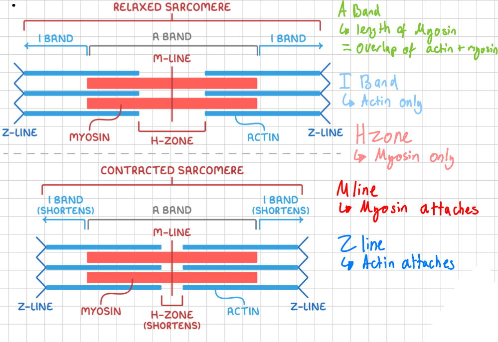
Myofibrils can be divided into units called sacromeres
Sacromeres contain actin and myosin in certain patters
Gives skeletal and cardiac muscle their striated appearance
What is the structure of a skeletal muscle? (Diagram)

What is the sliding filament model?
sliding filament theory explains how muscle contraction occurs
It is a simplified representation of the idea/process
The basis of the model surrounds actin and myosin filaments sliding over each other
How does the sliding filament model work?
Thin actin filaments slide over thick myosin filaments
The “z lines” (anchor points for actin) are drawn closer together and the sacromere is shortened, thus shortening the whole muscle
The myofilaments stay the same length, they overlap to shorten the muscle
When the muscle is relaxed, the actin and myosin are pulled back in the opposite direction, returning the muscle to its original state
What is the structure of the sacromere?
How does contraction work? Step 1
Sarcoplasmic reticulum releases calcium ions into sarcoplasm
How does contraction work? Step 2
Calcium ions will bind to troponin
→ causes troponin-tropomyosin complexes to move away from the myosin binding sites on actin
Once this happens, contraction can begin
How does contraction work? Step 3
Myosin heads contain ATP binding and ATPase enzymes
ATP os hydrolysed into ADP, feeeing up a phosphate molecule
→ energises the myosin head
How does contraction work? Step 4
Myosin attached to the myosin binding sites in actin and myosin threads
→ forms the cross-bridge between the actin and myosin threads
How does contraction work? Step 5
Phosphate released by myosin triggers the actual power contraction stroke
How does contraction work? Step 6
ADP pockets opens up on the myosin head
How does contraction work? Step 7
Myosin head rotates and releases the ADP
How does contraction work? Step 8
As the head rotates,, it pulls the thin filaments closer to the centre of the M line
How does contraction work? Step 9
When the motion is over, the myosin awaits another ATP so it can be released from the actin binding site
How does contraction work? Step 10
If ATP is available, the myosin detaches and begins another stroke of the cycle
How does contraction work? Step 11
Myosin heads will keep binding with actin sites until the ATP runs out or the calcium levels decrease
What is the function of the skeletal system?
Storage: minerals, salts and fats
Production: contains stem cells which can become blood cells
Protection: of organ systems
Leverage: allow muscles to attach and produce gross movements
Support: provides shape and supports its parts
What is the function of bones and general information
Axil skeleton: at the midline, 80 bones
Appendicular skeleton: Appendages (limbs) and their girdles (pelvic and shoulder) 126 bones
Function: to support its respective organs
What is the anatomy of a long Bone
Diaphysis: shaft
Epiphysis: extremity of bones
Articulate cartilage: covers epiphysis
Periosteum: covering around surface of bone
Compact bone
Spongy bone
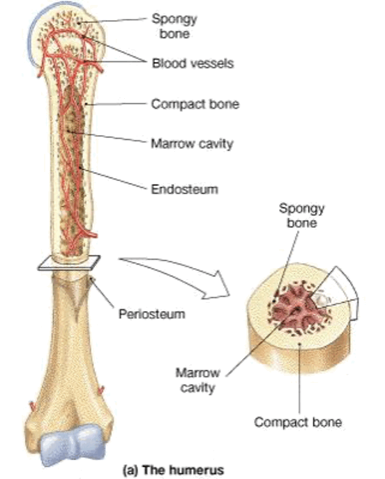
Why does your bone have cartilage?
It is soft and flexible to absorb shock
What is the structure of the Periosteum
Has 2 layers
→ fibrous layer
→ osteogeic layer
Which has 2 things
→ osteoblasts: produce bone
→ osteoclasts: break down bone
What is the structure of the Bone Tissue
Has pores that
→ living cells
→ channels for blood vessels
→ decrease weight of the bone
Has a degree of porosity
→ spongy (cancellous) bone
→ compact bone
What are the tissues of the skeletal system?
Bones are LIVING TISSUE
Compact bone: highly organised and composed of tubular osteons
Spongy bone: more porous and may be filled with red bone marrow
What is the structure of the compact bone?
Consists of small units known as osteons (similar in structure to myofibrils in muscles)
What is the structure of the osteon?
A central canal
Lamellae
Lucanae
Osteocytes
Canaliculi
What do each of the structures of the osteon do?
A central canal: space for blood vessels and nerves
Lamellae: circle of compact bony matrix
Lucanae: small spaces between lamellae
Osteocytes: cells that occupy the small spaces (Lucanae)
Canaliculi: small, canal-like, passages between lucanae (blood supply)
What is the structure of the spongy (cancellous) bone?
less organised than compact bone
Consists of random arrangement of trabecular (thin plates or bone)
Osteocytes take up places within trabecular, blood vessels and nerves pass through spaces
What is cartilage and where is it found and what does it do?
Found on the surface of bones, in trachea, end of nose etc
Provides flexibility and supports
Has very limited supply of nutrition and removal of wastes due to a lack of blood supply, therefore takes a long time to heal
What is the structure of cartilage?
Consists of collagen embedded in chondrin
Cartilage cells are known chondroblasts. Produce a matrix which gradually surrounds them until they are enclosed
When these chondroblasts are enclosed, they become chondrocytes (mature cartilage cells)
What are the types of Cartilage?
Hyaline
Fibrocartilage
Elastic cartilage
What is the hyaline cartilage?
generally found between joints (articular cartilage) where strength is important. Collagenous Fibres are closely packed
What is Fibrocartilage?
generally found in weight bearing regions, such as intervertebral discs
Coarse appearance (fibrous)
What is elastic cartilage?
generally found where flexible support is required
Eg; ears
Collagenous fibres are not closely packed (this makes it elastic)
What are articulations? (Joints)
where bones are joined together
they are classified by degree of movement
Immovable, slightly moveable, and freely moveable
What are fibrous joints?
immovable or fixed
Held together by connective tissue
Very strong
Examples
sutures of skull
Teeth and jaw
What are cartilaginous joints?
slightly moveable
Held in place by cartilage
Stronger than synovial
Examples
pubic symphysis
Intervertebral discs
What are Synovial Joints?
freely moveable
Ligaments, muscles and tendons control
Examples
shoulder joint
Knee joint
What are the 6 types of synovial joints?
Ball and socket
Hinge
Pivot
Gliding
Saddle
Condyloid
What is the ball-and-socket joint?
Spherical head of a bone fits into a cup-like depression of another bone
Allows for a very large range of motion
Examples:
shoulder joint
Hip joint
What is a hinge joint?
convex surface of one bone fits into concave of another
Allows movement in one plane-back and forth
Examples
elbow joint
Knee joint
What is the pivot joint?
pointed end of a bone articulates with a ring of bone/ligament
Allows for rotation
Examples:
1st and 2nd vertebrae (head rotation)
Radius and ulna
What is the gliding joint?
two bones that move alongside one another
Allows for limited movement in either side to side or back and forth
Examples
between carpal bones
Sternum and clavicle
What is the saddle joint?
concave and convex bones fit together (like a saddle)
Allows for a large range of motion back and forth and side to side
Examples:
thumb joining hand (metacarpal to carpal)
What is the Condyloid joint?
slightly convex bone fits into slightly concave bone
Allows for a limited range of motion both side to side and back to forth
Examples:
between phalanges and metacarpals
Radius and carpals
What is the structure of a synovial joint
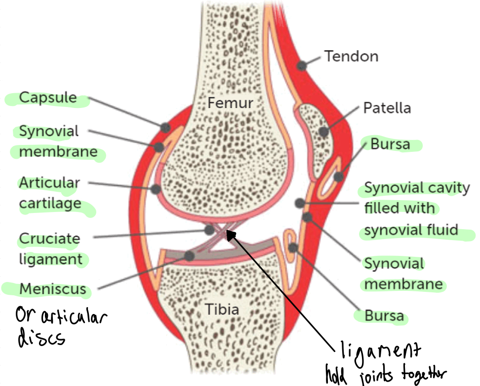
What is the articular capsule?
Surrounds the entire joint
What is the fibrous capsule?
outer layer made up of dense connective tissues attached to bones
flexible enough to allow movement, but strong enough to prevent dislocation of joint
What is the synovial membrane?
inner layer of capsule made up of loose connective tissue
lines the entire cavity and is well supplied with blood vessels
What is synovial fluid?
It is secreted by synovial membrane and fills the synovial cavity
What are the functions of the synovial fluid?
Lubricate
Provide nourishment
Contains phagocytes (white blood cells)
How does the synovial fluid lubricate?
it lubricates joints and keeps two surfaces of bones from rubbing one another
How does the synovial fluid provide nourishment?
It has minerals for cartilage cells
How does the synovial fluid contain phagocytes and what does that do?
to destroy any pathogens or debris from wear and tear
Injury to a synovial joint will increase the synovial fluid, leading to inflammation
What is the articular cartilage?
covers the surfaces of bones to reduce friction on surfaces.
Takes a long time to heal, white bits have lots of white blood cells
What are articular discs?
Meniscus in knee
Direct the synovial fluid into areas of greatest need such as articulating surfavces
What is the bursae?
sacs of synovial fluid that prevent friction between features such as tendons/joints/skin & bones
Membrane bound sac
In places of friction
What are ligaments?
Join bone to bone to add strength to joints
What movements can the joints do?
Fexion
Extension
Abduction
Adduction
Rotation
What is Flexion?
decreases the angle between bones
bicep curls
What is extension?
increases the angle between bones
kicking a footbsll
What is abduction?
movement away from the midline of the body
sidestepping
What is adduction?
Movement towards the midline of the body
putting arms by your side
What is rotation?
movement of bone around long axis
turning your head
What is Osteoporosis?
Loss of bone mass (density(
What are the causes/risk factors, symptoms and preventions/treatments for osteoporosis?
Causes/risk factors
age (over 30)
Menopause
Lack of calcium/vitamin D
Symptoms
increases risk of fractures
Preventions/treamtment
increase calcium intake
Regular excessive
Quit smoking
What is osteoarthritis?
Deterioration of cartilage in joints
What are the causes/risk factors, symptoms and preventions/treatments for osteoarthritis?
Causes/rick factors
age
Menopause
Symptoms
pains at joints
Growth of bone spurs
Restricted movement
Prevention/treatment
medication (pain relief)
Physiotherapy to strengthen muscles
Surgery