Cellular Response to Stress
 Call Kai
 Learn
Learn Practice Test
 Spaced Repetition
 Match
Match Flashcards
 Knowt Play
1/51

There's no tags or description
Looks like no tags are added yet.
Name | Mastery | Learn | Test | Matching | Spaced | Call with Kai | Chat |
|---|
No analytics yet
Send a link to your students to track their progress
52 Terms
Example of labile cells
Skin, GI, reproductive, urinary, exocrine epithelial cells, haematopoietic stem cells
Example of Stable cells
Liver, kidney, lung, pancreas epithelial cells, smooth muscle cell, fibroblast, endothelial cells
Example of permanent cells
Cardiac/skeletal myocytes, neurons
What is hyperplasia
Increase in the NUMBER of cells only in non-permanent cells
What is the difference between physiological and pathological adaptions
Physiologic: Due to normal stressor
Pathologic: Due to abnormal stressor
What is hypertrophy
Increase in cell SIZE; physiologic = from increased workload
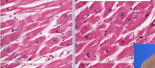
what is the difference between the two pictures
Left is normal; right is hypertrophy
What is atrophy
Decrease in size of cell that once was NORMAL size
When would physiologic and pathologic atrophy occur
Physiologic: Common during early development (thymus)
Pathologic: Loss of stimulus to organ
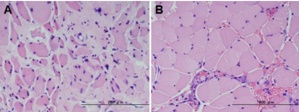
Describe this image
A = atrophy of muscle, B = normal muscle
What is metaplasia
Change of epithelium to another type (normal appearance but abnormal in location)
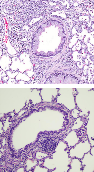
Compare these two images, which is abnormal
Top image shows transitional epithelium in bronchiole; bottom image is normal
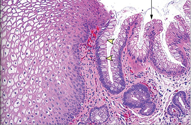
What is abnormal in this image
Esophagus showing change from stratified squamous into simple columnar with goblet cells (arrow) → Intestinal metaplasia
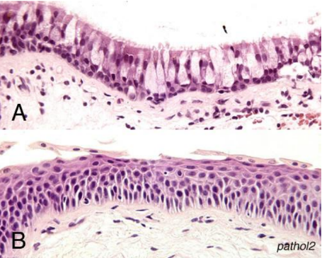
What pathologic change has occurred for A to change to B
Metaplasia
What are the causes of cell injury
Oxygen deprivation, physical agents, chemical agents, infectious agent, immunologic reaction, genetic abnormalities, nutritional imbalance
What are cellular responses to cell injury
Integrated stress response (ISR), unfolded protein response (ER stress), autophagy
What is integrated stress response
Stress → Activation of kinase enzymes → Phosphorylation of elF2a → Global protein translation stops + ATF4 activation → ATF4 lead to apoptosis/survival
What is ER stress
Protein folding demand exceed protein folding capacity of ER
What are the mechanisms of unfolded protein response
Reduce protein synthesis, increase chaperones, eliminate misfolded proteins
What is autophagy
Cell eats its own contents to recycle or clear debris
Steps of autophagy
Nutrient deprivation → Initiation → Elongation → Maturation of autophagosome → Fuse with lysosome → degradation
What are the signs of reversible cell injury
Generalized cell swelling, blebbing of plasma membrane, ribosome detachment from ER, chromatin clumping, fatty change
What is the difference between necrosis and apoptosis
Necrosis = accidental cell death; apoptosis = programmed cell death
What is the sequence of necrosis
Denaturation of cellular protein → Leakage of contents → Inflammation → Enzymatic digestion
What mechanisms lead to necrosis
Hypoxia/ischemia, ROS
What mechanisms lead to apoptosis
Radiation mutation, mutation, cell stress, infection
Process of necrosis caused by mitochondrial damage
O2 low → MT cannot make ATP → ATP dependent Na pump function decrease → Active transport system failure → cell swelling
How does membrane damage occur in necrosis
Low ATP from MT dysfunction → low phospholipid synthesis → Lipid bilayer and lysosome breakdown → Ca increase → Lipase/protease activation → Membrane breakdown
What are the signs of necrosis on histology
Eosinophilia and nuclear changes
Types of nuclear changes
Karyolysis, pyknosis, karyorrhexis
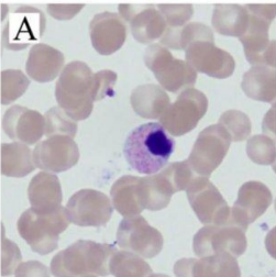
What does this show
Nuclear condensation → Pyknosis
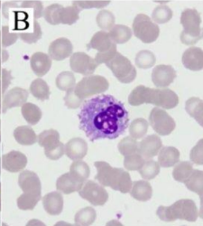
What does this show
Fading of chromatin basophilia → Karyolysis
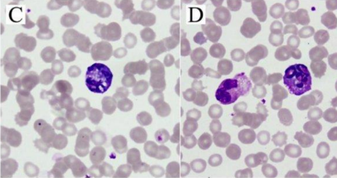
What does this show
Pyknotic nuclei become fragmented → Karyorrhexis
What are the types of necrosis
Coagulative, liquefactive, gangrenous, caseous, fat, fibrinoid
What is coagulative necrosis
Caused by ischemia; cells are dead but architecture is preserved
What is liquefactive necrosis
Tissue → Liquid; caused by microbes that stimulate leukocytes to accumulate and release enzymes to digest tissue
What is the difference between wet and dry gangrene
Dry = ischemia (coagulative necrosis); wet = ischemia + bacterial infection (liquefactive necrosis)
What is caseous necrosis
Cheesy debris; caused by TB infection
What is fat necrosis
Release of pancreatic lipase into peritoneum → Binds with Ca to form chalky white areas
What is fat saponification
FFA (from breakdown of TG into FFA and glycerol) + Ca → Soap
What is fibrinoid necrosis
Caused by immune reaction involving blood vessels
What are the steps of apoptosis
Cell shrinkage + chromatin condensation → Nuclear fragmentation and membrane blebbing → Apoptotic body formation
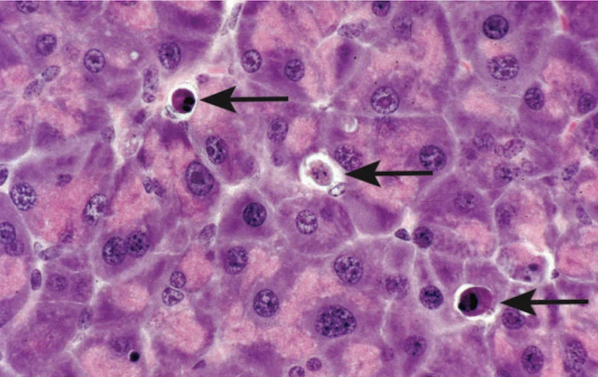
What do the arrows represent
Nuclear condensation and fragmentation → Sign of apoptosis
What are the physiologic and pathologic causes of apoptosis
Physiologic → Eliminate cells that aren’t needed
Pathologic → Eliminate cells beyond repair
Extrinsic apoptosis pathway
FasR and FasL binding → FADD + procaspase 8, 10 into DISC → Caspase 8 and 10 activate → Activation of caspase 3,6,7 → Apoptosis
Intrinsic apoptosis pathway
Stress → Activation of BH3 → BAX BAK release → BAX BAK make hole in MT inner membrane → release cytochrome C → APAF1 oligomerization → Apoptosome → Activate procaspase 9 → Caspase 3,6,7 activation
How is BAX BAK activated
Caspase 10 cleave BID → tBID → BAX BAK
Process of necroptosis
Extrinsic apoptosis → Inhibit caspase 8 → Necrosome activate → MLKL activate
Process of Pyroptosis
Inflammasome formation → Caspase 1 formation → IL-1B activation
Process of ferroptosis
Oxidative stress → Fe catalyze ROS → Lipid peroxidation → Membrane damage
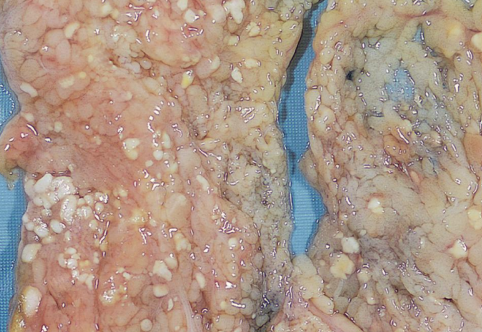
What type of necrosis is this
Fat necrosis
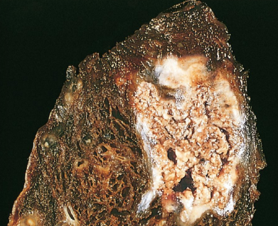
What type of necrosis is this
Caseous necrosis