Ch 6 Integumentary System
 Call Kai
 Learn
Learn Practice Test
 Spaced Repetition
 Match
Match Flashcards
 Knowt Play
1/67

There's no tags or description
Looks like no tags are added yet.
Name | Mastery | Learn | Test | Matching | Spaced | Call with Kai | Chat |
|---|
No analytics yet
Send a link to your students to track their progress
68 Terms
skin
largest organ in the body by weight
functions to protect from envrionment, intake vitamins, secretion, temperature regulation, sensory reception
integumentary system
the skin and it’s accessory structures (hair, nails, glands, sensory receptors)
2 layers: epithelial tissue (epidermis) overlying connective tissue (dermis)
epidermis
outer layer of the skin made of stratified squamous epithelial tissue
epithelial cells attached to basement membrane between epidermis and dermis
avascular - receives nutrients via diffusion from vessels in CT below to stratum basale
4-5 distinct layers: stratum basale, stratum spinosa, stratum granulosum, stratum lucidem (thick skin ONLY), stratum corneum
cells on the outer surface have protein keratin → waterproof
3 cell types: keratinocytes, melanocytes, dendritic cells
dermis
inner layer of skin made of densre irregular connective tissue (high in elastic fibers - stretchy)
thicker layer
where most of the accessories are rooted
vascular - gives nutrients to epidermis
hypodermis (subcutaneous layer)
located beneath the dermis
functions as the insulating layer → mostly adipose connective tissue!
not considered part of the skin, but is attached to dermis
cell cycle in epidermis
as cells grow, they are pushed toward the apical surface, away from nutrient supply (stratum basal) → apoptosis
→ the stratum basale produces 2 daughter cells: one moves through the layers over 25-45 days and dies at the surface, the other remains a stem cell to continue the cycle
→ as they migrate, older cells, keratinocytes, begin to flatten and die
→ as cells reach the outer surface, they become tightly packed, develop desmosomes, form outer layer: stratum corneum
thickest on palms and soles: lots of dead skin cells that are tightly packed and keratinized (waterproof)
3 cell types of epidermis
keratinocytes, melanocytes, dentritic cells
4-5 layers of epidermis
stratum basale, stratum spinosa, stratum granulosum, stratum lucidum (thick skin ONLY), stratum corneum
Stratum basale
deepest epidermal layer, up agains the dermis attached to the basement membrane
single row of STEM CELLS
actively mitotic
produces 2 daughter cells: one cell journeys to apical layer 25-45 days and then dies at the surface, one cell remains a stem cell
friction over time can accelerate cell production and keratin formation → calouses
stratum spinosum
several layers thick, second deepest epidermal layer
contains web-like system of intermediate pre-keratin filaments (“spindle-like”) attached to desmosomes that help to resist tension
abundant melansomoes and dendritic cells (immune cells!)
stratum granulosum
thin layer above stratum spinosum - 4-6 cells layers
cell appearance changes: cells flatten, nuclei and organelles disintegrate, keratinization begins, cells accumulate water-resistant glycolipids (slow-water loss)
cells above this layer begin to die
apoptosis
controlled cell suicide
changing of cells from stratum basale to stratum corneum
nucleus and organelles break down
plasma membrane thickens
allows cells to slough off as dandruff and dander
shed 50,000 cells every minutes
stratum lucidum
4th layer of epidermis, only found in thick skin: palms and soles
thin translucent band superficial to the stratum granulosum
a few rows of flat, dead keratinocytes
stratum corneum
20-30 rows of dead, flat, anucleate keratinized membranous sacs
though dead, its cells have a function - take the brunt of the harm so the living cells, tissues, and organs don’t
protect deeper cells from environment and water loss
protect from abrasion and penetration
barrier against biological, chemical, and physical assaults
keratinocytes
cells that produce fibrous protein keratin → waterproofing, especially outer layer
most abundant in epidermis
tightly connected by desmosomes
keratin
tough, fibrous, waterproof protein stored in the cells
melanocytes
cells in epidermis that produce dark pigment melanin which gives skin it’s specific color
absorbs UV light from sunlight and provides skin color via melanosomes (granules)
10-25% of cells in the deepest epidermis
located in stratum basale
distributed into keratinocytes to protect skin from damaging effects of UV light (DNA damage, fibroblast damage, skin cancer)

hereditary factors affecting skin color
same number of melanocytes in each person, but vary in the amount of melanin produced (genetically controlled)
varying distribution and size of melanin granules
albinos inherit mutation in melanin gene; lack melanin
envrionmental factors affecting skin color
sunlight, UV light from sunlamps, x-rays
physiological factors
oxygenation in blood of dermal blood vessels
vasodilation/vasoconstriction of dermal blood vessels
accumulation of carotene pigment from diet
jaudice
cyanosis
blueness of skin from deficiency of oxygen in the circulating blood
→ hemoglobin changes color when not attached to an oxygen molecule: turns from red to blue
can result from airway obstruction (drowning or choking)
lung diseases (emphysema or respiratory arrest)
cardiac arrest

jaundice
yellowing of skin and sclera due to excess of bilirubin (yellow pigment) in blood
cancer, hepatitis, cirrhosis, other compromised liver function
→ put babies in the sun

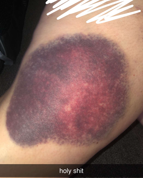
hematoma
aka bruising: mass of clotted blood showing through skin
pallor
pale or ashen color when there is so little blood flow through the skin that the white color of dermal collagen shows through
emotional stress, low BP, circulatory shock, color, anemia
→ constricted dermal blood vessels

erythema
abnormal redness of the skin due to dilated cutaneous vessels
exercise, hot weather, anger, or embarrassment

albinism
genetic lack of melanin that results in white hair, pale skin, and pink eyes

striae
silvery-white scars
“stretch marks”
extreme stretching causes dermal tears

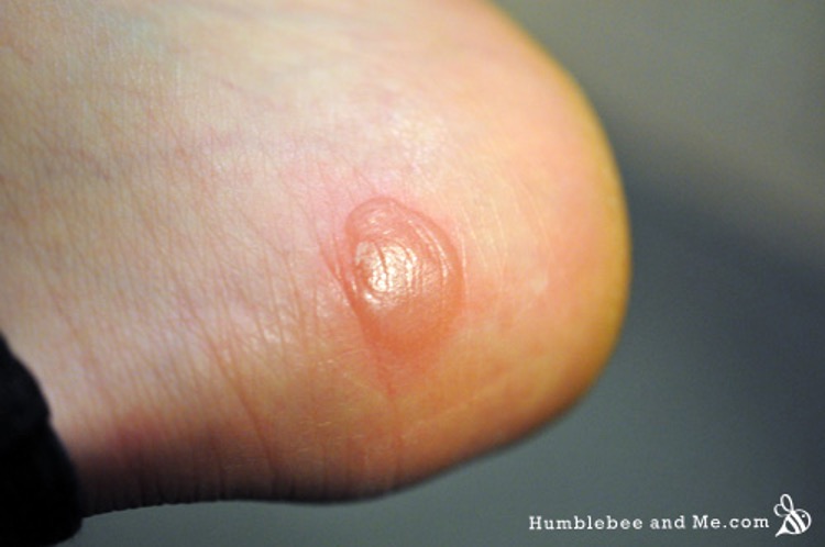
blister
from acute, short-term trauma
fluid filled pocket that separates epidermal and dermal layers
calluses
continued friction/pressure stimulates cells of the epidermis to become increasingly active
thickening process knows as hyperkeratinosis
dermis
second, largest layper of the skin
strong, flexible dense irregular connective tissue (made of elastic fibers)
cells: fibroblasts, macrophages, occasionally white blood cells (dendritic cells)
2 layers: papillary, reticular
contents: dermal blood vessels, smooth muscle tissue around hair follicles, nerve endings, skeletal muscle
cell types in dermis
fibroblasts, macrophages, whote blood cells (from blood vessels)
papillary layer
top layer of the dermis made of areolar CT, about 20% of dermis
collagen fibers and blood vessels
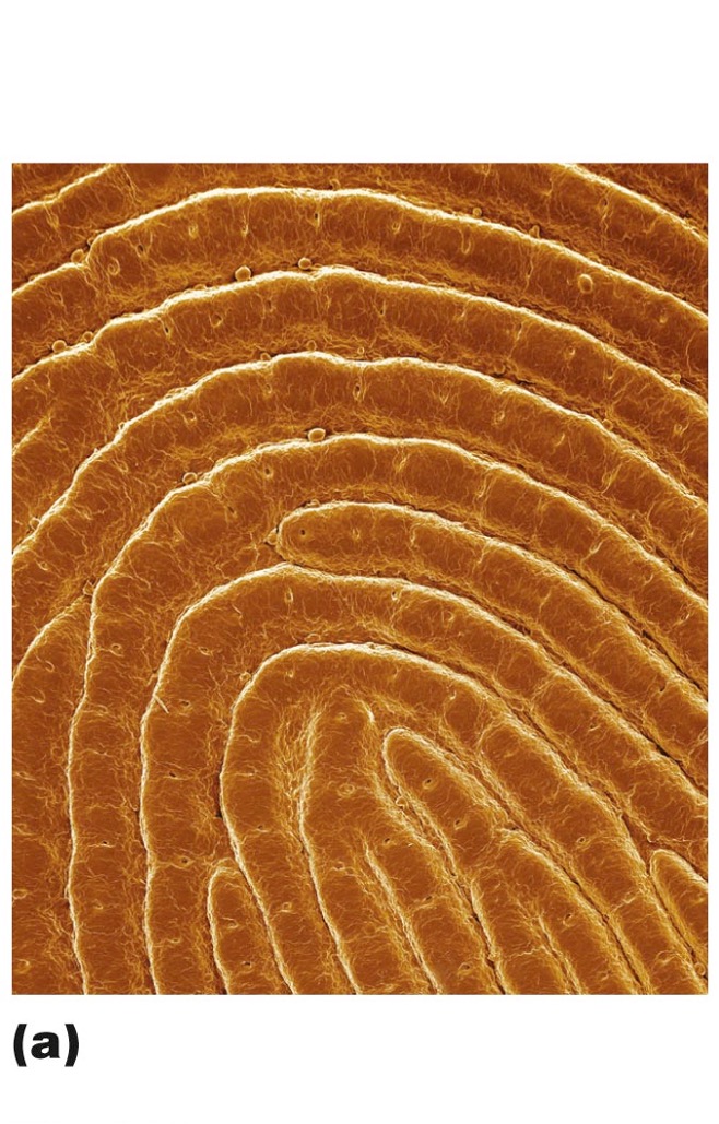
dermal papillae originate here → fingerprints
phagoctyes patrol for microorganisms
loose tissue
→ function: allow for diffusion of nutrients and oxygen from blood vessels of lower dermis to epidermis, also allows for white blood cells to climb to the epidermis surface to fight infection
dermal papillae
superficial peg-like projections from the papillary layer of the dermis
in thick skin → friction ridges in the epidermis → aka fingerprints!
function: enhance gripping ability, contribute to sense of touch
dermal blood vessels
provide nutrients and oxygen to overlying epidermis
diffuse through pappillary layer of areolar tissue
smooth muscle tissue of dermis
around hair follicles for movement in response to sensory perception and support
nerve endings in dermis
touch receptors: deep touch/pressue, light touch
free nerve endings: pain, temperature
skeletal muscle in dermis
anchors to the dermis only in the face muscles to form facial expressions!
accessory structures of the skin
hair follicles, nails, sensory receptors, skin glands (sweat and sebaceous)
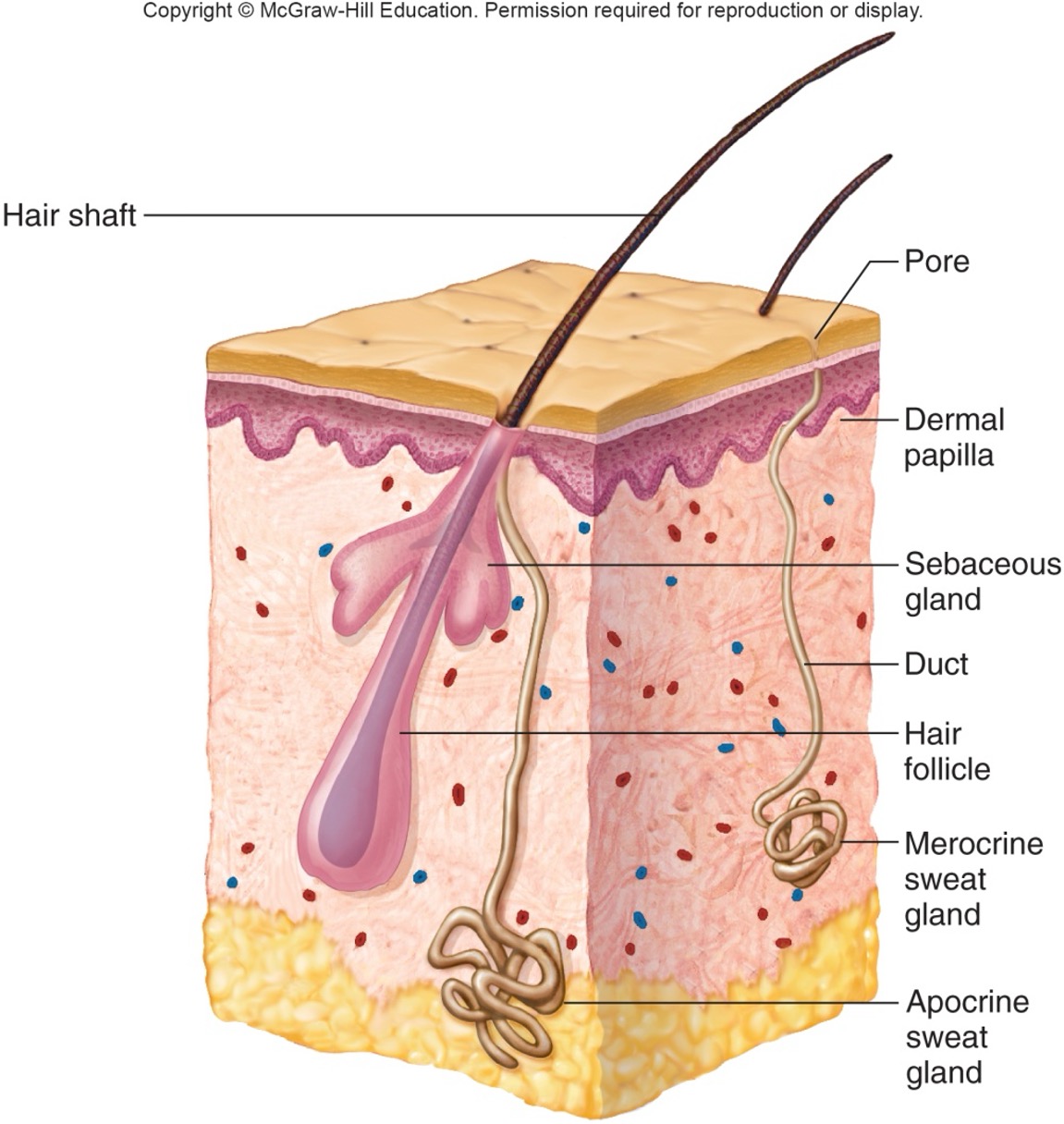
sebaceous glands
glands found in the dermis and a little in the hypodermis that produce sebum to keep the hair and skin soft and waterproof
usually found around hair follicles to be produced through the follicle at the epidermis
absent on palms and soles (for grip! also no hair)
excess sebum → acne

sweat gland
aka sudoriferous glands
widespread in the skin
originate in deeper dermis or hypodermis as ball-shaped coils
2 types: eccrine (merocrine glands) that produce sweat, apocrine glands in axillary and groin areas
modified sweat glands: ceruminous glands, mammary glands

eccrine (merocrine) glands
most numerous sweat gland that opens to a pore in the epidermis
responds to elevated body temperature by producing sweat to regulate and maintain homeostasis
abundant on forehead, neck, back
sweat
mostly water + salts and wastes secreted by sudoriferous glands (eccrine and apocrine)
sebum
fatty material produced in sebaceous glands that keeps hair and skin soft and waterproof
skin functions
overall: maintain homeostasis
protection (physical, chemical, biological)
barrier against harm
prevents some water loss
sensory reception
excretes wastes
produces vitamin D
regulate body temp
the skin as a physical barrier of protection
continuity of the skin!
flat, dead cells of stratum corneum surrounded by lipids → dead cells are hurt first
keratin and glycolipids block most water and water soluable substances
limited penetration of the skin: lipid soluable substances, plant oils, organic solvents, some drugs
what substances are allowed to penetrate the skin
lipid soluable substances
plant oils (poison ivy, etc)
organic solvents
some drugs: transdermal drug methods like nicotine patches, fentanyl patches, hormone patches
the skin as a chemical barrier of protection
skin secretions: low pH slows down bacterial multiplication, sebum and natural antibiotics kill bacteria
melanin: defense against UV radiation damage
how does the skin protect from harmful effects of UV radiation
melanocytes absorb the UV rays and distribute them into keratinocytes to protect skin cells from harsh and direct rays of UV light, which is a mutagen that can alter DNA, damage fibroblasts, and lead to skin cancer
dead skin cells of stratum corneum slough off and protect underlying new skin cells
the skin as a biological barrier of protection
dendritic cells of the epidermis and macrophages of dermis present foreign antigens to white blood cells
when body temperature rises
thermoreceptors signal hypothalamus
vasodilation of dermal blood vessels, vasoconstriction of deep blood vessels
sweat glands are activated
when body temperature falls
thermoreceptors signal hypothalamus
vasoconstriction of dermal blood vessels
sweat glands are inactive
muscles contract involuntarily (shivering)
methods of heat loss in the body
raditation, conduction, convection, evaporation
raditation
primary method of heat loss through the skin, infrared heat rays escape
conduction
heat moves from skin to cooler objects
convection
heat loss through the skin into circulating air currents
evaporation
sweat changes into gas, carries heat away
→ why we sweat when body temp is elevated!
hyperthermia
abnormally high body temperature
can occur on a hot, humid day, when sweat cannot evaporate
when air temperature is high, radiation is less effective (heat moves into body)
body may gain heat from hotter air
symptoms: skin becomes dry, person gets weak, dizzy, nauseous, with headache, rapid pulse
extreme vasodilation in attempt to regulate temperature can collapse cardiovascular system and can be fatal
hypothermia
abnormally low body temperature
result from prolonged exposure to cold or illness
shivering is involuntary skeletal muscle contraction, caused by hypothalamus
progessess to confusion, lethargy, loss of relfexes, and consciousness
without treatment, organs shut down → death
fever
temperature set point is purposefully elevated by the immune system to fight infection
phagocytes release pyrogens in response to presence of bacteria, viruses
hypothalamus increases set point and raises body temperature
elevated body temperature helps destroy pathogens
how: construct dermal blood vessels to reduce heat loss through the skin, slows down sweating, causes shivering (to produce more heat)
hypothermia is intentionally induced when
performing surgeries to prevent organ damage from decreased oxygen supply
cutaneous sensation
skin’s role in the nervous system
receptors in skin respond to external stimuli
touch receptors and deep pressure
metabolic function
skin produces vitamin D precursor from absorbed UV light
excretion
skin excretes some nitrogen containing wastes via urea
skin cancer
risk factors like excessive exposure to UV lights and chemicals can overwhelm the natural protective responses against skin cancer
3 types: basal cell carcinoma (common, least malignant), squamous cell carcinoma (spreads rapidly), and melanoma (least common, most deadly)
basal cell carcinoma
least malignant skin cancer type,
most common, 80% of skin cancers
most often in face
slow growing
squamous cell carcinoma
skin cancer that arises from keratinocytes of stratum spinosum
arise most often on head
grows rapidly, will spread if not caught
melanoma
skin cancer that arises from melanocytes
highly likely to metastasize
appears where there is pigment
1/3 come from preexisting moles
ABCD rule

life-span changes in skin
cell cycle slows → scaly skin, age spots
epidermis and dermis become thinner
loss of fat → cold
wrinkling, sagging
less sebum → dry skin
melanin production slows → white hair
hair thins, less follicles
impaired nail growth
sensory receptors decline
less effective body temp regulation
dimished vitamin D ability