anatomy exam 1
 Call Kai
 Learn
Learn Practice Test
 Spaced Repetition
 Match
Match Flashcards
 Knowt Play
1/131

There's no tags or description
Looks like no tags are added yet.
Name | Mastery | Learn | Test | Matching | Spaced | Call with Kai | Chat |
|---|
No analytics yet
Send a link to your students to track their progress
132 Terms
anatomy
study of the structure of body parts and their relationship to one another
physiology
study of the function of body parts
gross (macroscopic) anatomy
study of body structures visible to naked eye
regional anatomy
looks at structures in a particular body area
gross anatomy
system anatomy
looks at 1 system (i.e. nervous system)
gross anatomy
surface anatomy
looks at internal structures as they relate to overlying skin (i.e. visible muscle masses)
gross anatomy
microscopic anatomy
structures too small to be seen with naked eye
cytology
study of cells
histology
study of tissues
developmental anatomy
structural changes throughout life span
embryology
developmental changes that occur before birth
what skills you need to study anatomy
anatomical terminology
observe
manipulate
palpate (feel w/ hands)
auscultate (listen w/ stethoscope)
principle of complementarity of structure and function
anatomy and physiology are inseperable
function reflects structure
levels of structural organization (chemical, cellular, tissue, organ, organ system, organismal)
chemical(smallest): atoms and molecules
cellular: cells and organelles
tissue: groups of similar cells
organ: 2+ tissue types
organ system: organs that work together
organismal(largest): all organ systems, entire organism
necessary life functions
maintaining boundaries between internal/external environments
movement of body parts (muscle cells can contract), of cells, and of substances
responsiveness/excitability to sense changes and respond to environment
digestion to break down food
metabolism: all chemical reactions that occur in cells (anabolism: making molecules)(catabolism: breaking down molecules)(making ATP)
excretion of waste
reproduction of cells and offspring
growth
survival needs
nutrients (carbs, proteins, fats, minerals and vitamins)
oxygen for metabolic reactions
water
normal body temp
appropriate atmospheric pressure
homeostasis
maintenance of internal conditions despite changing environment
dynamic process (constantly monitored and regulated by nervous and endocrine systems)
homeostatic controls
receptor (sensor): responds to stimuli and sends info/input to control center along AFFERENT PATHWAY
control center: receives input, determines set point and appropriate response, sends info/output to effector along EFFERENT PATHWAY
effector: recieves output, responds with either negative or positive feedback

negative and positive feedback
negative: more common, reduces/shuts off original stimulus
positive: exaggerates original stimulus
feedforward (anticipatory) response
occurs in anticipation of a change to internal environment
*think Pavlovs dogs
homeostatic imbalance
disturbance of homeostasis
serosa/serous membrane
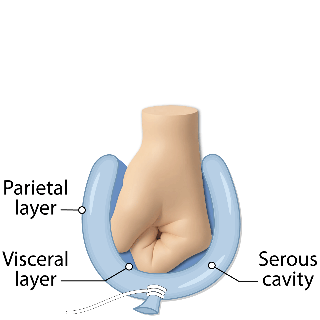
thin, double-layered membranes in ventral body cavity
parietal seosa: lines cavity walls
visceral serosa: covers organs in cavity
serous fluid: secreted by both parietal and visceral serosa, cavity between serosas filled with fluid
named for the specific cavity and associated organs (parietal/visceral pleurae and pleural fluid in lungs)(parietal/visceral peritoneum and peritoneal fluid in abdominopelvic) cavity)(parietal/visceral pericardium and pericardial fluid in heart area)
oral/digestive cavity
mouth
has teeth and tongue
continuous with digestive organs cavity
open cavity (exposed to environment)
nasal cavity
in nose
part of respiratory system
open cavity
orbital cavities
house eyes
open cavities
middle ear cavities
in skull
has bones that transmit sound vibrations for hearing
open cavities
synovial cavities
secretes synovial fluid to lubricate joints
closed cavities (not exposed to environment)
4 tissue types
epithelial (skin)(protects)
connective (connects tissues)
muscle (contracts)(skeletal, cardiac, smooth)
nervous (internal communication)
steps to view tissue samples under microscope
fixed: preserved w/solvent
sectioned: cut into thin slices that light electrons can be transmitted through
stained: to enhance contrast (colored syes in light microscopy)(heavy metal salts in electron miscoscopy)
epithelial tissue/epithelium
covers body surface/lines body cavities
2 main forms:
skin, lining or urogenital/digestive/respiratory systems
forms glands of the body
functions: protection, absorption, filtration, excretion, secretion, sensory reception
epithelial tissue characteristics
polarity (apical surface (top, most smooth, some have microvilli) and basal surface (bottom, attaches to basal lamina)
specialized contacts: tight junctions and desmosomes bind adjacent epithelial cells to form sheets
supported by connective tissue (reticular lamina and basement membrane)(reinforces and defines boundary)
avascular but innervated (no blood vessels, supplied by nerve fibers)
regeneration: high regeneration capacities due to high damage
classification of epithelial tissue
cell layers: simple (1 layer of cells)(absorbtion, secretion, filtration), stratified (2+ cell layers)(durable, protection)
cell shapes: squamous (squished), cuboidal ( box like), columnar (tall, column like)
stratified is named based on the top layer cell shape
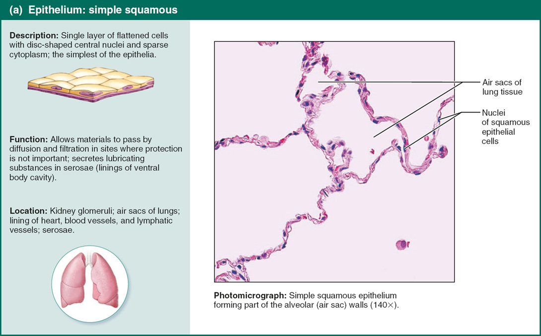
simple squamous
where rapid diffusion occurs (alveoli in lungs coir CO2 and O2 exchange)

simple cuboidal
in kidneys
cilliated

simple columnar
have goblet cells in between
in digestive tract
have cilia to move things along
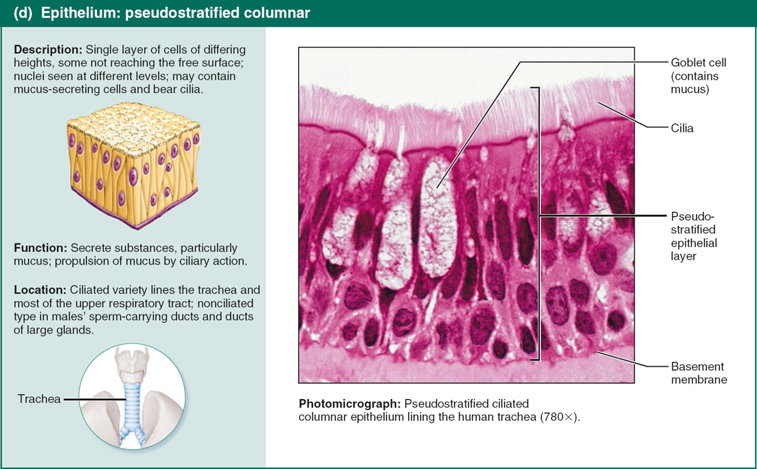
pseudostratified columnar
cells very in height and appear to be stratified
secretion/movement of musuc with cilia
in respiratory system

stratified squamous
most widespread
where high wear and tear occurs
in mouth and skin
stratified cuboidal
rare
usually 2 cell layers
sweat and mammary glands
stratified columnar
rare
at transition areas between 2 epithelia types

transitional epithelium
can transition into something bigger/smaller
lining of bladder, uterus, urethra so it can expand
resembles all 3 cell types
gland
1+ cells that make/secrete a secretion (fluid)
gland classification
site of product release (endocrine=ductless gland that produces hormones, secrere hormones into fluid)(exocrine=secrete products into body surfaces)
number of cells making gland: (unicellular (i.e. goblet cells), produce mucin to form mucus for lubrication)(multicellular), have ducts

multicellular exocrine glands
a duct and secretory unit
surrounded by connective tissue
classified by:
structure: simple (unbranched ducts) or compound (branched ducts) and tubular, alveolar, or tubuloalveolar
mode of secretion: merocrine (secreted by exocytosis, most common) or holocrine (accumulates products until rupture)
*know simple tubular/branched tubular and compound tubloalveolar

connective tissue
most abundant tissue type
bindibng and support, protection, insulation, storing reserve fuel, transporting substances
4 main classes of connective tissue
connective tissue proper
cartilage
bone
blood
common characteristics of connective tissue
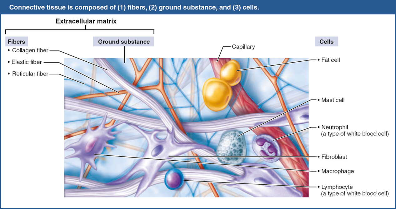
cells are suspended in extracellular matrix
cells arise from mesenchyme (embryonic tissue)
elements that all connective tissues have
ground substance (gel-like material that fills space between cells)(has interstitial/extra fluid, cell adhesion proteins/”glue”, and proteoglycans (large protein sugars))
fibers (provide support)
cells
connective tissue fibers (collagen, elastic, reticular)
fibers provide support
collagen fibers: strongest, most common, tough
elastic fibers: allow for stretch/recoil
reticular fibers: thin, branching fibers that offer more ;give”
connective tissue cells (blast vs cyte)
“-blast” cells are immature that secrete ground substance and fibers
“-cyte” cells are mature that maintain matrix health
EX: fibroblasts/cytes in connective tissue proper, chondroblasts/cytes in cartilage, osteoblasts/cytes in bone
structural elements of connective tissue (adipocytes, leukocytes, mast cells, macrophages)
adipocytes: store energy as fat, adipo=fat
leukocytes: white blood cells, respond to injury, leuko=white
mast cells: detect foreign material and initiate inflammatory response
macrophages: breakdown material
connective tissue proper: loose connective tissue
areolar, adipose, reticular

areolar loose connective tissue
areolar think air (loose)
universal packing material (think house insulation)
supports/binds other tissues
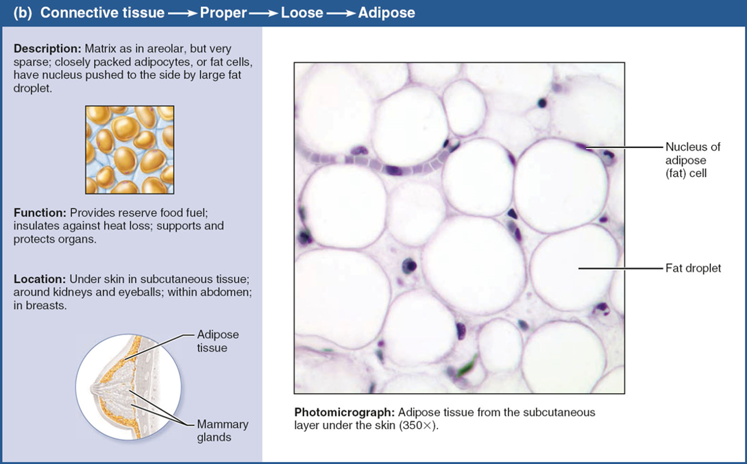
adipose loose connective tissue
looks like oil (fat) in water
nutrient storage
white fat (adipocyte cells)
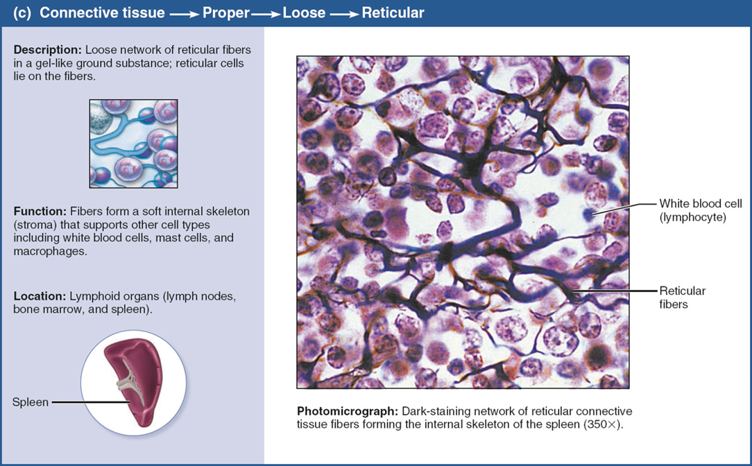
reticular loose connective tissue
looks like a tree
like fibers
around the spleen
forms stroma that supports blood cells
connective tissue proper: dense connective tissue
regular
irregular
elastic

dense regular
consistent structure
can withstand high tension/stretching
parallel, wavy collagen fibers
poorly vascularized
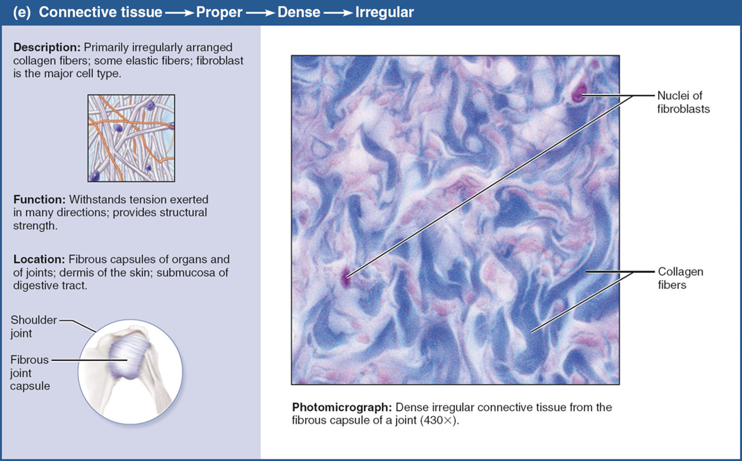
dense irregular
irregularly arranged
in dermis (skin) so skin can move in all directions

dense elastic
like accordion
ligaments
can stretch
connective tissue: cartilage
matrix secreted from chondroblasts/cytes
80% water
tough and fleexible
avascular
hyaline, elastic, and fibrocartilage

hyaline cartilage
most abundant
looks glassy
tough
at tips of long bones
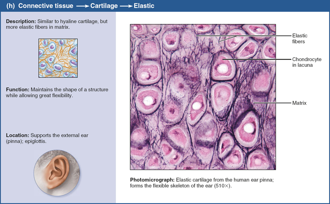
elastic cartilage\
in ears

fibrocartilage
very strong
in vertebreal disks and knee

connective tissue: bone/osseus tissue
supports/protects body structyres
osteoblasts produce matric
osteocytes maintain matrix
vascularized

connective tissue: blood
fluid, consists of blood cells surrounded by plasma (fluid matrix)
red blood cells, white blood cells, platlets
transports
muscle tissue
highly vascularized
allows for most movement (myofilaments bring about contraction)

skeletal muscle
AKA voluntary muscle b/c consciously controlled
cells are muscle fibers (multiple nuclei and banded)
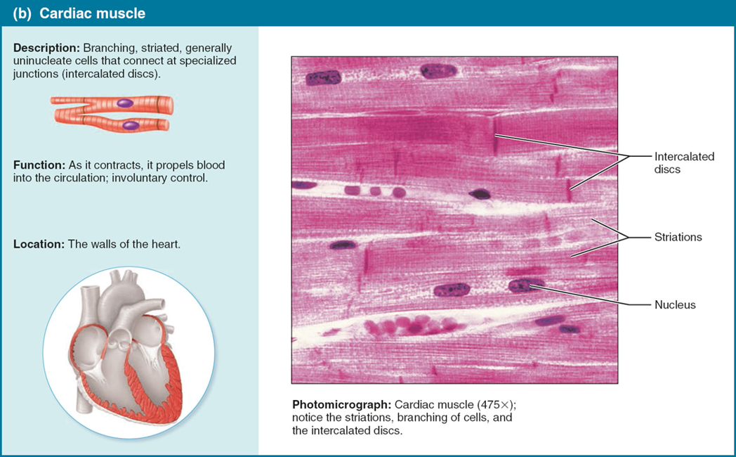
cardiac muscle
only in walls of heart
involuntary
banded cells with 1 nucleus
intercalated discs: joints where branched cardiac muscle cells are joined

smooth muscle
in walls of hollow organs (NOT HEART)
involuntary
not banded
spindle shaped w/ 1 nucleus
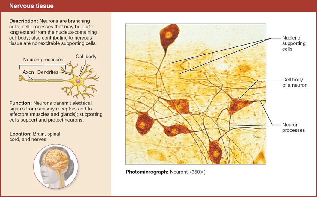
nervous tissue
regulates/controls body functions
made up of 2 specialized cells:
neurons: respond to stimuli via dendrites and trasmit signals along axons
glial cells support, insulate, protect neurons
membranes
cover and line
3 types: cutaneous, mucous, serous
cutaneous membrane
AKA skin
keratinized(tough protein) stratified squamous attached to thick connective tissue
dry membrane
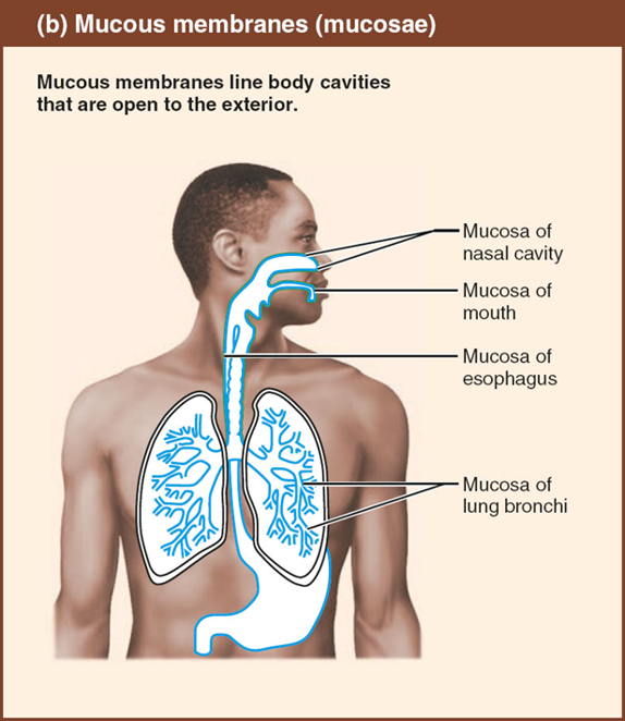
mucous membranes/mucosae
lines body cavities o
open to exterior
moist
many mucosae secrete mucus

serous membranes/serosae
line closed ventral body cavities
moist
simple squamous epithelium on areolar connective tissue
parietal (lines internal body cavities)/visceral (lines internal organs)
integumentary system
skin
sudiferous (sweat) glands
sabaceous (oil) glands
hairs
nails
subcutaneous tissue
epidermis
outermost protective shield of the body
made of epithelial tissue (keratinized stratified squamous epithelium)
avascular
karatinocytes
a cell of the epidermis
produce keratin (protein that allows skin to protect)
major epidermis cells
melanocytes
cells of the epidermis
produce melanin pigment thats packaged into melanosomes (spider shaped cells)
dendritic cells
cells of the epidermis
insert foreign substances and key activators of immune system
dendritic means branching
tactile epithermal cells
cells of the epidermis
sensory receptors for touch
layers of the epidermis
CLGSB
come lets go sun bathe
corneum, lucidum, granulosum, spinosum, basale
thick skin has all 5 layers
thin skin has 4 layers (no lucidum)
stratum corneum
most superficial
¾ of the epidermal
protects, prevents water loss
stratum lucidum
only in thick skin
clear layer (lucid=clear)
stratum granulosum
flat cells
keratinization begins (keratin fibers form)
stratum spinosum
pre-keratin filaments allows them to resist tension
stratum basale
deepest layer
attached to dermis
single layer
where mitosis occurs
the dermis
under epidermis
makes up most of skin
strong, flexible, dense connective tissue
vascular
fibroblasts, macrophages, sometimes mast and white blood cells
has nerves, blood vessels, lymphatic vessels, hair follicles, oil glands, sweat glands
2 layers: papillary and reticular
papillary dermis
thin, areolar connective tissue w/ loose collagen and elastic fibers and blood vessels
phagocytes (macrophages) patrol
dermal papillae )sends fingerlike projections into epidermis)
fruction ridges: formed by dermal papillae on top of dermal ridges to enhance grippability
reticular dermis
80% of dermal
dense irregular connective tissue
dermal vascular plexus: network of blood vessels between reticular layer and subcutaneous tissue
cleavage (tension) lines: collagen fibers running parallel to skin surface (where surgeons make inscisions)
flexure lines: dermal folds near joints
striae: white scars from extreme straching of skin (stratch marks)
blisters: fluid-filled pockets that separate epidermal and dermal layers from traumas
subcutaneous tissue/hypodermis
covers muscle
mostly adipose tissue w/ areolar connective tissue
what gives skin its color
melanin
carotene
hemoglobin
melanin
only pigment made in skin made from malanocytes
shield DNA from UV sunlight
more sun = more malanin produced
2 forms: reddish yellow to brownish black
cyanosis
blue skin
low oxygen
red/purple/yellow/green marks
bruises from blood leakage from damaged blood vessel
yellowness/jaundice
liver disorder
pallor/blanching
paler skin from emotional stress, low blood pressure, or anemia
redness of skin
embarassment, fever, inflammation, allergy
from hemoglobin
brown/black patches
from melanin
hyperpigmented, thickened skin folds
carotene
think carrot
yellow to orange pigment
in palms and soles
hemoglobin
pinkish hue of fair skin due to decreased melanin levels
hair/pili
flexible strands of dead, keratinized cells produced by hair follicles
hard keratin