CH3 Histology
 Call Kai
 Learn
Learn Practice Test
 Spaced Repetition
 Match
Match Flashcards
 Knowt Play
1/97

There's no tags or description
Looks like no tags are added yet.
Name | Mastery | Learn | Test | Matching | Spaced | Call with Kai | Chat |
|---|
No analytics yet
Send a link to your students to track their progress
98 Terms
Embryology
Formation of Tissues
Zygote
Single cell produced after fertilization
contains 46 chromosomes
Cleavage
Cell divisions during this stage produce a hollow ball of cells, takes about a week to complete
Blastocyst
Hollow ball of cells
Trophoblast
outer layer of the blastocyst that will form the placenta
Ectoderm
cells that will form the epidermis of skin and epidermal derivatives
nervous tissue and sense organs
pituitary gland
adrenal medulla
enamel of teeth
Lens of eye
Mesoderm
cells that will form dermis of skin and epithelial lining of blood vessels, lymph vessels, and serous membranes
muscle tissue
connective tissue
adrenal cortex
heart, kidneys, ureters, internal reproductive organs, spleen
Endoderm
cells that will form the epithelial lining of respiratory tract, GI tract, tympanic cavity, auditory tube, urinary tract, reproductive tract
liver (most of)
gallbladder
pancreas
palatine tonsils (portion of)
thyroid gland
parathyroid glands
thymus
Epithelial tissue
covers exposed surfaces
lines internal passageways and chambers
produces glandular secretions
Connective tissue
fills internal spaces
provides structural support
stores energy
Muscular tissue
contracts to produce movement
includes skeletal muscle, cardiac muscle, and smooth muscle
Neural tissue
conducts electrical impulses
carries information
Epithelial, connective, muscular, neural
Four primary tissue types
Epithelial tissue
No intercellular space, tightly packed cells
Avascularity
no blood vessels
Sheets and layers
sheets of cells
single or multiple layers thick
regeneration of surface cells via stem cells
polarity - structural and functional differences between top and bottom of cells
exposed apical surgace
have an attached basal surface
apical/basal surfaces are structurally and functionally different
Functions of epithelial tissue
provides physical protection
from abrasion, dehydration, and destruction
controls permeability
sensory cells
general (touch)
neuroepithelia (sense organs)
produces secretions
unicellular glands
glandular epithelia
Apical surface structures
Microvilli, stereocilia, cilia
Microvilli
Short, can move;
increases surface area
line urinary and digestive track
Stereocilia
long, rigid
inner ear and male reproductive tract
Cilia
long, can move
move substances over apical surface
line respiratory tract
Intercellular connections
Give epithelium strength and stability
holds cells together
prevents passage of chemicals, water, pathogens
Gap junctions, CAMs, tight junctions, hemidesmosome, and proteoglycans
also bind cells to basal lamina
Epithelial renewal
must be replaced frequently due to exposure/use
disruptive enzymes
toxic chemicals
pathogens
mechanical abrasion
replaced through time via continual division of stem cells near basal lamina
Simple and stratified
Two types of layering
Simple epithelium
one layer of cells
protected internal compartments of body
Stratified epithelium
two or more layers of cells
areas of mechanical or chemical stresses
Squamous epithelium
relatively flat, “pancakes”

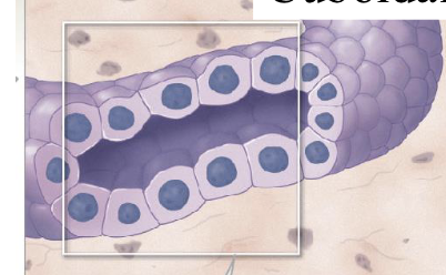
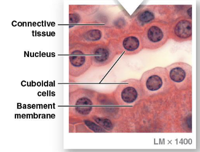
Cuboidal epithelium
shaped like cubes
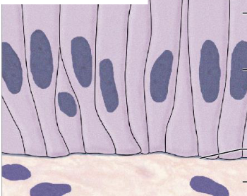
Columnar epithelium
longer than wide; columns
Simple squamous epithelium
Thin delicate layer
reduces friction, controls vessel permeability, performs absorption and secretion
lining body cavities
lining heart and blood vessels
alveoli

Stratified squamous epithelium
physical barrier, some surfaces keratinized
mucosal linings are nonkeratinized
lining of orfices
surface of skin

Simple cuboidal epithelium
secretion, absorption
thyroid gland
renal tubules
ducts
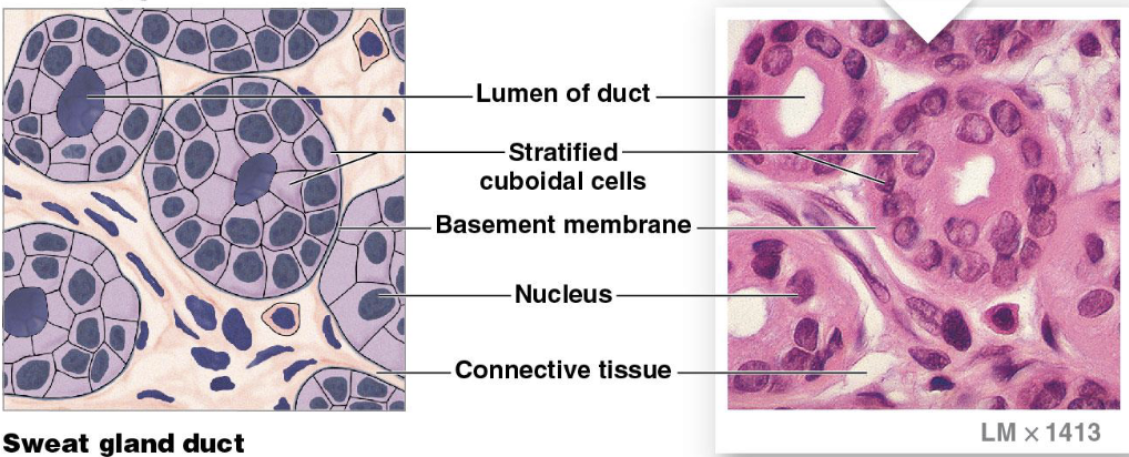
Stratified cuboidal epithelium
secretion, absorption
ducts of sweat glands
ducts of mammary glands
Lumen: center of duct/tube
Simple columnar epithelium
highly polarized
high secretion, high absorption
stomach, intestine, gallbladder, uterine tubes, renal collecting ducts
most have microvilli/cilia

Stratified columnar epithelium
high activity areas
protection
pharynx, epiglottis, anus, mammary glands, salivary glands, and urethra

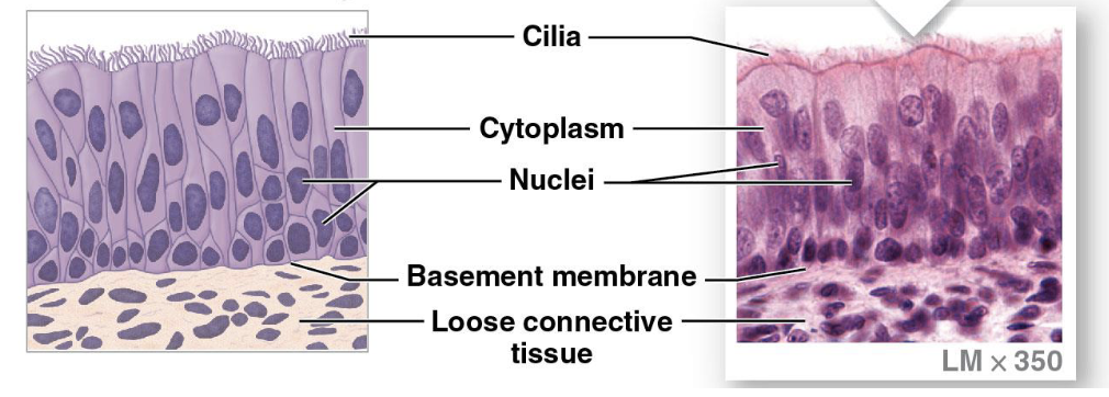
Pseudostratified ciliated columnar epithelium
protection, secretion
have cilia, actually a simple epithelium
appears as stratified but is actually simple
nasal cavity, trachea, bronchi, male reproductive tract
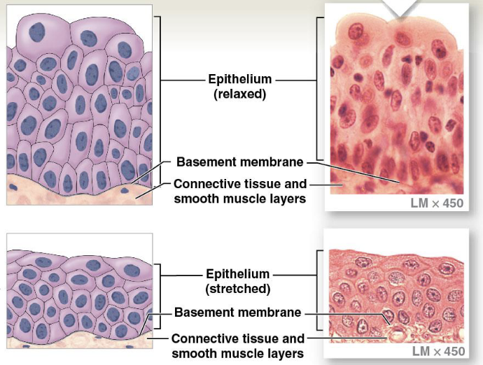
Transitional epithelium
“stretchy”, expansion, recoil
mainly cuboid, but mix of others
renal pelvis, ureters, bladder
Glandular epithelium
Classified based on: type of secretion released, structure of gland, mechanism of secretion
Exocrine and endocrine
Types of glands
Exocrine glands
ducts to an epithelial surface, body opening, or space
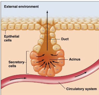
Endocrine glands
into ECF then to blood stream or lymph, hormones

Serous, mucous, mixed
Types of exocrine glands
Unicellular
Mucins
goblet cells (mucous cell)
isolated from other glandular cells
Multicellular
secretory sheets and clusters, mixed
Gland cells
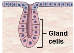
Duct cells

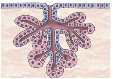
Compound tubular gland
Ex. mucous glands in oral cavity

Compound alveolar gland
Ex. mammary glands

Compound tubuloalveolar gland
Eccrine secretion
secretory product is packaged into secretory vesicles
exocytosis
can be continuous
fastest secretion rate
ex. salivary glands
Apocrine secretion
product released during shedding of apical portion of cell
non-continuous
production in waves
ex. mammary glands
Holocrine secretion
lysis of entire cell
typically oily products
slowest production rate
ex. sebaceous gland cells
Embryonic connective tissue
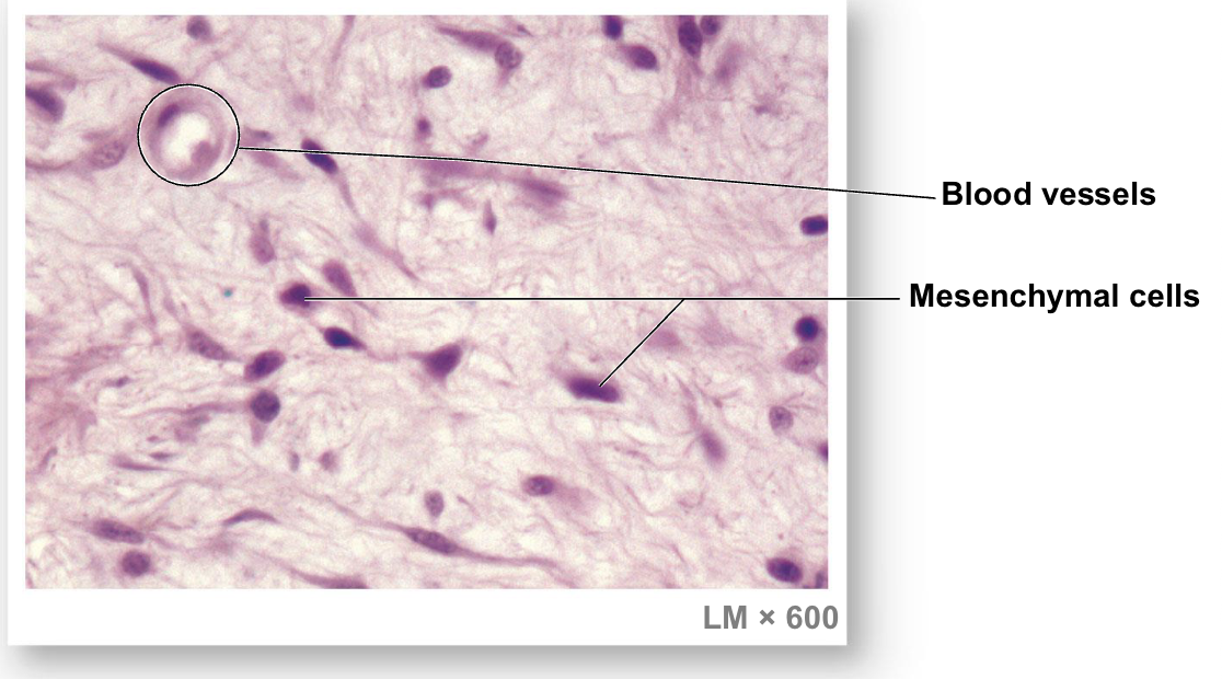
Connective tissue
Specialized cells, extracellular protein fibers, ground substance
establishing body’s structural framework
transport fluid and dissolved materials
protect organs
support, surround, and connect other tissues
store energy
defend body from mic0ororganisms

Matrix
extracellular component of protein fibers and “ground substance” (fluid portion)
Matrix of bone is calcified
connective tissue cells embedded within it
collagen, reticular, and elastic fibers, ground substance
Fibers of connective tissue
Collagen fibers
high tensile strength, tendons and ligaments
Reticular fibers
structural mesh within organs
Elastic fibers
protein called elastin: “rubbery”, lines vasculature
Ground substance
Thick syrupy consistency, mix of proteins and carbohydrates
Connective tissue proper
matrix of loose fibers and dense fibers
Fluid connective tissue
watery matrix of dissolved proteins
blood and lymph
Supporting connective tissue
dense matrix of fibers and may have insoluble calcium salts
cartilage and bone
Loose connective tissue proper
Fibers create loose, open framework
areolar tissue
adipose tissue
reticular tissue
Dense connective tissue proper
fibers are densely packed
dense regular
dense irregular
elastic
Blood
contained in cardiovascular system
Lymph
contained in lymphatic system
Cartilage
solid rubbery matrix
hyaline cartilage
elastic cartilage
fibrous cartilage
Bone
solid, crystalline matrix
Areolar tissue
“Stuffing” of body
light connection that allows movement between skin and muscle
cushions organs
within deep dermis of skin

Adipose tissue
padding
white fat: energy storage
brown fat: thermogenesis
Deep to the skin

Reticular tissue
“structural sponge”
matrix is stiffest of loose CT
provides supporting framework
liver, kidney, spleen, lymph nodes, bone marrow

Dense regular connective tissue
ligaments
tendons
aponeuroses
high tensile strength
provides firm attachment, conducts pull of muscle, reduces friction, stabilizes relative positions of bones
between skeletal muscles and skeleton, between bones or stabilizing positions of internal organs, covering skeletal muscles

Dense irregular connective tissue
organ sheaths
provides strength to resist forces applied from many directions, prevents over-expansion
capsules of viceral orangs, periostea and perichondria, nerve and muscle sheaths, dermis

Elastic connective tissue
“rubbery”
springy and resilient
stabilizes positions of vertebrae and penis, cushions shocks, permits expansion and contraction of organs
Between vertebrae of spinal column, ligaments supporting penis, ligaments supporting transitional epithelia, in blood vessel walls

FCT matrix
Matrix of watery mixture of dissolved proteins
blood and lymph
Blood
erythrocytes (red blood cells): gas transport
leukocytes (white blood cells): immune function
thrombocytes (platelets): blood clotting
plasma: matrix
Lymph
monitors for signs of infection, mainly lymphocytes (T and B cells), but some phagocytic cells
Cartilage
Contains chondrocytes
gel matrix made of chondroitin sulfate
cells reside in lacunae
avascular
have a fibrous perichondrium
cellular layer: maintenance
fibrous layer: connection to other structures
most cartilage cannot repair severe damage
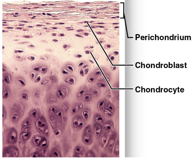
Hyaline cartilage
tracheal rings
costal cartilage
joints
provides stiff but somewhat flexible support

Elastic cartilage
most flexible cartilage
provides support but tolerates distortion without damage and returns to original shape
auricle of external ear, epiglottis, auditory canal, cuneiform cartilages of larynx

Fibrous cartilage
firmest, strongest cartilage
resists compression, prevents bone-to-bone contact, limits relative movement
pads within knee joint, between pubic bones of pelvis, intervertebral discs

Periosteum
External surface of bone
Endosteum
Internal surface of bone
Lamellae
mix of collagen fibers and calcium salts in bone
Membrane
Epithelia + connective tissue
sheet of epithelial cells
and underlying connective tissue
mucous
serous
cutaneous
synovial

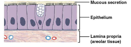
Mucous membrane
coated with secretions of mucous glands, line most of digestive and respiratory tracts and portions of urinary and reproductive tract
Serous membrane
Line the peritoneal, pleural, and pericardial body cavities

Cutaneous membrane
covers skin/outer surface of the body

Synovial membrane
line joint cavities and produce the fluid within the joint

Sarcoplasm
Muscle tissue cytoplasm
Sarcolemma
Muscle tissue cell
Skeletal muscle tissue
cells are long, cylindrical, striated, and multinucleate
voluntary or reflex arcs
combined with connective tissues and neural tissue in skeletal muscles
moves or stabilizes the position of the skeleton
hypertrophy not hyperplasia via myosatellites

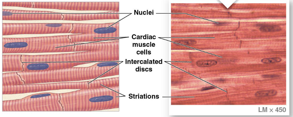
Cardiac muscle tissue
cells are short, branched, and striated, usually with a single nucleus, interconnected by intercalated discs
myogenic
branched: “mesh”
scar tissue
location: heart
circulates blood, maintains blood pressure
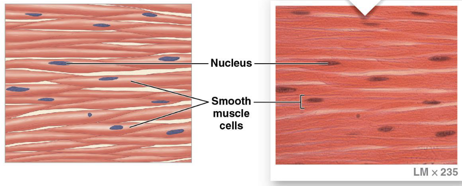
Smooth muscle tissue
cells are short, spindle-shaped, and nonstriated, with a single central nucleus
can replicate
reflex arcs are myogenic
found in walls of blood vessels and in digestive, respiratory, urinary, and reproductive organs
moves food, urine, and reproductive tract secretions
involuntary
Neuroglia and neurons
Neural cell types
Neuroglia
supporting cells of neural tissue, can divide
Neuron
specialized to conduct electrical signals (action potential)
longest cells in body
incapable of cell reproduction

Dendrite
receives stimuli on neuron
Axon
transmits a signal (action potential) down neuron