Unit 1 Biochemistry
1/13
There's no tags or description
Looks like no tags are added yet.
Name | Mastery | Learn | Test | Matching | Spaced |
|---|
No study sessions yet.
14 Terms
Role and structure of carbohydrates- Multiple Mark
Monosaccharides: Single sugar units (glucose and fructose). Energy sources. Composed of carbon, hydrogen, and oxygen in a 1:2:1 ratio
Disaccharides: Formed by the joining of two monosaccharides through a glycosidic bond in a dehydration reaction. Examples: sucrose (glucose + fructose), lactose (glucose + galactose), and maltose (glucose + Glucose).
Polysaccharides: Large polymers like starch (plants), glycogen (animals), and cellulose (plant cell walls). Used for energy storage and structural purposes.
4 Different polysaccharides: Storage: Starch, glycogen Structural: Cellulose, Chitin
Role and structure of proteins- Multiple Mark
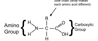
Composed of amino acids linked by peptide bonds. Amino acids: Have amino, carboxyl, and R-groups
Structure levels:
Primary: Amino acid sequence.
Secondary: Alpha helices and beta sheets. Coils and folds in the polypeptides. Held together by hydrogen bonds
Tertiary: 3D folding due to R-group interactions.
Quaternary: Multiple polypeptide chains assembled.
Functions: Enzymes, hormones, antibodies, and structural support.
Different amino acids have different properties
Can be polar (hydrophilic)
Can be non-polar (hydrophobic)
Charged (acidic-basic)
Role and structure of lipids (5 types)- Multiple Mark
1) Fatty Acids
Role: Building blocks of other lipids like triglycerides and phospholipids
Structure: Single hydrocarbon chain with a carboxyl functional group at one end
2) Triglycerides (Fats)
Most common type
Role: Eenergy storage in animals, providing insulation
Structure: 1 Glycerol + 3 Fatty acids
3) Phospholipids
Role: Major component of cell membranes, forming the lipid bilayer that separates the cell's interior from its external environment.
Structure: 1 Glycerol + 2 Fatty acids + 1 Phosphate. Attached to glycerol backbone
4) Steroids
Role: Signaling molecules (testosterone and estrogen) and components of cell membranes (cholesterol)
Structure: Four carbon rings
5) Waxes
Role: Provide protective coatings on leaves, feathers, and skin, preventing water loss. Insoluble, non-polar, soft solids over a wide range of temps.
Structure: Long chain fatty acids linked to alcohol or carbon rings
Biological macromolecules, examples, and functional groups- Multiple Mark
1) Proteins
Monomer: Amino acid
Example: Enzymes (catalyze reactions), structural proteins (collagen), transport proteins (like hemoglobin), antibodies (for defense)
Functional Groups: Amino (-NH2), carboxyl (-COOH), and R groups (variable side chains)
2) Lipids
Monomer: Fatty acids and glycerol
Example: Fatty Acids, Triglycerides, oils, waxes, steroids
Functional Group: Carboxyl (-COOH), ester (R-COO-R'), and hydroxyl (-OH) groups.
3) Carbohydrates
Monomer: Monosaccharides (glucose)
Example: Sugars, starches, cellulose
Functional groups: Hydroxyl (-OH) and carbonyl (C=O) groups
4) Nucleic Acids
Monomer: Nucleotides
Examples: DNA and RNA
Functional groups: Phosphate (-PO4), nitrogenous bases (containing amino groups), and hydroxyl (-OH) groups
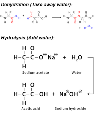
Condensation/Dehydration and Hydrolysis Reactions- 1 Mark
In many reactions, water is produced or added
Enzymes and Activation Energy- 1 Mark
Enzymes are biological catalysts.
Ea: Amount of energy needed to strain and break the reactant bonds
Factors affecting the rate of enzyme activity- Next 3 slides- Multiple Mark
How do changes in substrate concentration affect the rate of enzyme activity?
An increase in substrate concentration will increase the rate of enzyme activity because there is more substrate available to bind to the enzyme and produce product. Rate will increase to a point where there is no more enzyme available. The rate of reaction will increase with increased substrate concentration until the enzyme reaches its saturation level.
Dependent Variable
Rate of reaction (1/5)
Independant Variable
Substrate (Hydrogen peroxide) concentration
Control
Volume and concertation of enzyme (catalyze) temp. (room temp.)
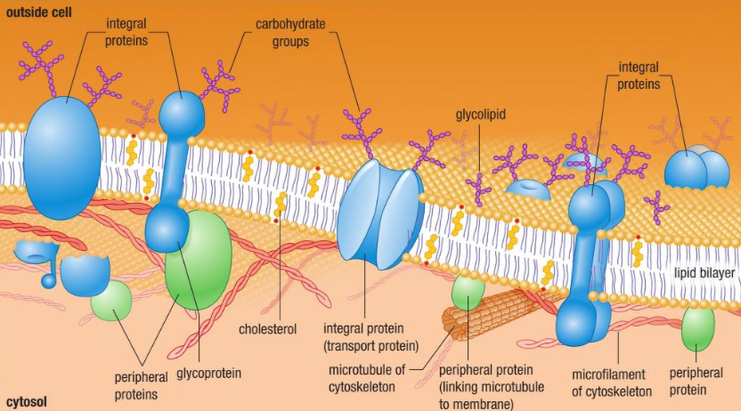
Fluid Mosaic Model- 1 Mark
Describes the structure of cell membranes
A model that refers to how the lipid bilayer tends to act more like a liquid than a solid and contains a number of different components
Transport (Diffusion & Osmosis)- 1 Mark
Diffusion is the movement of molecules from a region of high concentration to low concentration.
Osmosis is the movement of water molecules through a semi-permeable membrane from a region of high water concentration to low water concentration.
Cell membrane structure-type of molecules and function- Multiple Mark
Phospholipids: Basic structure (lipid bilayer), hydrophilic head, and hydrophobic tails allows formation of the bilayer
Integral proteins: Embedded in the lipid bilayer, span the entire lipid bilayer
Peripheral proteins: Loosely attached to the membrane surface
Functions: Transport, signaling, enzymatic activity, cell adhesion
Cholesterol: 2 different functions, at diff temps
Cold: Occupies spaces between lipids, prevents bilayer from becoming too vicious
Hot: Reduces fluidity of membrane
Carbohydrates: Attached to proteins or lipids on the outer surface. Important for cell recognition, cell signaling, and cell adhesion
Cell Membrane Diagram