Functions of the Brain PSYC C1000
1/17
Earn XP
Description and Tags
Name | Mastery | Learn | Test | Matching | Spaced |
|---|
No study sessions yet.
18 Terms
lesion
tissue destruction. A brain lesion is a naturally or experimentally caused destruction of brain tissue.
A new generation of scientists selectively lesion(destroy) tiny clusters of normal or defective brain cells, observing any effect on brain function.
EEG
(electroencephalogram)
an amplified recording of waves of electrical activity sweeping across the brain’s surface. These waves are measured by electrodes placed on the scalp.
Right now, your mental activity is emitting telltale electrical, metabolic, and magnetic signals that would enable neuroscientists to observe your brain at work. Electrical activity in your brain’s billion of neurons sweeps in regular waves across its surface.
MEG
(magnetoencephalogram)
a brain-imaging technique that measures magnetic fields from the brain’s natural electrical energy.
To isolate the brain’s magnetic fields, researches create special rooms that cancel out other magnetic fields. Participants sit underneath a head coil that resembles a hair salon hairdryer. While participants complete activities, tens of thousand of neurons create electrical impulses. The speed and strength of magnetic fields enable researches to understand how certain tasks influence brain activity.
PET
(positron emission tomography) a brain-imaging technique for detecting brain activity that displays where a radioactive form of glucose goes while the brain is performing a given task.
PET depicts brain activity by showing each brain area’s consumption of its chemical fuel, sugar glucose… the PET scan can track the gamma rays released by this “food for thought” as a task is performed.
MRI
(magnetic resonance imaging)
a technique that uses magnetic fields and radio waves to produce computer-generated images of soft tissue. MRI scans show brain anatomy.
In MRI brain scans, the persons’ head is put in a strong magnetic field, which aligns the spinning atoms of brain molecules. Then, a radio-wave pulse momentarily disorients the atoms. When the atoms return to their normal spin, they provide a detailed picture of soft tissues, including the brain.
fMRI
(functional MRI)
a technique for revealing blood flow and, therefore, brain activity by comparing successive MRI scans. fMRI scans show brain function as well as structure.
By comparing successive MRI scans, researchers can watch as specific brain areas activate, showing increased oxygen-laden blood flow.
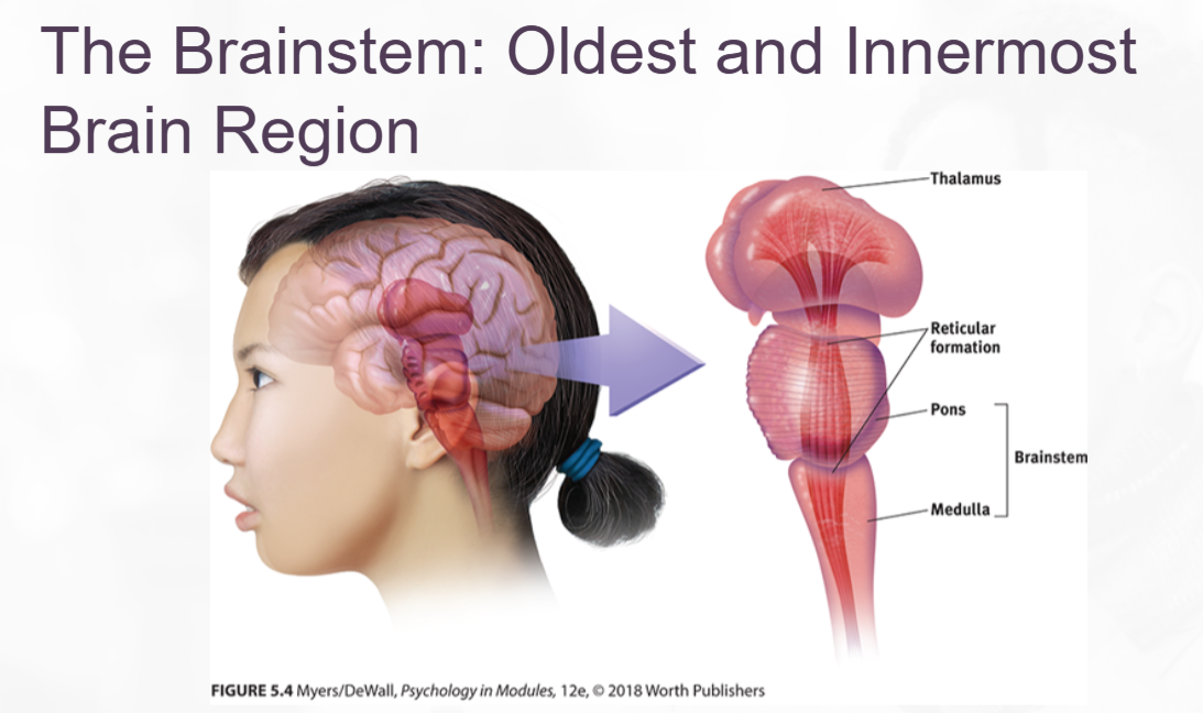
brainstem
the oldest part and central core of the brain, beginning where the spinal cord swells as it enters the skull; the brainstem is responsible for automatic survival functions.
The brainstem is the brain’s oldest and innermost region.
A crossover point, where most nerves to and from each side of the brain connect with the body’s opposite side.
medulla
the base of the brainstem; controls heartbeat and breathing
At it’s base is the medulla, the slight swelling in the spinal cord just after it enters the skull.
pons
part of the brainstem.
Just above the medulla sits the pons, which helps coordinate movements and control sleep.
thalamus
the brain’s sensory control center, located on top the brainstem; it direct messages to the sensory receiving areas in the cortex and transmits replies to cerebellum and medulla.
Sitting atop the brainstem is the thalamus, a pair of egg-shaped structures that acts a s the brain’s sensory control center.
Structures of the Brainstem
reticular formation
a nerve network that travels through the brainstem into the thalamus; filters information and plays an important role in arousal
Inside the brainstem, between your ears, lies the reticular(“netlike") formation, a nerve network extending from the spinal cord to the thalamus.
Electrically stimulating a cat’s reticular formation without damaging nearby sensory pathways instantly produced an alert, awake animal. When Magoun severed a cat’s reticulum formation without damaging nearby sensory pathways, the effect was equally dramatic: The cat lapsed into a coma from which it never wakened.
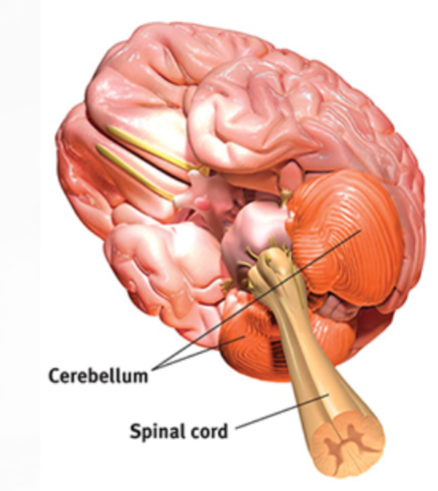
cerebellum
the “little brain” at the rear of the brainstem; functions include processing sensory input, coordinating movement output and balance, and enabling nonverbal learning and memory.
limbic system
neural system (including the amygdala, hypothalamus, and the hippocampus) located below the cerebral hemispheres; associated with emotions and drives.
Between the oldest and the newest brain areas lies the limbic system(limbus means “border”)
amygdala
two lima-bean-sized neural clusters in the limbic system; linked to emotion
…enables aggression and fear… surgically removed a rhesus monkey’s amygdala, turning the normally ill-tempered animal into the most mellow of creatures. [Electrically stimulate an amygdala] in one spot and the cat prepares to attack… move the electrode only slightly within the amygdala, cage the cat with a small mouse, and now it cowers in terror.
hypothalamus
a neural structure in the limbic system laying below(hypo) the thalamus; it directs several maintenance activities(eating, drinking, body temperature) helps govern the endocrine system via the pituitary gland, and is linked to emotion and reward.
Note the interplay between the nervous and endocrine systems: the brain influences the endocrine system, which in turn influences the brain.
Olds and Milner realized they had stumbled upon a brain center that provides pleasurable rewards… Just how rewarding are these reward centers? Enough to cause rats to self-stimulate these brain regions more than 1000 times per hour.
hippocampus
a neural center located in the limbic system; helps process explicit(conscious) memories—-of facts and events— for storage.
Humans who lose their hippocampus due to surgery or injury lose their ability to to form new memories of facts and events.
Cerebellum placement