BIO 168 Ch. 9 & 10 exam
1/98
There's no tags or description
Looks like no tags are added yet.
Name | Mastery | Learn | Test | Matching | Spaced | Call with Kai |
|---|
No analytics yet
Send a link to your students to track their progress
99 Terms
Muscle tissue
They make up nearly half of body’s mass
Can transform chemical energy (ATP) into directed mechanical energy, which is capable of exerting force
Prefixes for muscle
Myo-, mys-, and sarco-
Ex: sarcoplasmm - muscle cell cytoplasm
3 types of muscle tissue
skeletal
smooth
cardiac
Where are muscle fibers found?
Elongated muscle cells found in skeletal and smooth muscle, but not cardiac
4 Important functions of skeletal muscle in the body
Movement
Posture + body position
Stabilize joints
Generate heat
Skeletal muscle: connective tissue sheaths
Each skeletal muscle, as well as each muscle fiber, is covered in connective tissue
Support cells and reinforce whole muscle
Epimysium
Most external
Dense irregular connective tissue surrounding entire muscle; may blend with fascia
Perimysium
Intermediate
fibrous connective tissue surrounding fascicles (groups of muscle fibers)
Endomysium
Most internal
fine aerolar connective tissue surrounding each muscle fiber
Skeletal muscle attachments
Direct (fleshy) attachments: epimysium fused to periosteum of bone or perichondrium of cartilage
Indirect attachments: connective tissue wrappings extend beyond muscle: tendons and aponeurosis
Fascicles
A discrete bundle of muscle cells, segregated from the rest of the muscle by a connective tissue sheath
Surrounded by perimysium
Muscle fiber (cell)
An elongated multinucleate cell; it has a straited (banded) appearance
Surrounded by endomysium
Contains glycosomes for glycogen storage and myoglobin for O2 storage
Specialized structures within sk muscle cells: myofibrils, SR, T-tubules
Sarcolemma
Muscle fiber plasma membrane
Sarcoplasm
Muscle fiber cytoplasm
Myofibrils
Densely packed, rod-like elements. A single muscle fiber can contain 1000s. Accounts for ~80% muscle cell volume
Features: striations, sarcomeres, myofilaments, molecular comp of myofilaments
Myofibril striations
Stripes formed from repeating series of dark and light bands along length of each myofibril
A band - darker region
H zone - lighter region in middle of dark A band
M Line - line of protein (myomesin) that bisects H zone vertically
I band - lighter regions
Z disc - coin shaped sheet of proteins on midline of I band
Sarcomere
Smallest contractile unit (functional unit) of muscle fiber
Contains A band with half of an I band at each end. Consists of area between z-discs
Individual sarcomeres align end to end along myofibril
Actin myofilaments
Thin filaments
Extend across I band and partway in A band
Anchored to z discs
Have active sites for myosin head attachment during contraction
Two F actin strands twist together to form a thin filament
Myosin filaments
Thick filaments
Extend length of A band
Connected at M line
Heavy chains intertwine to form tail
Light chains form myosin globular head
Tropomyosin and troponin
Regulatory proteins bound to actin
Cover and expose myosin binding sites on actin
Troponin binds to calcium ions to remove tropomyosin from actin
Titin
Elastic myofilament
Holds thick filaments in place; helps recoil after stretch; resists excessive stretching
Dystrophin
Links thin filaments to proteins of sarcolemma
Nebulin, myomesin, and C proteins
Bind filaments or sarcomeres together
maintain alignment of sarcomere
Sarcoplasmic Reticulum (SR)
Network of smooth ER tubules surrounding each myofibril
Most run longitudinally
Terminal cisterns from perpendicular cross channels at A-I band junction
SR functions in regulation of intracellular Ca2+ levels
Stores and releases Ca2+
T-tubules
Tube formed by protrusion of sarcolemma deep into cell interior
Increase muscle fibers surface area greatly
Lumen continuous with extracellular space
Allow electrical nerve transmissions to reach deep into interior of each muscle fiber
Tubules penetrate cells interior at each A-I band junction between terminal cisterns
Triad
Area formed from terminal cistern of one sarcomere, T-tubule, and terminal cistern of neighboring sarcomere
When an electrical impulse passes by, T tubule proteins change shape, causing SR proteins to change shape, causing release of calcium into cytoplasm
Sliding filament model of contraction
The activation of cross bridges to generate force
Shortening occurs when tension generated by cross bridges on thin filaments exceeds forces opposing shortening
Contraction ends when cross bridges
Cross bridge attachments form and break several times, each time pulling thin filaments a little closer toward center of sarcomere in a racheting action
Causes shortening of muscle fiber with: I bands shorten, Z discs become closer, H zones disappear, A bands move closer to each other
T or F: During contraction, sarcomeres shorten
True
T or F: During contraction, A bands shorten
False
T or F: During contraction, I bands shorten
True
T or F: During contraction, the distance from one Z disc to the next Z disc decreases
True
T or F: During contraction, the M line disappears during a maximal contraction
False
T or F: During contraction, the H zone disappears during maximal contraction
True
T or F: During contraction, the Thin filaments slide past the thick filaments
True
T or F: During contraction, the thick and thin filaments overlap each other more
True
T or F: During contraction, the thick and thin filaments both shorten
False
How do motor neurons stimulate skeletal muscle fibers to contract?
Decision to move is activated by the brain, signal is transmitted down spinal cord to motor neurons which then activate muscle fibers
Neurons and muscle cells are excitable cells capable of action potentials (APs). They are capabale of changing resting membrane potential voltages
AP crosses from neuron to muscle cell via the neurotransmitter acetylcholine (ACh)
Ion channels
Play major role in changing of membrane potentials
Two classes:
Chemically gated: opened by chemical messengers such as neurotransmitters. Ex: ACh receptors on muscle cells
Voltage-gated: open or close in response to voltage changes in membrane potential
Anatomy of motor neurons and NMJ
SK muscles are stimulated by somatic motor neurons
Axons (long extensions of motor neurons) travel from CNS to sk muscle
Each axon divides into many branches as it enters muscle
Axon branches end on muscle fiber, forming NMJ or motor end plate. Each muscle fiber has one NMJ with one motor neuron
Axon terminal (end of axon) and muscle fiber are separated by gel-filled space called synaptic cleft
Stored within axon terminal are membrane bound synaptic vesicles that contain ACh
Infolding of sarcolemma called junctional folds contain millions of ACh receptors
NMJ consists of axon terminals, synaptic cleft, and junctional folds
4 big steps that must occur for skeletal muscle to contract
Events at NMJ
Muscle fiber excitation
Excitation-contraction coupling
Cross bridge cycling
6 Events of the muscular junction
AP arrives at axon terminal of the motor neuron
Calcium ions enter the axon terminal through voltage-gated channels
Influx of calcium ions causes the release of ACh from the axon terminal
ACh diffuses across the synaptic cleft from the axon terminal to the sarcolemma
Chemically gated channels open, ions pass into and out of the muscle fiber creating a graded potential
ACh is broken down by acetylcholinesterase
Generation of an AP across the sarcolemma
Resting sarcolemma is polarized, meaning a voltage exists across membrane
Inside of cell is negative compared to outside
AP is caused by changes in electrical charges
Generation of end plate potential
Depolarization
Repolarization
Generation of an AP across the sarcolemma: 1. End plate potential
ACh released from motor neuron binds to ACh receptors on sarcolemma
Causes chemically gated ion channels (ligands) on sarcolemma to open
Na+ diffuses into muscle fiber. Some K+ diffuses outward but not much.
Because Na+ diffuses in, interior of sarcolemma becomes less negative (more positive)
Results in local depolarization called EPP
Generation of an AP across the sarcolemma: 2. Depolarization
Generation and propagation of an AP
If EPP causes enough change in membrane voltage to reach critical level called threshold, voltage-gated Na+ channels in membrane will open
Large influx of Na+ through channels into cell triggers AP that is unstoppable and will lead to muscle fiber contraction
AP spreads across sarcolemma from one voltage-gated Na+ channel to next one in adjacent areas, causing that area to depolarize
Generation of an AP across the sarcolemma: 3. Repolarization
Restoration of resting conditions
Na+ voltage-gated channels close, and voltage gated K+ channels open
K+ efflux out of cell rapidly brings cell back to initial resting membrane voltage
Refractory period: muscle fiber cannot be stimulated for a specific amount of time, until repolarization is complete
Ionic conditions of resting state are restored by Na+-K+ pump
Na+ that came into the cell is pumped back out, and K+ that flowed outside is pumped back into cell
Excitation-Contraction (E-C) Coupling
Events that transmit AP along sarcolemma (excitation) are coupled to sliding of myofilaments (contraction)
AP is propagated along sarcolemma and down into T tubules, where voltage sensitive proteins in tubules stimulate Ca2+ release from SR. This leads to contraction
AP is brief and ends before contraction is seen
Cross bridge cycling
Cross bridge formation requires Ca2+
At low intracellular Ca2+ concentration: tropomyosin blocks active sites on actin so that myosin heads cannot attach to it. Muscle fiber remains relaxed. In response, voltage-sensitive proteins in T tubules change shape, causing SR to release Ca2+ to cytosol
At higher intracellular Ca2+ concentrations, Ca2+ binds to troponin. Troponin changes shape and moves tropomyosin away from myosin-binding sites. Myosin heads is then allowed to bind to action, forming cross bridges. Cycling is initiated, causing sarcomere shortening and muscle contraction
When nervous stimulation ceases, Ca2+ is pumped back into SR, and contraction ends
4 Steps of Cross Bridge Cycling
Cross bridge formation
The power stroke
Cross bridge detachment
Cocking of myosin head
4 Steps of Cross Bridge Cycling: 1. Cross bridge formation
Energized myosin head attaches to actin myofilament, forming a crossbridge
4 Steps of Cross Bridge Cycling: 2. Power stroke
Myosin head pivots and bends, pulling actin toward M-line, leaving myosin head in low energy state. ADP and P are released
4 Steps of Cross Bridge Cycling: 3. Cross bridge detachment
ATP binds to myosin, causing it to detach from actin. Cross bridge breaks
4 Steps of Cross Bridge Cycling: 4. Cocking of myosin head
Myosin hydrolyzes ATP to ADP & P. This causes the myosin head to return to its prestroke high energy position.
Whole skeletal muscle contraction
Same principles apply to contraction of both single fibers and whole muscles
Contraction produces muscle tension, the force exerted by a contracting muscle on an object
Load is the opposing force to muscle contraction, the force exerted on the muscle by the weight of the object to be moved
Force and duration of muscle contractions vary in response to stimuli of different frequencies and intensities
The motor unit
Each muscle is served by at least one motor nerve.
Motor nerve contains axons of up to hundreds of motor neurons
Axons branch into terminals, each of which forms NMJ with single muscle fiber
Motor unit consists of the motor neuron and all muscle fibers (four to several hundred) it supplies. Smaller the fiber number, the greater the fine control
Muscle fibers from a motor unit are spread throughout the whole muscle, so stimulation of a single motor unit causes only weak contraction of entire muscle
Muscle Twitch
Simplest contraction resulting from a muscle fiber’s response to a single action potential from motor neuron
Muscle fiber contracts quickly, then relaxes
Twitch can be observed and recorded as a myogram. Tracing: line recording contraction activity
3 Phases: latent period, Period of contraction, Period of relaxation
3 phases of muscle twitch: 1. Latent period
Events of excitation-contraction coupling
No muscle tension seen
Events of excitation-contraction coupling are occurring
3 phases of muscle twitch: 2. Period of contraction
Cross bridge formation
Tension increases
Cross bridge cycling is occurring, from the onset of tension development to the peak of tension
3 phases of muscle twitch: 3. Period of relaxation
Ca2+ reentry into SR
Tension declines to zero
Muscle contracts faster than it relaxes
Number of cross bridges declines as calcium ions are actively transported into the SR
Muscle twitch differences
Differences in strength and duration of twitches are due to variations in metabolic properties and enzymes between muscles
Ex: eye muscles contraction are rapid and brief, whereas larger, fleshy muscles (calf muscles) contract more slowly and hold it longer
Muscle Tone
Constant, slightly contracted state of all muscles
Due to spinal reflexes. Groups of motor units are alternately activated in response to input from stretch receptors in muscles
Keeps muscles firm, healthy, and ready to respond
Isometric contraction
No shortening; muscle tension increases but does not exceed load
Load is not moved and muscles does not shorten or lengthen
Isotonic contraction
Muscle changes in length and moves load. They can be two types:
Concentric contractions: muscle shortens and does work. Ex: biceps contract to pick up book
Eccentric contractions: muscle lengthens and generates force. Ex: laying a book down causes biceps to lengthen while generating a force
Providing energy for muscle contraction
ATP supplies the energy needed for the muscle fiber to:
Move and detach cross bridges
Pump calcium back into SR
Pump Na+ out of and K+ back into cell after excitation-contraction coupling
Available stores of ATP depleted in 4-6 seconds
ATP is the only source of energy for contractile activities; therefore, it must be regenerated quickly
ATP is regenerated quickly by three mechanisms…
Direct phosphorylation of ADP by creatine phosphate (CP)
Anaerobic pathway: glycolysis and lactate formation
Aerobic pathway: glycolysis and aerobic respiration in mitochondria
Direct phosphorylation of ADP by creatine phosphate (CP)
Creatine phosphate is a unique molecule located in muscle fibers that donates a phosphate to ADP to instantly form ATP
Creatine Kinase is an enzyme that carries out transfer of phosphate
Muscle fibers have enough ATP and CP reserves ti power cell for about 15 secs
Creatine phosphate + ADP → creatine +ATP
No oxygen use and 1 creatine per ATP
Anaerobic pathway: glycolysis and lactate formation
ATP can also be generated by breaking down and using energy stored in glucose
Glycolysis: first step in glucose breakdown. Does not require oxygen, glucose is broken into 2 pyruvate molecules, 2 ATPs are generated for each glucose broken down.
Low oxygen levels prevent pyruvate from entering aerobic respiration phase.
Normally, it should enter mitochondria but there is no oxygen available. Bulging muscles impair oxygen delivery.
Anaerobic glycolysis and pyruvate is referred to as lactate. Lactate is then diffused into the blood stream to be used as fuel in the liver, kidneys, and heart
Anaerobic respiration doesn’t produce as much ATP as aerobic respiration, but produces ATP way faster
Aerobic pathway: glycolysis and aerobic respiration in mitochondria
Produces 95% of ATP during rest and light to moderate exercise. Slower than anaerobic pathway
Consists of a series of chemical reactions that occur in mitochondria and require oxygen. Breaks glucose into CO2, H2O, and large amount of ATP (32)
Fuels used include glucose from glycogen stored in muscle fiber, then bloodborne glucose, and free fatty acids. Fatty acids are main fuel after 30 minutes of exercise
Energy systems used during exercise
Aerobic edurance: length of time muscle contracts using aerobic pathways. Light to moderate activity which can continue for hours
Anaerobic threshold: point at which muscle metabolism converts to anaerobic pathway
Muscle fatigue
Inability to maintain muscle tension
Ionic imbalances (K+, Ca2+, Na+) disrupt membrane potential of muscle cell
Increased inorganic phosphate from CP and ATP breakdown interferes with calcium release in SR
Decreased ATP
Increased Magnesium (interferes with T tubule proteins
Decreased glycogen: lack of ATP is rarely a reason for fatigue, except in severely stressed muscles
Excess Postexercise Oxygen Consumption (EPOC)
Referred to as oxygen debt. For a muscle to return to its pre-exercise state, you require extra oxygen:
Oxygen reserves are replenished
Lactate is reconverted to pyruvate
Glycogen stores are replaced
ATP and creatine phosphate reserves are resynthesized
How is muscle fiber classified?
Two characteristics
Speed of contraction: slow or fast. According to speed at which ATPases split ATP. Pattern of electrical activity of motor neurons
Metabolic pathways used for ATP synthesis: Oxidative fibers (aerobic), Glycolytic fibers (anaerobic)
Can be classified as Slow oxidative fibers, fast oxidative fibers, or fast glycolytic fibers. Most muscles contain a mix of fiber types, resulting in a range of contractile speed and fatigue resistance.
All fibers in one motor unit are the same type
Genetics dictate individual’s percentage of each
Slow oxidative fibers
Red Slow Ox 🐮 Low intensity, edurance activites like maintaining posture or running marathon
High myoglobin, mitochondria, capillaries, red color
Aerobic ATP synthesis
Slow speed of contraction, myosin ATPase acitivity, rate of fatigue
Low glycogen stores
Fast oxidative glycolytic
FOG; Medium intensity activities like sprinting or walking
High mitochondria, capillaries, myoglobin, pink color
Aerobic pathway (some anaerobic glycolysis)
Intermediate fatigue, glycogen stores
Fast contraction, myosin ATPase activity
Fast Glycolytic fibers
Too Fast White Sugar; Short term intense or powerful movements like hitting a baseball (bicep or eye muscles)
Fast speed of contraction, myosin ATPase activity, fatigue (most fatigueable)
Few/low capillaries, mitochondria, myoglobin content, white in color
Anaerobic glycolysis
High glycogen stores
Load and recruitment: Load
Muscles contract fastest when no load is added
The greater the load, the shorter the duration of contraction and the slower the contraction
Load and recruitment: Recruitment
The more motor units contracting, the faster and more prolonged the contraction
Smooth muscle
Found in walls of most hollow organs like respiratory, digestive, urinary, reproductive, circulatory (except in smallest of blood vessels) except heart
packed into sheets of tightly packed fibers
Logitudinal layer: run parallel to axis; contraction causes organ to shorten
Circular layer: run around circumference; contraction causes lumen of organ to constrict
Mix and squeeze substances of hollow organs
Spindle shaped with only one nucleus and no striations
Only has endomysium
contains varicosities instead of NMJ. controlled by ANS
Contraction of smooth muscle
They have less elaborate SR and no T tubules. Calcium instead comes from extracellular origins. Sarcolemma contains caveolae pouches that allow calcium influx
Electrically connected by gap junctions
Few thick filaments but a lot of myosin heads so still powerful
No troponin (replaced by calmodulin) in thin filaments but does have tropomyosin
Filaments arranged diagnolly and are anchored by dense bodies
When contracted, it squishes
Contraction regulated by ANS, hormones, and chemical signals
Prime mover (agonist)
Major responsibility for producing specific movement
Antagonist
Opposes or reverses particular movement
Synergist
Helps prime movers, adds extra force to same movement, reduces undesirable movement
Fixator
Type of synergist that immobilizes bone or muscle origin, provides stable base
What term describes the biceps brachii muscle during forearm flexion?
Prime Mover
Which term describes the biceps brachii muscle during forearm extension
Antagonist
A prime mover and its antagonist are located ________ of the joint across which they act.
On opposite sides
Muscle origin
Muscles attachment point on the bone that moves the least; usually proximal/medial attachment
Muscle insertion
Muscle attachment point on bone that moves the most; Usually distal or lateral attachment
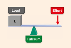
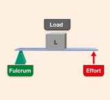
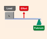
Lever system components
Lever
Fulcrum
Load
Effort
Lever
Rigid bar (bone) that moves on a fixed point (fulcrum = joint)
Fulcrum
Fixed point; joint
Load
Resistance (anyhting that adds weight like bone, tissues, added weight) moved by effort
Effort
Force (supplied by muscle contraction) applied to lever to move resistance
Mechanical advantages (good things) of levers
Moves a heavier load over a smaller distance
Requires less muscular effort to move the load
Achieved with a power lever that is strong
Effort is applied to farther from the fulcrum than the load to be moved
Mechanical disadvantages (bad things) of levers
Moves a lighter load over a great distance
Effort is applied closer to the fulcrum than the load to be moved
Achieved with a speed lever that is fast
Greater muscular effort is required to move the load
First class lever
Seesaw or scissors
Fulcrum is between load and effort
Ex: neck extension, posterior neck muscles provide effort, atlantoccipital joint is fulcrum and facial skeleton is load
Can be both advantage or disadvantage
Second class lever
Wheelbarrow or standing on toes
Load is between fulcrum and effort
Ex: effort is calf muscles, joints of the ball of foot are fulcrum, body weight is load
Uncommon in body; Mechanical advantage
Third class lever
Tweezers, foreceps, most skeletal muscles
Effort is applied between fulcrum and load
Ex. effort exterted on proximal radius of forearm, fulcrum is elbow joint, load is hand and distal forearm
Mechanical disadvantage (ideal for fast, large movements
Speed in mechanical disadvantage (speed levers)
Force is lost but speed and range of movement are gained
Speed in mechanical advantage (power levers)
Slower, but more stable. Used where strength is priority