Outcome 2: Fetal Lie, Growth, & Wellbeing
 Call Kai
 Learn
Learn Practice Test
 Spaced Repetition
 Match
Match Flashcards
 Knowt Play
1/43

There's no tags or description
Looks like no tags are added yet.
Name | Mastery | Learn | Test | Matching | Spaced | Call with Kai | Chat |
|---|
No analytics yet
Send a link to your students to track their progress
44 Terms
initial survey
when beginning a 2nd/2rd trimester OB exam, in both SAG and TRV planes we look for..
# of fetuses
FHR
fetal lie and presentation
any obvious, large abnormalities
fetal lie, presentation, and situs
fetal lie:
the relationship b/w fetus and the pt/maternal body
fetal presentation:
reference to the part of the fetus that’s overlying the internal cervical os
can further describe fetal lie (ie. frank breech, complete breech, etc.)
often used interchangeably w/ fetal lie
fetal situs:
position and orientation of the heart, stomach, liver, spleen, aorta, and IVC relative to midline fetus
we observe heart and stomach on LEFT side of chest and abdo
uterine growth during pregnancy
maternal uterus expands upwards and outwards to accommodate the fetus, placenta, and amniotic fluid
symphysis-fundal height (SFH):
distance b/w symphysis pubis and uterine fundus; measured in dr’s office
size correlated to GA (ie. 24cm SFH = 24 wk GA)
weights of uterus:
before preg. = ~50-70g
12wk = ~140g
20 wk = ~300g
36wk = ~1000g

fetal lie types
longitudinal:
fetus parallel w maternal body
cephalic = head down
breech = head up
transverse:
fetus perpendicular w/ maternal body
oblique:
fetus diagonal w/ maternal body
mobile/variable:
fetal lie changes throughout exam
—
you MUST check fetal lie first to know what side of baby is their left side before confirming that their heart and stomach are on the correct side of their body

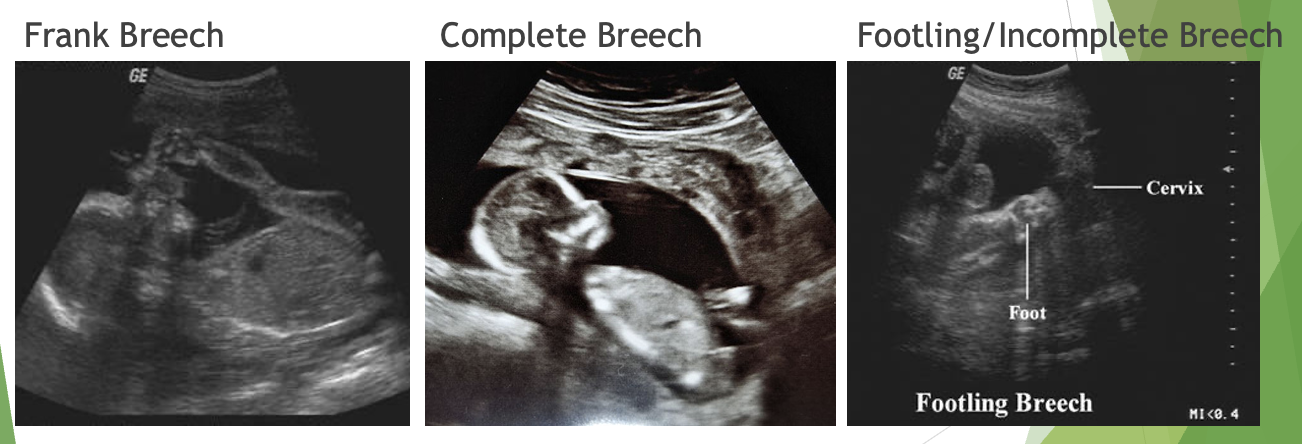
breech lie types
frank breech:
fetal bum presenting
legs and knees are extended
feet by fetal head
complete breech:
fetal bum presenting
legs and knees are flexed
feet by fetal bum
footling/incomplete breech:
fetal feet presenting
either 1 foot (single footling) or both feet (double footling) closest to internal cervical os


fetal lie: transverse
we need to also comment on where fetal head is in relation to mom


fetal heart and abdomen - fetal situs
if we look closely, we can actually see which chambers are atria and which ones are ventricles
the atria are further back in the chest, more round, and thinner walls
the ventricles are more anterior in the chest, pointed, with thicker walls
the ventricles point towards the left side of the chest, so the half of the chest with the majority of chambers of the heart is the left side.
terminology
appropriate-for-gestation age (AGA):
a fetus whose size is within the normal range for its GA (bw 10%-90%ile)
large-for-gestational age (LGA):
a fetus whose size is above a predefined threshold for its GA (>90%ile)
small-for-gestational age (SGA):
a getus whose size is below a predefined threshold for its GA (<10%ile)
normal but smaller than avg.
fetal growth restriction (FGR) or intrauterine growth restriction (IUGR):
a fetus that hasn’t achieved its growth potential
abnormally restructed in growth
macrosomia:
a baby who measures >4000g at birth
fetal growth and biometry
biometry:
measurements of fetal body structures in an attempt to establish the GA and ensure appropriate interval growth
—
the accurate estimation of GA underlies all OB management decisions
1st trimester biometry:
crown-rump-length (CRL)
1st trimester estimated gestational age = X ± 1 week
we can’t use CRL when it’s >84mm (~14wks) bcuz that’s the machine’s limit
2nd and 3rd trimester biometry
biparietal diameter (BPD):
linear measurement of the TRV diameter of fetal skull through transthalamic plane
leading edge of the near field to leading edge of far field
head circumference (HC):
ovoid measurement along the outer perimeter of the fetal skull in transthalamic plane
doesn’t include skin
abdominal circumference (AC);
ovoid measurement along the outer perimeter of the fetal abdominal soft tissue in a TRUE TRV plane at the lvl of the stomach and umb. vein
includes skin
femur length (FL):
linear measurement of the axial length of the ossified portions of the femur
—
gives us an estimated GA and fetal weight

thalamic view for BPD and HC
view for biparietal diameter and head circumference:
TRV image of fetal head at lcl of paired thalami and cavum septi pellucidi (CSP), w/ symmetry of the thalami and cerebral hemispheres and w/o visualization of the posterior fossa
sonographic landmarks:
paired thalami = paired ovoid structures in middle of brain (* in above image)
cabum septi pellucidi (CSP) = fluid-filled space anterior to thalamus (anechoic space b/w arrows in above image)
falx = linear structure separating cerebral hemispoheres (left side arrow in image)
ovoid fetal head w/ symmetrical hemispheres

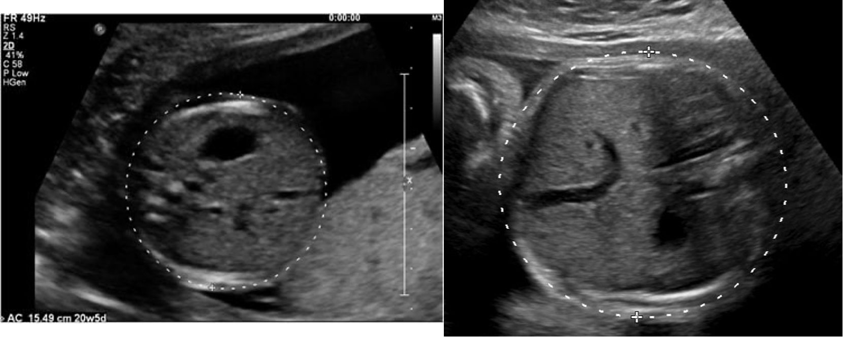
abdominal circumference (AC)
view:
stomach and curved, intrahepatic portion of portal vein must be seen
fetal kidneys NOT seen
round abdomen w/ ribs symmetric on either side of abdomen
true TRV plane

femur length (FL)
image one of the femur bones in LAX, horizontal on screen
measure blunt ends of echogenic femur bone (diaphysis), don’t include pointed ends or epiphysis
—
as the pregnancy continues, the epihysis locations on either end of the femur bone will be more clearly seen
hypoechoic on u/s

occipitofrontal diameter (OFD) - optional measurements
linear measurement of the length diameter of the fetal skull through transthalamic plane
measured from mid bone of anterior skull to mid bone of the posterior skull
perpendicular to BPD
contributes to cephalic index (CI)measurement, not routinely measured
cephalic index (CI) - optional measurements
a ratio calculated using BPD and OFD
used to indicate fetal head shape
CI = BPD/OFD x 100
—
mesocephalic (normal) = 70-85%
brachycephalic (abnormally wide) = >85%
dolichocephalic (abnormally long) <70%
—
not routinely measured

humerus length (HL) - optional measurements
linear measurement of axial length of the ossified portions of the humerus
same method as FL, measured in LAX and the blunt ends of the bone
not routinely measured

orbital measurements (OOD, IOD, OD) - optional measurements
orbital measurements:
linear measurements of fetal orbits (eye sockets) acquired in TRV view of fetal head at lvl of orbits
—
outer orbital distance (OOD):
aka binocular distance (BOD)
linear measurement from outer edge of one orbit to outer edge of other orbit
inner orbital distance (IOD):
aka interocular/interorbital distance
linear measurement of the distance b/w inner edges of both orbits
orbital diameter:
linear measurement of the diameter of one orbit
—
normally
IOD = OD
OOD = IOD x 3 = OD x 3
fetal size vs. fetal growth
fetal size:
indicated the estimated fetal weight and corresponding estimated GA at any given point in time
fetal growth:
indicated the estimated fetal weights and corresponding estimated GA compared over diff. points in time
fetal growth = fetal size + time
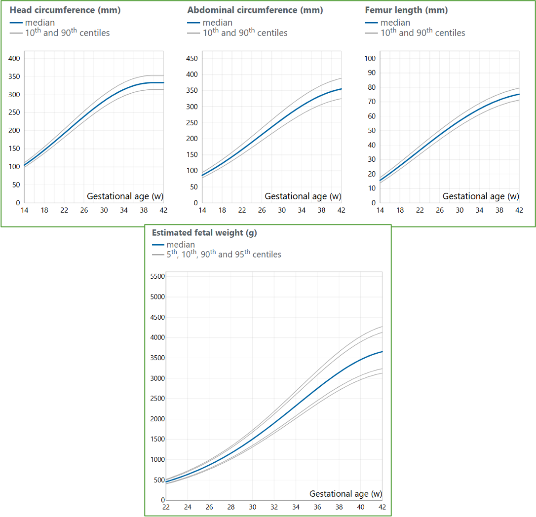
fetal growth charts
canadian standard:
fetal growth chart are created from research studied that collect fetal biometric measurements from large populations of pregnancies
a percentile curve is generated, showing the expected distribution of measurements at each GA
most u/s departments in North America use the Hadlock charts, but there isn’t necessarily a single Canadian fetal growth chart every Canadian u/s department uses
controversy:
whether the estimated weight of a fetus should be compared w/ norms from the overall population (ie. Canadian pop.) or to customized norms (ie. ethnicity)
—
BPD, HC, AC, FL, and EFW growth charts are embedded into the u/s machines
growth chart trajectory includes the median (avg, 50%) and extremes
fetal growth charts cont.
weight gain:
weight gain = diff. b/w two estimated weights (of 2 diff. u/s exams)
EXAMPLE:
Patient A arrives for an ultrasound exam at 28w0d. After the ultrasound is complete, biometry measurements gave us an EFW of 1005 grams. Based on growth chart (seen above), this fetus measures exactly 50%ile.
Patient A arrives for another ultrasound exam exactly 4 weeks later, at 32w0d. After the ultrasound is complete, biometry measurements gave us an EFW of 2000 grams. Based on growth chart, this fetus measures nearly 90%ile.
Weight Gain = Difference in two weights = 2000g – 1005g = 995 grams of weight increase in 4 weeks time.
—
biometry measurements and estimated fetal weight should only be assessed after 2 wks has passed since previous measurements were acquired

large-for-gestational age (LGA)/macrosomia
LGA:
>90%ile of estimated fetal weight
macrosomia:
>4000g birthweight
—
risk factors:
maternal obesity
hx of previous LGA infant
prolonged pregnancy (>40wk GA)
excess pregnancy weight gain
multiparity
advanced maternal age
complications:
inc. morbidity/mortality
shoulder dystocia
fractures
facial/brachial plexus palsies
traumatic delivery
asphyxia
meconium aspiration
neonatal hypoglycemia
sono signs:
EFW >90%ile for GA
usually affects abdomen circumference (AC) in 3rd tri
potential for macrosomia

diabetic patients
diabetes mellitus is a chronic condition where the body can’t properly use blood sugar (glucose) » leading to high blood sugar lvls
inc. lvls of maternal insulin leads to overgrowth of fetal abdomen, but normal head
~28-32 weeks GA, fetuses will have larger than normal AC measurement » often causing LGA/macrosomia
complications:
inc. morbidity/mortality
shoulder dystocia
fractures
facial/brachial plexus palsies
traumatic delivery
asphyxia
meconium aspiration
neonatal hypoglycemia

small-for-gestational age (SGA)/fetal growth restriction (FGR/IUGR)
SGA:
<10%ile of EFW
FGR/IUGR:
a fetus is one that hasn’t achieved its growth potential
abnormally restricted growth
—
risk factors:
small parents
placental abnormalities
chromosomal abnormalities/aneuploidy
congenital malformations
infections
maternal abnormalities
multiples
complications:
inc. morbidity/mortality
respiratory distress syndrome
acidosis
NICU admission
sono signs:
EFW <10%ile for GA
constitutionally small; may also notice small pt/parents
sono signs of FGR (bottom images):
delayed/absent growth on serial u/s exams
abnormal umb. artery doppler
oligohydramnios
grade 3 placenta
low BPP score

fetal wellbeing
healthy status of a fetus
ability to detect signs of fetal distress early helps supports maternal and fetal health & allows time to prepare for safe delivery and identifies potential interventions that may be considered
indications:
FGR
oligohydramnios
multiples
dec. fetal movement
prior stillbirth
maternal HTN
diabetes
renal disease
assessments:
ultrasound assessments..
biophysical profile
dynamic structures
fetal doppler
other..
non-stress test (NST)
cardiotocography (CTG)
contraction stress test (CST)

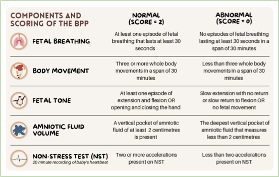
biophysical profile
assessment of fetal wellbeing, scored out of 8
commonly performed after 28wks in 3rd tri.
fetus has 30 mins to demonstrate each parameter to receive all points
parameters used (each /2 = /8):
fetal movement
fetal tone
fetal respiration
amniotic fluid
additional parameter (making score /10):
non-stress test (NST)
—
clinical indications:
diabetes
HTN
renal disease
asthma
multiples
current concerns
past pregnancies w/ concerns
past due date (>41 wks GA)
primarily tests for:
asphyxia
low cord pH
cerebral palsy
stillbirth

fetal movement - biophysical profile
3 or more discrete gross body/limb movements within 30 mins
“movement” indicated overall body or limb activity
sonographically:
movement such as kicking, rolling, stretching of body
usually seen in fetal limbs

fetal tone - biophysical profile
one or more episodes of active extension and flexion of a fetal extremity OR opening/closing of hand within 30 mins
“tone” indicated specifically muscle tone
ie. actively extending a limb or trunk, and then returning it to a flexed position
sonographically:
extension and flexion of fetal spine
extension and flexion of limbs
opening and closing of hand

fetal respiration - biophysical profile
one or more episodes of fetal breathing lasting at least 30 secs, within 30 mins
hiccups are considered breathing activity
sonographically:
alternating squeezing of chest (ribs) and abdo wall, in inhale/exhale like motions
diaphragm and abdominal contents moving up and down in chest/abdomen
hiccups are shorter, quicker versions of these movemtns
best seen in coronal view of fetal chest/abdo

amniotic fluid - biophysical profile
AFV = single pocket of amniotic fluid at least 1cm x2cm in AP dimension
SDP = normal is >2×2cm
sonographically:
can’t measure through fetal tissue of amniotic cord
pockets of fluid are typically smaller in 3rd tri
non-stress test (NST)
measurement of fetal movement, heart rate, and uterine contractions for fetal wellbeing
inc. fetal HR w movement indicated appropriate O2 lvls in fetus
completed at dr’s office w/ cardiotocograph machine
results:
reactive (normal) = fetal HR goes up 2 or more times in testing period (2 points)
nonreactive (abnormal) = no change in FHR when baby moves (no points)
cardiotocography (CTG)
measurement of fetal movement, FHR, and uterine contractions during labour
uses cardiotocograph machine to look for signs of fetal distress
contraction stress test (CST)
hormone (oxytocin) is administered to pt. to stimulate stress of baby by creating uterine contractions
observing the change in FHR befire and after contraction can indicate fetal wellbeing or distress
normally, HR should lower then return to normal after contraction
CST may be performed to further test for signs of fetal distress after an abnormal BPP or NST
abnormal CST may suggest C-section over vaginal birth
>34wks GA (late 3rd tri)

dynamic structures
aka “things that change”
fluid-filled structures in fetus and fetal environment that are routinely assessed at every OB u/s
another way fetal wellbeing can be assessed is observing these structures
includes:
lateral cerebral ventricles
cavum septi pellucidi (CSP)
cisterna magna
4C heart
stomach
kidneys
bladder
amniotic fluid
fetal hypoxia and asphyxia
hypoxia:
when the fetus is deprived of an adequate supply of O2
one of the leading causes of fetal mortality
asphyxia:
severe form of O2 deprivation (worse form of fetal hypoxia)
one of the leading causes of fetal mortality
—
causes:
FGR (can also be a consequence)
umb. cord prolapse
cord occlusion/thrombosis
placental insufficiency/infarction
maternal smoking
—
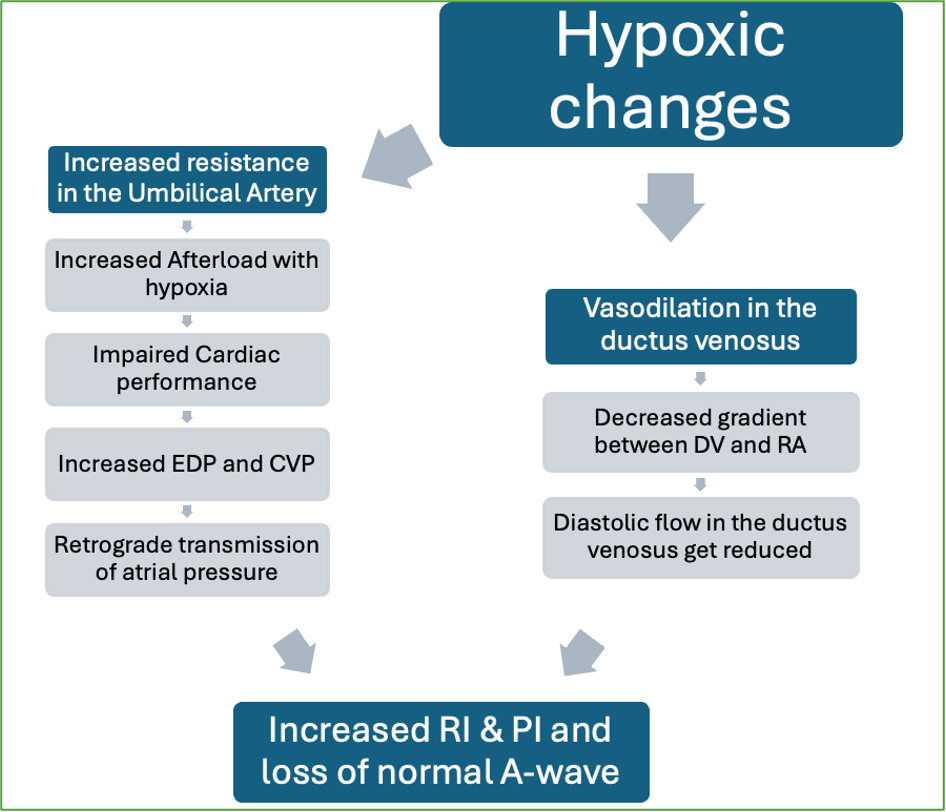
fetal doppler is used to assess for risk of FGR bcuz waveform appearance, measurements, S/D ratio, RI, and PI can indicate hypoxic changes
fetal doppler
PW and colour
helpful in evaluating fetal wellbeing and fetal risk of FGR
vessels that can be evaluated:
umb. artery/arteries
umb. vein
ductus venosus
middle cerebral artery (MCA)
maternal uterine artery
fetal doppler rules:
careful technique and proper machine settings must be used
adhere to ALARA principle
only used in high-risk fetsuses
umbilical artery (UA) doppler
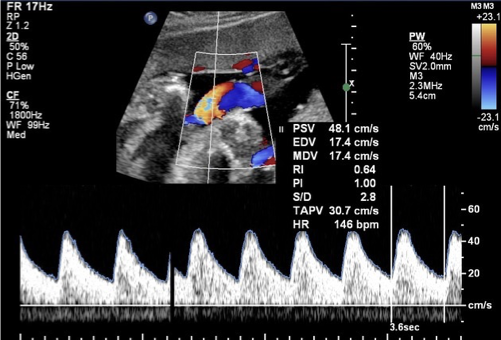
typically performed >24wks GA
normal = low resistance
abnormal = high resistance waveform, absent or reversed flow
normal S/D ratio..
24wks = 3.23
40 wks = 2.19


umbilical vein doppler
continuous, monophasic flow towards fetus
normal mean velo. = 10-15cm/s
abnormal = pulsatile flow


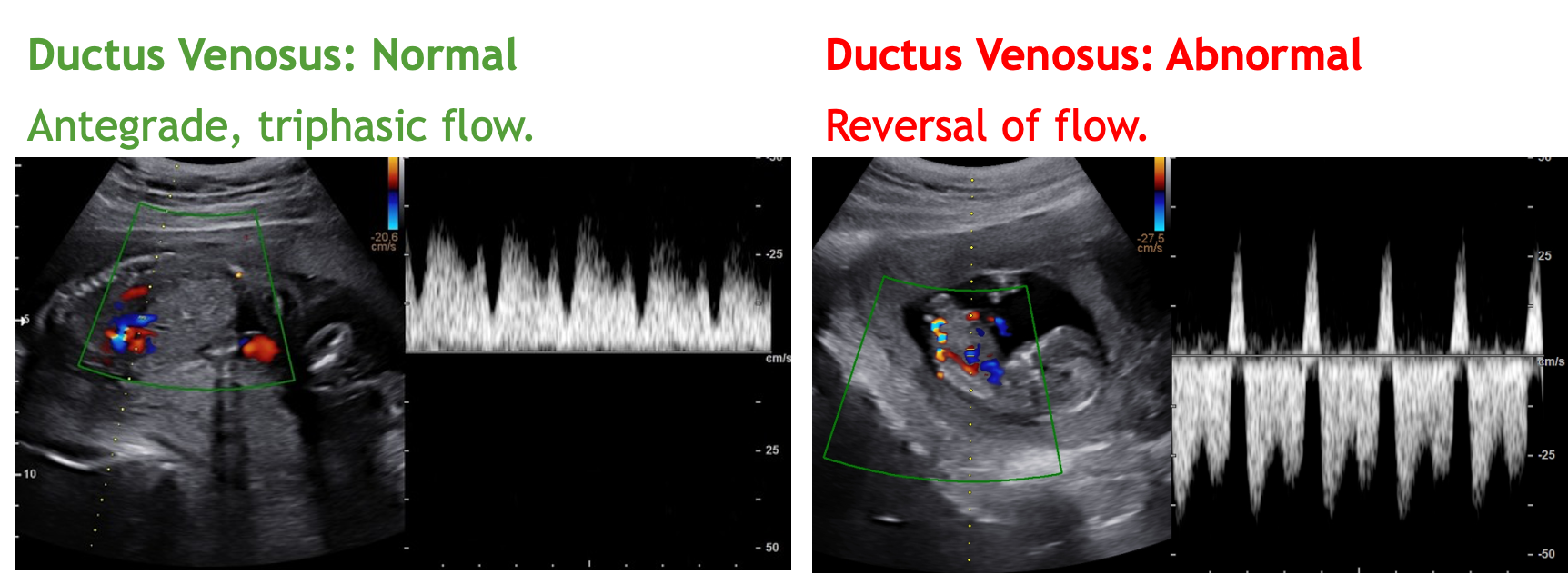
ductus venosus doppler
connects umbilical vein to the IVC » becomes ligamentum venosum
venous structure
triphasic, antegrade, continuous flow towards fetal heart
abnormal = absent or reversed flow

middle cerebral artery (MCA) doppler
MCA is near circle of willis
helps assess for FGR, fetal hypoxia, and fetal anemia (fetal distress)
imaged in TRV view of fetal brain @ lvl of cerebellum
not commonly performed
normal = high resistance waveform
abnormal = low resistance waveform
sonography:
obtain TRV view fetal head at lvl of thalami/cerebellum
place colour to identify rt and lt MCAs
perform PW doppler on bilateral vessels 3x


middle cerebral artery (MCA) doppler chart
normal values change over time
peak values occur ~28-30wks GA
maternal uterine artery doppler
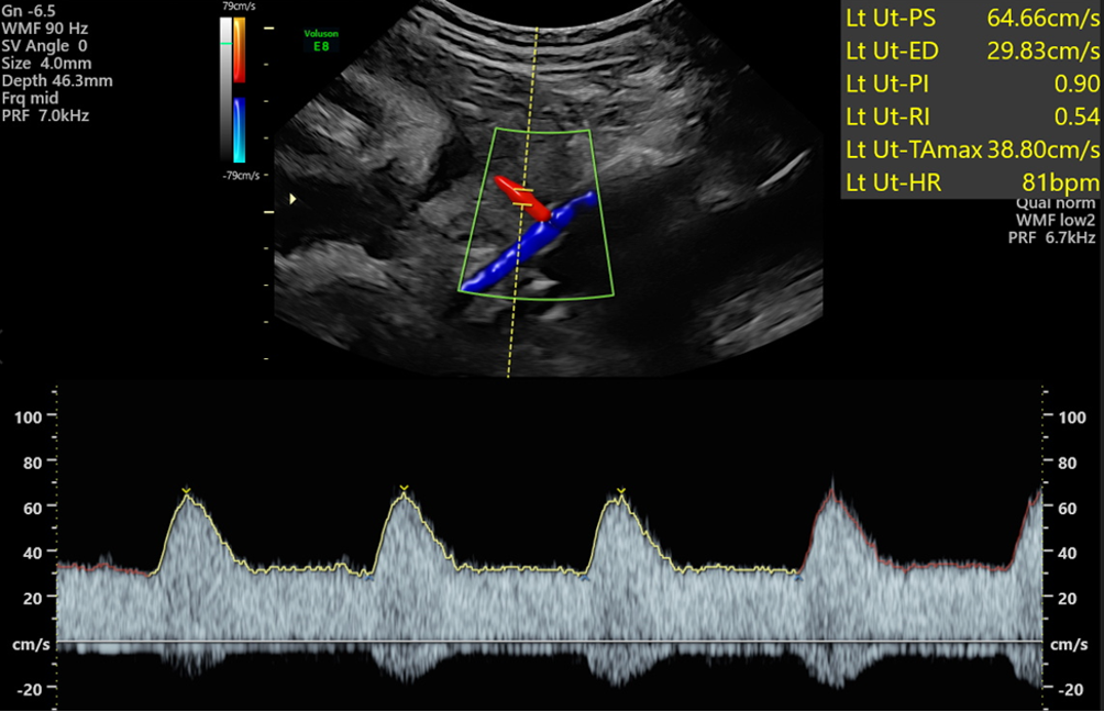
lateral sides of cervix/lower uterine segment contains uterine arteries
normal = low resistance waveform w RI <0.55
abnormal = inc. PI, diastolic notch, and RI >0.55
abnormal waveforms indicate abnormal vasculature of the placenta or placental insufficiency
sonography:
pt. has full bladder
SAG cervix, sweep laterally and angle slightly
uterine arteries found on lateral sides of cervix, crossing paths w external iliacs
assess and image bilateral uterine arteries 3x

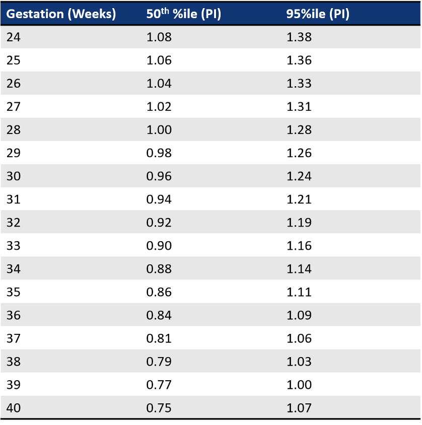
maternal uterine artery doppler chart
normal values change over time
values steadily dec. throughout pregnancy
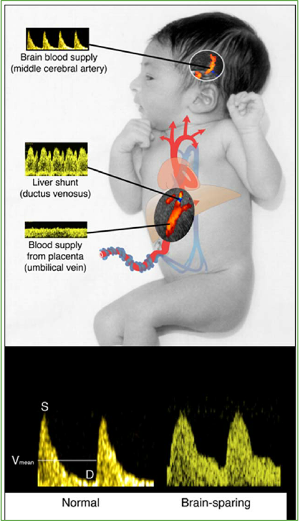
brain sparing effect
redistribution of oxygenated blood to vital fetal organs (brain, heart, adrenals) when a fetus is in distress (placental insufficiency, hypoxia, growth restriction)
MCA doppler waveform changes from the normal high-resistance wave to an abnormal low-resistance wave » helping get as much oxygenated blood to the brain as possible, inc. fetal survival
brain sparing = blood shunted during fetal distress = low resistance MCA waveform
seen as vasodilation in the brain on OB u/s
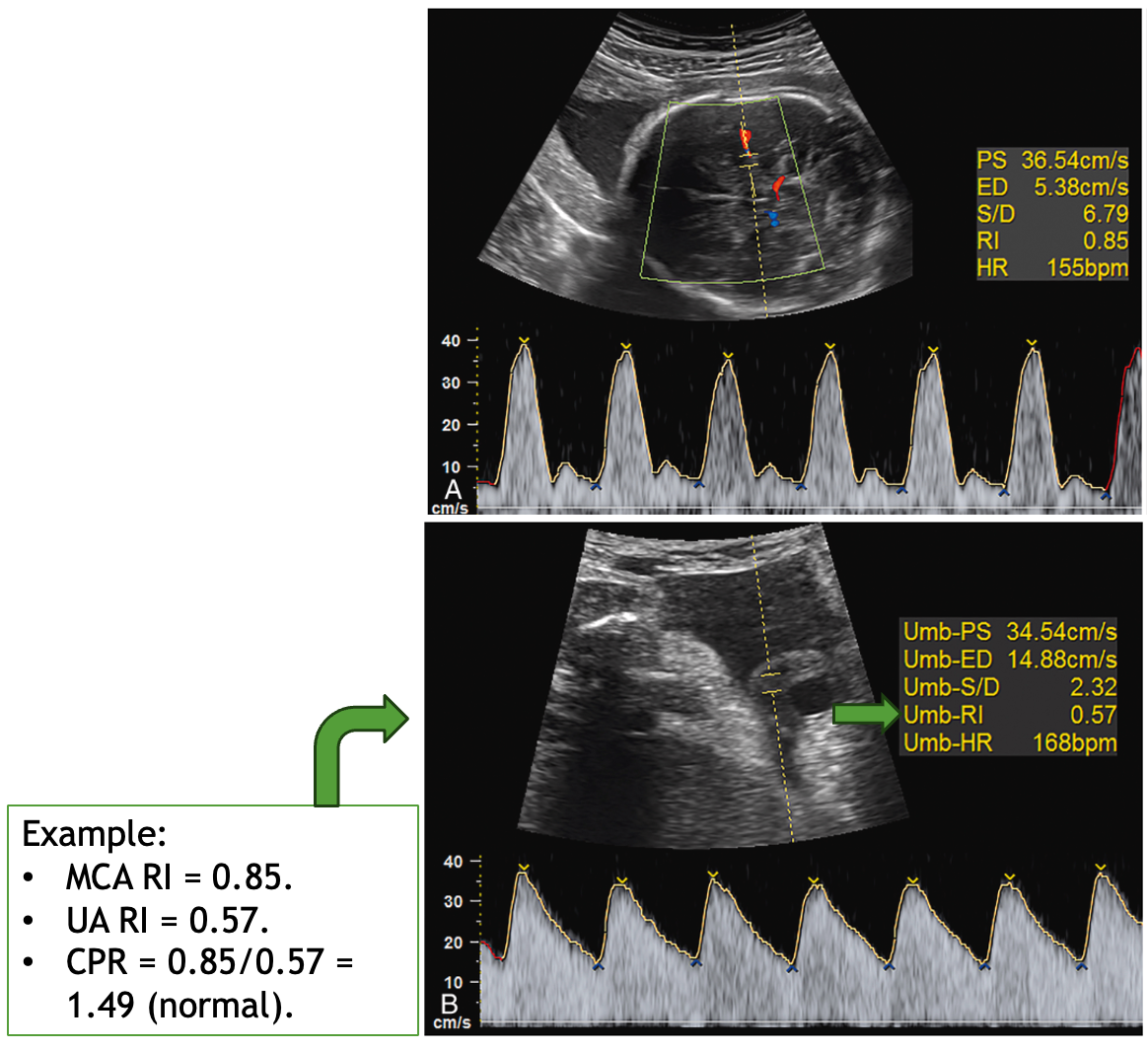
cerebroplacental ratio (CPR)
ratio of resistance index (RI) or pulsatility index (PI) of middle cerebral artery (MCA) compared to umb. artery
CPA = (MCA RI/PI)/(UA RI/PI)
normal = CPR >1.0
abnormal = CPR <1.0