Cell Attachment and Connective Tissue Vocabulary Flashcards
 Call Kai
 Learn
Learn Practice Test
 Spaced Repetition
 Match
Match Flashcards
 Knowt Play
1/79

Earn XP
Description and Tags
Comprehensive vocabulary flashcards covering cell attachments, epithelial classification, cytoskeleton components, and connective tissue extracellular matrix based on Ten Cate Chapter 4.
Name | Mastery | Learn | Test | Matching | Spaced | Call with Kai | Chat |
|---|
No analytics yet
Send a link to your students to track their progress
80 Terms
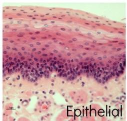
Epithelial Tissue
A general tissue type consisting of aggregated polyhedral cells with a small amount of extracellular matrix, functioning in protective lining, glandular secretions, and transport.
Connective Tissue
A general tissue type containing various fixed and wandering cell types with a large amount of extracellular matrix, functioning to support and protect body structures.

Muscle Tissue
A distinct contractile tissue containing a moderate amount of extracellular matrix and connective tissue components.
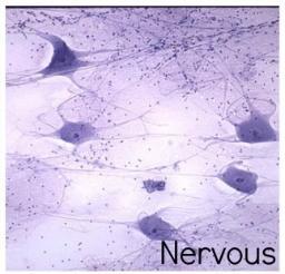
Nervous Tissue
A tissue type specialized for conducting nerve impulses, containing very little extracellular matrix.
Apical Pole
The surface face of an epithelial cell or cell sheet that faces the lumen or open space.
Basal Pole
The surface face of an epithelial cell or cell sheet that faces toward the underlying connective tissue.
Avascularity of Epithelium
The characteristic of epithelial tissue lacking blood vessels, relying instead on diffusion from blood vessels located in adjacent connective tissue.
Basement Membrane
A thin sheet of extracellular matrix anchoring the epithelium to underlying connective tissue, measuring 50–100 nm thick and visible under light microscopy.

Basal Lamina
The term used when referring to the basement membrane structure as viewed specifically under electron microscopy.
Lamina Lucida
The zone of the basement membrane closest to the epithelial cell layer containing laminin, entactin, and integrins.
Lamina Densa
The intermediate zone of the basement membrane composed primarily of Collagen IV.
Lamina Fibroreticularis
The layer of the basement membrane adjacent to connective tissue, which is sometimes absent and typically contains Collagen III and VII.
Simple Epithelium
An epithelial tissue classification where a single layer of cells contacts the basement membrane.
Stratified Epithelium
An epithelial tissue classification containing multiple layers of cells, where not all cells contact the basement membrane.
Pseudostratified Columnar Epithelium
A type of epithelium appearing layered due to cell nuclei at varying heights, though all cells touch the basement membrane.
Urothelium
The specialized transitional epithelial classification lining regions of the urinary tract.

Microfilaments
Cytoskeletal components composed of actin, measuring 6–8 nm in diameter, providing structural shape and contractility.
Actin
The protein subunit that polymerizes to form microfilaments, supporting cell structures like microvilli and filopodia.
Myosin Tracks
Pathways formed by actin microfilaments that allow motor interactions to provide cell contractility.
Intermediate Filaments
Non-contractile, structural cytoskeletal components around 10 nm in diameter that anchor cell structures and vary by tissue type.
Vimentin
A specific type of intermediate filament protein characteristic of cells derived from the mesenchyme.

Cytokeratin
The specific type of intermediate filament protein prominent in epithelial cells.
Microtubules
Tubular cytoskeletal structures around 25 nm in diameter composed of tubulin that act as monorail transport systems.
Tubulin
The protein monomer subunit that forms cylindrical microtubules.

Kinesins
Motor proteins that move along microtubule tracks as part of the intracellular transport monorail system.
Dynein
Motor proteins associated with microtubules involved in movement within structures like cilia and flagella.
Tight Junctions (Zonula Occludens)
Intercellular attachments formed by homotypical interactions between transmembrane proteins, maintaining cell polarization and controlling paracellular movement.
Claudins, Occludin, and JAM
Transmembrane proteins present in tight junctions that bind intracellularly to actin microfilaments.

Zonula Adherens (Cell to Cell Adhesive Junctions)
Adhesive cell-to-cell junctions characterized by intercellular gaps of 20 nm, linking cell membranes via cadherin and catenin complexes.

Cadherins in Zonula Adherens
Transmembrane adhesion proteins located on the cell exterior that require Ca2+ to hold adjacent cells together.
Nectin-Afadin Complex
A molecular junction complex involved in cell-to-cell adhesive junctions that plays a key role in contact inhibition.
Focal Adhesion Junctions
Cell-to-matrix junctions where transmembrane integrins replace cadherins to anchor the cell to extracellular matrix components.
Integrins in Focal Adhesions
Transmembrane receptors in focal adhesions that interact with ECM proteins such as collagen and fibronectin, functioning in cell signaling.
Desmosomes
Cell-to-cell adhesive junctions with an intercellular gap of 25 nm that attach to intermediate filaments to help resist shearing forces.
Desmoglein and Desmocollin
External cadherin proteins in desmosomes that undergo heterotypical interactions between adjacent cell membranes.
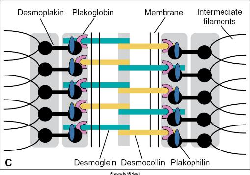
Desmoplakin, Plakoglobin, and Plakophilin
Internal plaque catenin proteins in desmosomes that appear as dark electron-dense bands under electron microscopy.
Hemidesmosomes
Cell-to-matrix adhesion structures that tightly attach epithelial cells to the basal lamina using integrins to resist shear forces.

Collagen XVII
A transmembrane collagenous component found in hemidesmosomes that contributes to anchoring the cell to the basal lamina.
Gap Junctions
Cell-to-cell aqueous channels separated by 2–3 nm that allow direct pass-through of ions, small water-soluble molecules, and electrical signals.
Connexon
An assembly of 6 connexin protein subunits forming a single 2 nm channel pore in a gap junction.

Connexins
Individual transmembrane protein subunits that hexamerize to create a connexon pore.
Junctional Complex
A structural grouping of cell attachments comprising Tight Junctions, Zonula Adherens, Desmosomes, and Gap Junctions.
Terminal Bar
A collective band of junctional complexes visible on lateral cell surfaces under a light microscope.
Extracellular Matrix (ECM)
The non-cellular network composed of protein fibers and ground substance surrounding and supporting cells in tissues.
Mesenchymal Progenitor Cells
Multipotent stem cells derived from mesoderm that give rise to various connective tissue cell lineages.
Permanent Connective Tissue Cells
Fixed resident cell types in connective tissue including fibroblasts, adipocytes, macrophages, and mast cells.
Transient Connective Tissue Cells
Wandering cells that migrate into connective tissues, such as plasma cells, lymphocytes, neutrophils, and eosinophils.
Fibroblasts
The principal motile cell type of connective tissue responsible for synthesizing extracellular matrix fibers and ground substance.

Fibronexus
A specialized transmembrane cytoskeletal-to-ECM connection linking actin microfilaments to extracellular fibronectin via integrins.
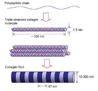
Collagen
The most abundant protein in the human body, possessing a triple-helical molecular structure with 28 characterized types.
Proline and Lysine in Collagen
Amino acids abundant in collagen whose hydroxylated forms facilitate crucial hydrogen bonding during triple helix assembly.
Collagen Type I and III in Dental Tissues
Dentine ECM consists mostly of Collagen Type I with some Type III, while pulp ECM contains a mixture of Collagen Types I and III.
Collagen Type IV
The non-fibrillar collagen type forming a 'chicken wire' meshwork within the basement membrane.
Collagen Microfibril Stagger
The parallel alignment of 5 collagen microfibrils staggered by approximately 1/4 of their length, producing a characteristic banded fibril structure.
Scurvy
A condition caused by Vitamin C deficiency, leading to impaired prolylhydroxylase and lysyl hydroxylase function and unstable collagen assembly.
Prolylhydroxylase and Lysyl Hydroxylase
Enzymes that modify proline and lysine residues during collagen synthesis, requiring Vitamin C as a cofactor.
Elastin
A connective tissue fiber core protein assembled on a glycoprotein scaffold that imparts elastic recoil properties.
Fibrillin-1 and Fibrillin-2
Glycoproteins forming microfibril scaffolds onto which elastin fibers accumulate during elastic fiber formation.
Marfan's Syndrome
A genetic connective tissue disorder caused by a mutation in the gene encoding fibrillin-1.
Adipocytes
Connective tissue cells containing a single large lipid droplet, a thin rim of cytoplasm, a flattened nucleus, and an individual basal lamina.
Adipose Tissue Functions
Functions of fat tissue including energy storage as lipid, thermal insulation, mechanical cushioning, and hormone synthesis.
Ground Substance
The highly hydrated, non-fibrillar gel-like extracellular material composed primarily of proteoglycans and glycoproteins that provides compressive strength.
Proteoglycans
Molecules synthesized by fibroblasts consisting of a core protein covalently bound to negatively charged glycosaminoglycan (GAG) chains.

Glycosaminoglycans (GAGs)
Hydrophilic, highly negatively charged carbohydrate chains (such as hyaluronic acid, chondroitin sulfate, and keratin sulfate) responsible for fluid sequestration.
Hyaluronic Acid
An unbranched glycosaminoglycan chain acting as an axis for proteoglycan aggregation and fluid retention in the extracellular matrix.
Perlecan
A non-aggregating proteoglycan found specifically in basal laminae.
Syndecan
A transmembrane non-aggregating proteoglycan involved in cell signaling and extracellular growth factor binding.
Glycoproteins
Proteins containing attached carbohydrate chains that facilitate cell migration, adhesion, and matrix alignment, generally having a smaller carbohydrate component than proteoglycans.
Fibronectin
An extracellular matrix glycoprotein critical for cell migration, cell adhesion, and wound healing.
Tenascin
An extracellular matrix glycoprotein that plays a key role in directing cell migration during tissue development and repair.
Thrombospondin
An extracellular matrix glycoprotein involved in cell attachment, cell migration, and collagen fiber alignment.
Loose Connective Tissue
The most abundant connective tissue type, containing abundant ground substance and cells with relatively fewer fibers, underlying epithelia and forming stroma.

Stroma
The supportive loose connective tissue framework surrounding functional parenchymal elements, blood vessels, and nerves within organs.
Dense Irregular Connective Tissue
A connective tissue proper type composed of densely packed, randomly arranged collagen fibers with few cells, offering multi-directional strength.

Dense Regular Connective Tissue
A connective tissue proper type with collagen fibers organized in parallel in the same direction, located in tendons and ligaments to provide directional support.

Epidermis
The epithelial outer layer of the skin that acts as a protective surface covering.
Dermis
The underlying connective tissue layer of the skin beneath the epidermis.
Hypodermis
The deep subcutaneous layer beneath the dermis containing adipose and loose connective tissues.
Functions of Epithelial Tissue
The three primary functions of epithelium: 1) Covering, lining, and protecting surfaces, 2) Absorption, and 3) Secretion.
Functions of Connective Tissue
Primary functions including mechanical/protective tissue support, interstitial fluid/water storage, early organ repair, and immune system defense.