Cardiac Anatomy
 Call Kai
 Learn
Learn Practice Test
 Spaced Repetition
 Match
Match Flashcards
 Knowt Play
1/32

There's no tags or description
Looks like no tags are added yet.
Name | Mastery | Learn | Test | Matching | Spaced | Call with Kai | Chat |
|---|
No analytics yet
Send a link to your students to track their progress
33 Terms
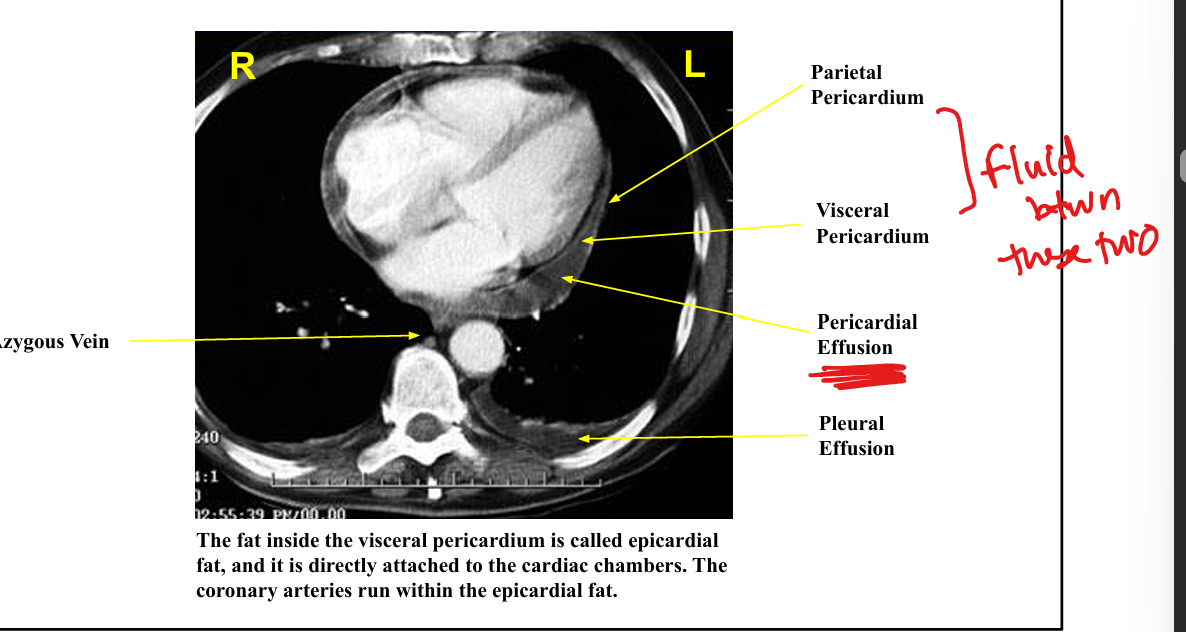
Layers of the pericardium
Visceral pericardium on heart parietal pericardium outside small lubricating fluid.

Sources of deoxygenated blood to RA
SVC, IVC, coronary sinus.

Gross orientation: PA bifurcation
Main PA splits into R and L PA, the right PA travels below aortic arch.

Gross orientation: LA
LA is posterior, directly in front of esophagus.

CXR rib spaces: 1st-2nd space stab on right side wound hits
Aorta

CXR rib spaces: 5th/6th space on medial left stab wound hits
Right ventricle
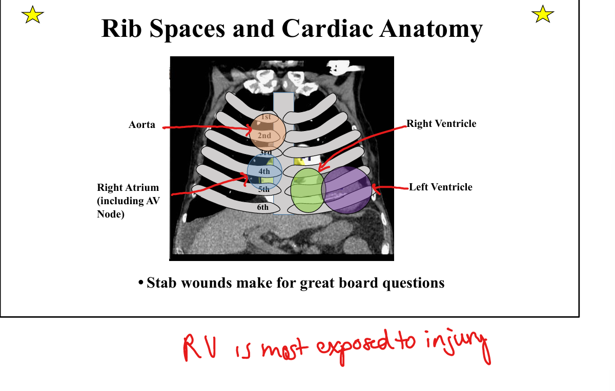
CXR rib spaces: 3rd+4th space on right side stab wound hits
Right atrium including AV node.

CXR rib spaces: 5th-6th spaces on left side stab wound hits
Left ventricle
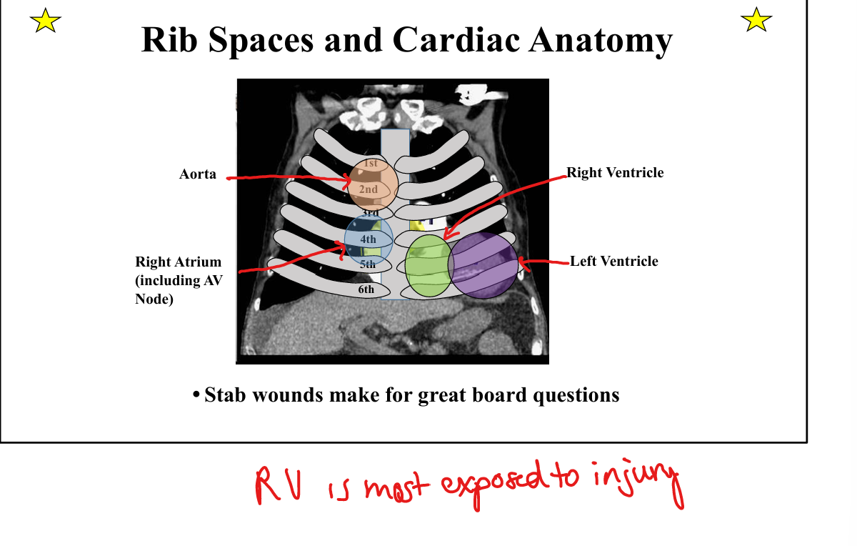
Knowledge check: Most exposed chamber to penetrating trauma?
Right ventricle.
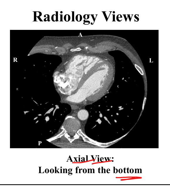
Coronal vs Sagital vs Axial View
Coronal: Look the crown in the eyes (from the front)
Sagital: S for from the side
Axial: from the bottom (axx)

Pulmonic vs aortic valve orientation
Pulmonic valve is anterior and left of aortic valve.

RV relationship to sternum
RV sits directly behind sternum.

LA relationship to esophagus
LA sits directly in front of esophagus, enlargement → dysphagia.

Epicardial fat definition
Fat within visceral pericardium directly attached to cardiac chambers; coronary arteries run within it.
Valve types: Semilunar
Aortic (LV→aorta) and pulmonic (RV→PA)
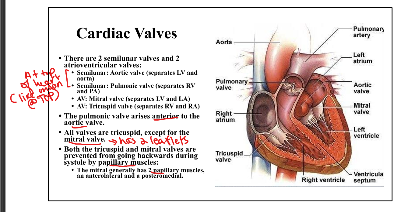
Valve types: AV
Mitral (LA→LV) and tricuspid (RA→RV)
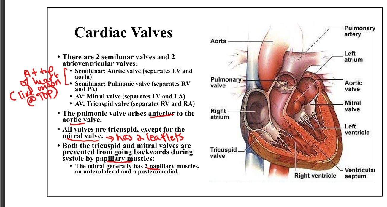
Papillary muscle function + on which valves
Prevent AV valve prolapse during systole. Mitral and Tricuspid

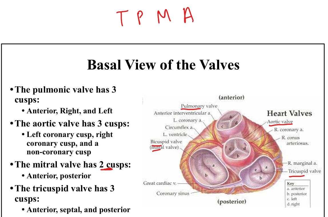
AV groove arteries
RCA in right AV groove, LCX in left AV groove.

Interventricular septum arteries
Anterior: LAD, Posterior: PDA.
RCA course
Travels in AV groove from right coronary cusp → RV → posterior IV septum.

LCA branches
LAD + LCX.

LAD course
Down anterior IV septum → apex → distal inferior wall.
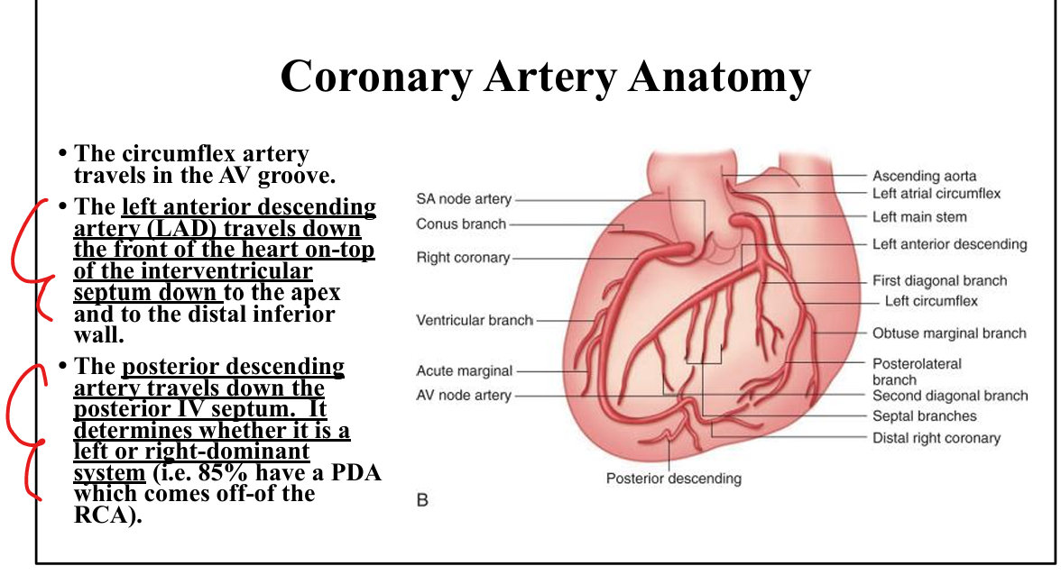
PDA course
Down posterior IV septum, determines dominance

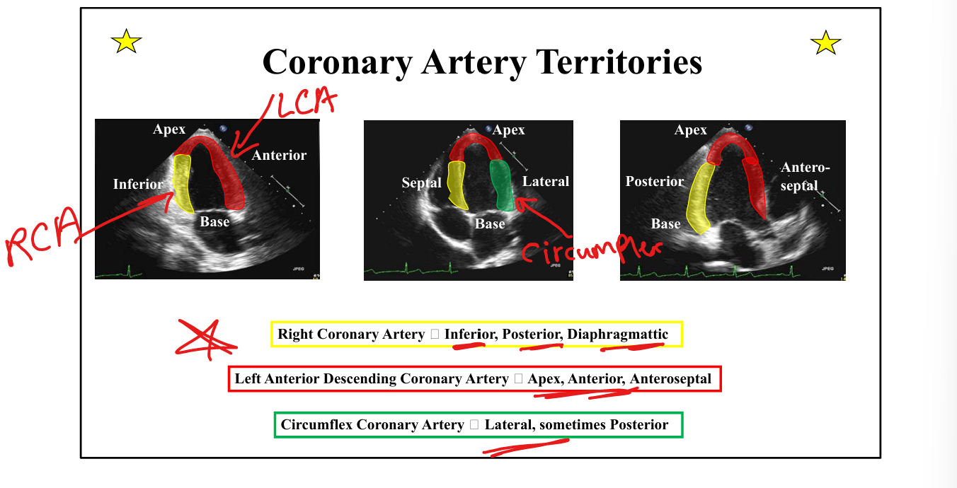
Coronary territories: RCA
Inferior, posterior, diaphragmatic

Coronary territories: LAD
Apex, anterior wall, anteroseptal.
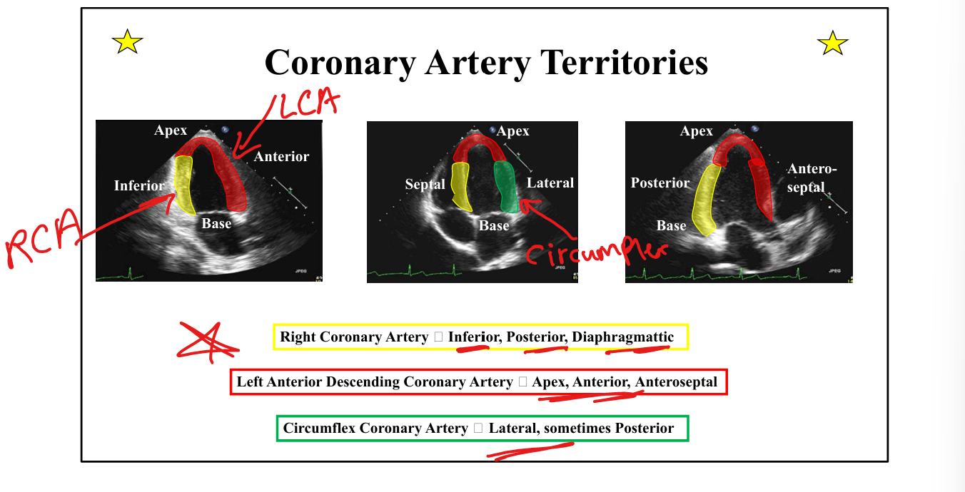
Coronary territories: LCX
Lateral wall, sometimes posterior
Papillary muscle blood supply (posteromedial +anterolateral)
Posteromedial papillary muscle: single supply (RCA or LCX)
Anterolateral: dual supply (LAD + LCX)

Coronary veins
Great cardiac, middle cardiac, small cardiac → converge into coronary sinus.
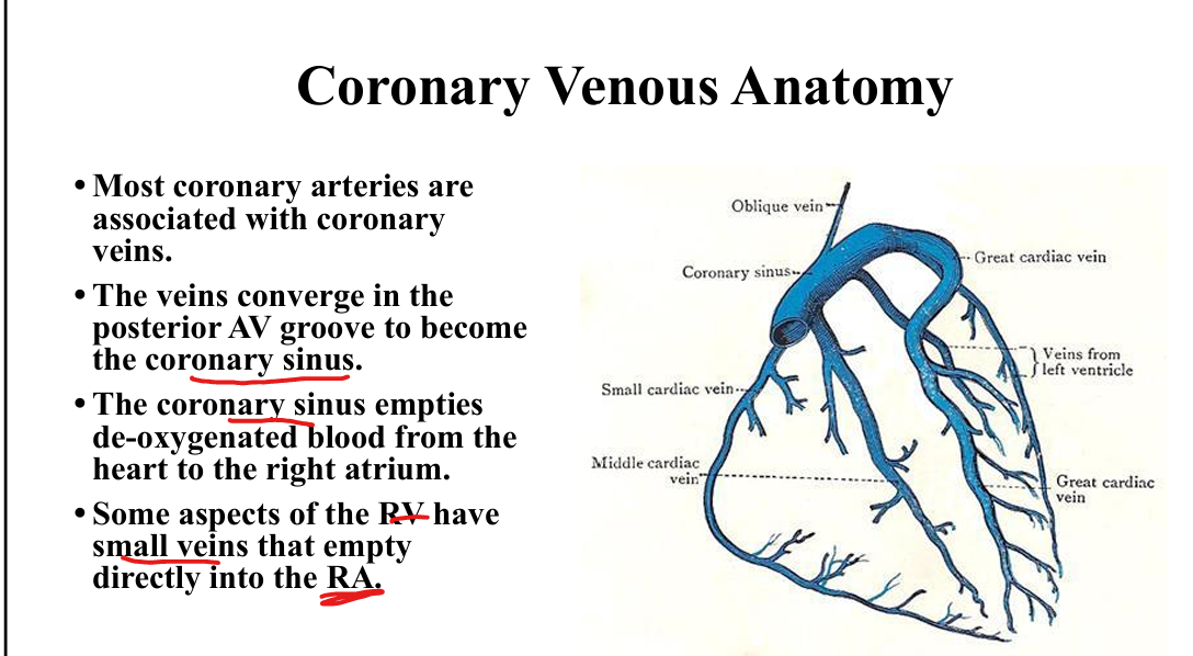
Coronary sinus drainage
Drains into RA

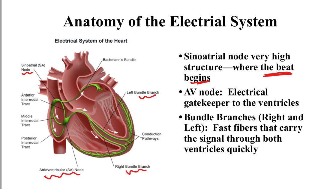
SA node location + function
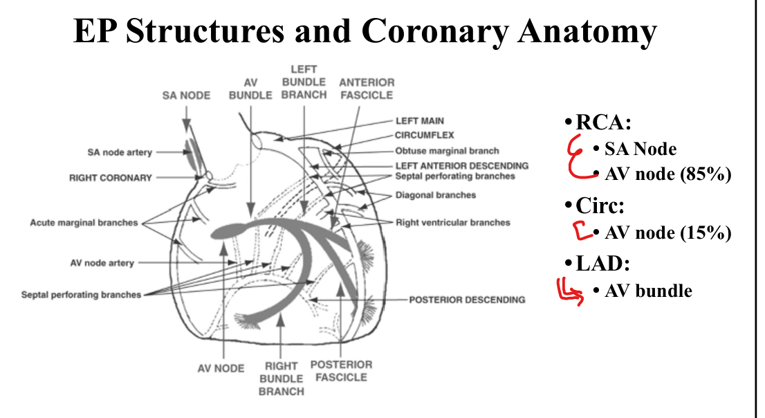
High in RA near SVC. Starts beat
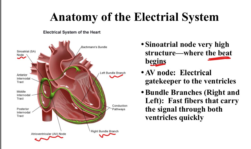
AV node location + function
Between valves, near crux. Electrical gatekeeper to ventricles

Bundle branches function
Fast fibers that carry the signal through both ventricles quickly
Blood supply to conduction system
RCA supplies SA node + AV node (85%), LCX supplies AV node (15%), LAD supplies AV bundle