MSK DSA 8-9
 Call Kai
 Learn
Learn Practice Test
 Spaced Repetition
 Match
Match Flashcards
 Knowt Play
1/44

There's no tags or description
Looks like no tags are added yet.
Name | Mastery | Learn | Test | Matching | Spaced | Call with Kai | Chat |
|---|
No analytics yet
Send a link to your students to track their progress
45 Terms
head of fibula articulates w/
lateral condyle of tibia [NOT femur]
3 structures connecting tibia and fibula
proximal [superior] tibiofibular joint
small plane-type synovial joint
allows slight gliding movements
helps dissipate forces transmitted up from ankle
interosseous membrane
distal [inferior] tibiofibular joint
syndesmosis [fibrous joint]
provides strong stability for ankle joint
mortise
“socket”
formed by medial and lateral malleoli of tibia and fibula
grips talus securely
talus
sits on calcaneus
articulates w/ tibia/tubula
transmits body weight
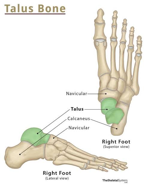
calcaneus
largest, heel bone
achilles tendon insertion

navicular
boat-shaped
medial side
supports arch

medial cuneiform
largest cuneiform
articulates w/ 1st metatarsal

intermediate cuneiform
smallest
wedge-shaped
stabilizes 2nd metatarsal

lateral cuneiform
btwn cuboid & intermediate
behind 3rd metatarsal

cuboid
lateral
cube-shaped
groove for fibularis longus tendon

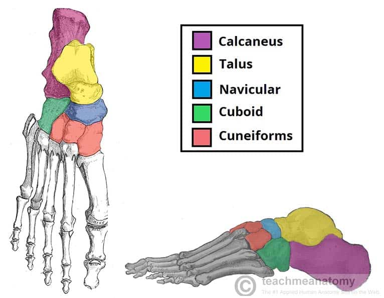
Tarsal bones [7]
calcaneus
talus
navicular
cuboid
3 cuneiform bones
only talus articulates w/ tibia & fibula at ankle joint
ossification takes place in all tarsal bones by 5th yr
metatarsals [5]
bases promixal
base of 1st metatarsal = attachment of fibularis longus muscle
base of 5th metatarsal = palpable landmark at midpoint of lateral aspect of foot
attachment of fibularis brevis muscle
heads distal
sesamoid bones
lateral ligaments of ankle
stabilize ankle laterally; prevent excessive inversion
anterior talofibular ligament [ATFL]
calcaneofibular ligament [CFL]
posterior talofibular ligament [PTFL]
anterior talofibular ligament [ATFL]
lateral malleolus → talus
weakest, most commonly injured
calcaneofibular ligament [CFL]
lateral malleolus → calcaneus
resists inversion when foot is in neutral
posterior talofibular ligament [PTFL]
lateral malleolus → posterior talus
strongest
medial ligament of ankle
deltoid ligament
stabilizes ankle medially; resists excessive eversion
helps maintain medial longitudinal arch
medial malleolus → talus, navicular, & calcaneus
made of 4 separate ligaments
distal tibiofibular ligaments
[syndesmotic ligaments]
bind tibia & fibula together at ankle mortise
important in high ankle sprains
avulsion fracture of tuberosity of 5th metatarsal
when foot suddenly & violently inverted, tuberosity of 5th metatarsal may be avulsed by tendon of fibularis brevis
associated w/ severely sprained ankle
pain & edema at base of 5th metatarsal
plantar fasciitis
repeated minor trauma → ossification in posterior attchmnt of aponeurosis
calcaneal spur [heel spur] may form
subtalar joint
AKA talocalcaneal joint
plane synovial joint
allows inversion/eversion
talocalcaneonavicular joint
ball-and-socket joint
supported by spring ligament
transverse tarsal [midtarsal] joint
[Chopart’s joint]
collective term for talonavicular part of talocalcaneonavicular joint
imp for inversion/eversion of foot
tarsometatarsal joints
[lisfranc’s joint']
plane synovial joints [gliding]
strengthen transverse arch
Lisfranc injury
occurs when bikers get foot caught in pedal clips or as result of high-E car accident
intermetatarsal joints
gliding type synovial joints
metatarsophalangeal joints
condyloid synovial joints
permit flexion/extension & ab/adduction of toes
lateral deviation of great toe at this joint
bunion
interphalangeal joints
hinge synovial joints
permit flexion/extension of toes
long plantar ligament
calcaneus → cuboid
helps maintain longitudinal arch of foot
short plantar ligament [plantar calcaneocuboid ligament]
deep to long plantar ligament
anterior surface of calcaneus → cuboid
plantar calcaneonavicular [spring] ligament
calcaneus → navicular
supports head of talus
plays imp role in maintaining longitudinal arch of foot
bunion
localized swelling at medial side of 1st metatarsophalangeal joint [inflammatory bursa]
*unusually associated w/ hallux valgus
medial longitudinal arch
highest & most important
talar head is “keystone” of arch
lateral longitudinal arch keystone
cuboid
deep veins
located underneath deep fascia, course along arteries
superficial veins
located subcutaneously, eventually drain into deep veins through series of perforating veins [including great & small saphenous veins]
perforating vein
connects deep & superficial venous systems
plantar venous arch
formed by plantar digital veins [formed by venous plexuses within plantar regions of toes] & plantar metatarsal veins
dorsal venous arch
formed by common digital veins [formed by dorsal digital veins & intercapitular veins] uniting w/ dorsal metatarsal
drains into medial and lateral marginal veins
medial drains into great saphenous vein
lateral drains into small saphenous vein
some veins from arch form anterior tibial vein [empties into popliteal vein]
popliteal vein
formed by anterior tibial, posterior tibial, and fibular veins
becomes femoral vein after piercing adductor magnus
becomes external iliac vein after leaving thigh & running underneath inguinal ligament
femoral vein
common site for deep vein thrombosis [DVT]
iliofemoral DVT carries greater risk of pulmonary embolism
used to place central venous catheter or line
used to deliver recreational drugs
internal iliac vein
receives from inferior & superior gluteal veins
drains into inferior vena cava
→ drains into right atrium
long [great] saphenous vein
longest vein in body
superficial, continuation of medial marginal vein of foot
passes anterior to medial malleolus
short [small] saphenous vein
superficial, continuation of lateral margin vein of foot
moves up posterior side of leg
virchow’s triad
contribute to venous thromboembolism [DVT formation]
increased tendency for intravascular clot formation [hypercoagulability]
increased structural changes in walls of affected vessels [changes in endothelial blood vessel lining]
decreased vascular blood flow [venous stasis]
DVT can be diagnosed by
venous decompression duplex ultrasound
doppler
CT
MRI
Serum D-dimer levels