Chapter 20- anatomy
 Call Kai
 Learn
Learn Practice Test
 Spaced Repetition
 Match
Match Flashcards
 Knowt Play
1/63

There's no tags or description
Looks like no tags are added yet.
Name | Mastery | Learn | Test | Matching | Spaced | Call with Kai | Chat |
|---|
No analytics yet
Send a link to your students to track their progress
64 Terms
gonads for F and M
Primary sex organs
– Female: ovaries
– Male: testes
functions of gonads
-Produce sex cells (gametes)
• Oocyte (female) and sperm (male)
– Produce sex hormones
• Estrogen, progesterone, testosterone
• Hormones affect the maturation, development, and activity of the reproductive system
Puberty
• Initiated when hypothalamus increase secretion of GnRH (gonadotropin-releasing hormone)
• GnRH stimulates release of gonadotropins (FSH and LH) from anterior pituitary
• FSH and LH stimulate the gonads to produce sex hormones
• Increased sex hormones start the process of gamete maturation and sexual maturation
Perineum
• Diamond-shaped area
– Bordered by pubic symphysis, ischial tuberosities and coccyx
• Urogenital triangle and anal triangle
female anatomy
1. Ovary
2. Fimbriae
3. Uterine tubes
(oviduct)
4. Uterus
5. Vagina
6. Vulva
7. Clitoris
ovaries
• Tunica albuginea
• Ovarian cortex
– Follicles (oocytes
surrounded by follicle cells)
• Ovarian medulla
– Loose connective tissue
– Contains blood vessels, lymphatic vessels, and nerves
Oogenesis and Follicle Development
• Oogenesis: Maturation of primary oocyte to secondary oocyte
• Follicle development: Primordial follicle → Primary follicle →
Secondary follicle → Mature follicle →Ovulation
28 day ovarian cycle
Three Phases:
Follicular phase (days 1-13)
– FSH and LH stimulate maturation of follicle and oocyte
Ovulation (day 14)
– LH surge causes expulsion of oocyte from ovary
Luteal phase (days 15-28)
– Remaining follicular cells turn into corpus luteum
– Corpus luteum secretes estrogen and progesterone, preparing uterus for possible implantation
– Corpus luteum breaks down into a white scar called corpus albicans
– Decrease in hormone release
leads to menstruation
Uterine Tube (fallopian tubes or oviducts)
Regions
• Fimbriae
• Ampulla
– Most common site of fertilization
Layers of the wall
• Mucosa
– Ciliated simple columnar epithelium
• Muscularis
• Serosa
Ectopic Pregnancy
• Implantation outside of the uterus
• Tubal pregnancy
– Fertilized oocyte implants in the uterine tube
– Uterine tube is unable to expand as embryo grows
– Embryo cannot survive past week 8
• Occasionally implantation occurs in the
abdominal cavity
uterus
Posterior and superior to the bladder
Functions
– Site of implantation
– Support, protect, and nourish the embryo and fetus
Gross Anatomy
– Fundus (#1)
– Body (#2)
– Cervix (#3)
External os (#4) is filled with a mucus plug that thins during ovulation
Cervical Cancer
Risk factors:
– Human papilloma virus (HPV) infection
– HIV infection
– Increased age
• Papanicolaou (Pap) smear
– Test to detect cervical cancer
• Treatments:
– Cone biopsy
– Hysterectomy
uterine wall
Three Layers
Perimetrium (#1)
• Serosa
Myometrium (#2)
• Muscularis
• Three smooth muscle layers
Endometrium (#3)
• Mucosa with two layers…
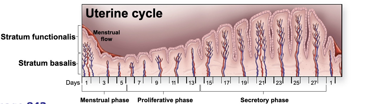
• Stratum functionalis
– Shed as menses
• Stratum basalis
28 day uterine (menstrual) cycle
Days 1-5: Menstrual Phase
– Functional layer sloughed
• Days 6-14: Proliferative Phase
– Development of functional layer
• Days 15-28: Secretory Phase
– Further vascularization of the functional layer
– Further development of uterine glands
• Endometrium is displaced onto external surface of organs within the abdominal cavity
• Displaced endometrium can still grow in response to hormones but menses cannot be shed and expelled out of vagina
• The displaced endometrium causes pain, scarring, and deformities
• Treated with hormones or surgery
vagina
Anatomy
• Fibromuscular tube
• Distensible wall
– Mucosa
– Muscularis
– Adventitia
• Fornix, rugae, vaginal orifice, vestibular glands, hymen
Functions
• Birth canal
• Receives the penis during intercourse
• Passageway for menstruation
External Genitalia (Vulva)
Mons pubis
• Labia majora
• Labia minora
• Vestibule
• Clitoris
• Urethral opening
• Vaginal orifice
Mammary gland
• Mammary glands
– Prolactin - create milk
– Oxytocin - eject milk
• Lobes and lactiferous ducts
– 10-20 per breast
• Nipple
• Areola
– Pigmented
– Many sebaceous glands
• Suspensory ligaments
breast cancer
• Affects 1 in 8 women
• Can occur in men
• Risk factors:
– Genetic influences
Family history
Breast cancer genes
– Increased exposure to estrogen over a long period of time
Early menarche with late menopause
Never having been pregnant
• Early diagnosis is key
– Self breast exam
– Mammograms
Menopause
• Normally occurs between the ages of 45 and 55
• Cessation of ovulation and menstruation
• Circulating levels of estrogen and progesterone decline
• Reduction in size of uterus and breasts
• Bone mass declines
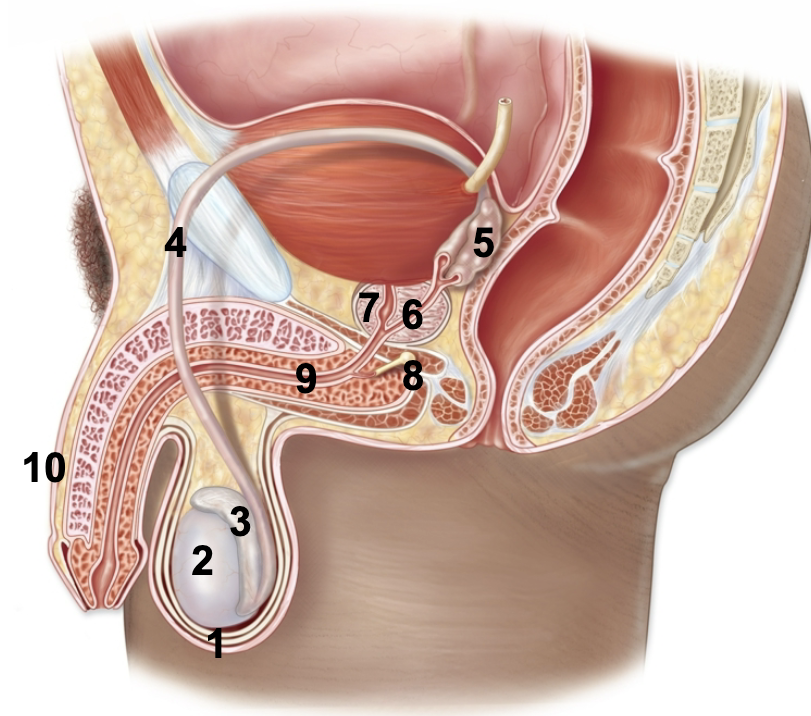
male anatomy
1. Scrotum
2. Testis
3. Epididymis
4. Ductus (vas) deferens
5. Seminal vesicles
6. Ejaculatory duct
7. Prostate gland
8. Bulbourethral gland
9. Urethra
10.Penis
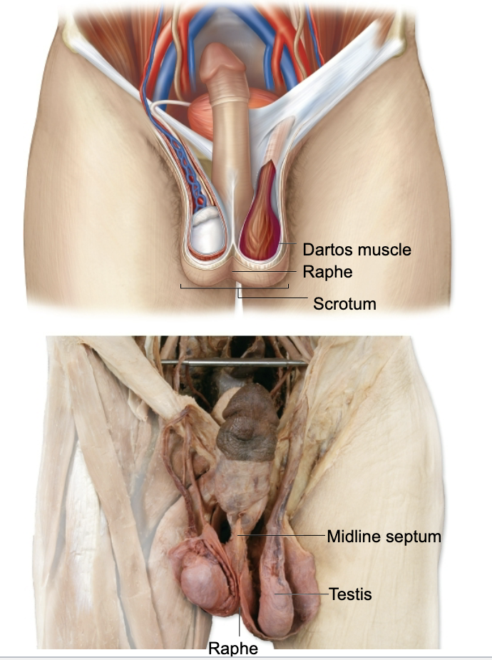
Scrotum
Function
• Provides cooler temperature needed for sperm production
Anatomy
• Raphe
• Median septum
• Dartos muscle
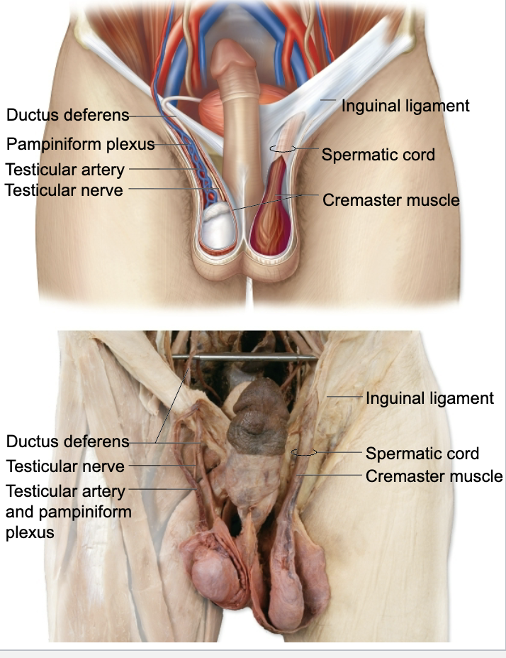
Spermatic Cord
Location
• Connective tissue tube that runs through the inguinal ligament
Contents
• Ductus deferens
• Cremaster muscle
• Testicular nerve
• Testicular vessels
– Testicular artery
– Pampiniform plexus
testes
Function
• Produce sperm and androgens (testosterone)
Anatomy
• Tunica vaginalis
• Tunica albuginea
• Septa
• Testicular lobules
• Seminiferous tubules
• Efferent ductules
Seminiferous Tubules
Sustentacular (Sertoli) cells
– Line seminiferous tubules
– Assist with sperm development
• Interstitial cells
– Between seminiferous tubules
– Produce androgens
• Spermatogenesi
Sperm
• Spermatogenesis
– Begins at puberty
– Occurs in seminiferous tubules
– Controlled by FSH and testosterone
– 100-200 million sperm produced each day
• Sperm become motile in
the epididymis
Male Ducts
• Epididymis
– Sperm are stored in the
tail of the epididymis
• Ductus (Vas) Deferens
– Wall contains mucosa,
muscularis and
adventitia layers
• Ejaculatory Duct
• Urethra
– Prostatic urethra
– Membranous urethra
– Spongy urethra
Accessory Glands
Seminal fluid is produced by:
Seminal vesicles
• Fructose (nourishes sperm)
• Prostaglandins (widens cervix)
• Bicarbonate (neutralizes vaginal acid)
Prostate gland
• Citric acid (nutrient for sperm)
• Seminalplasmin(antibiotic that combats UTI in males)
• Prostate specific antigen
(liquefies semen after ejaculation)
Bulbourethral glands
• Mucus (coats urethra, lubricant for sexual intercourse)
Semen
• Seminal fluid from the accessory glands combines with sperm from the epididymis to make up semen
• When released during intercourse, semen is called ejaculate
• Average ejaculation contains around 1 teaspoon of fluid and 200-500 million sperm
Prostate Disorders
• Benign Prostatic Hyperplasia
– Noncancerous enlargement of the prostate
– Very common: >90% of men over 80 suffer from it
– Symptoms
• Nocturia: urination at night
• Polyuria: more-frequent urination
• Dysuria: painful urination
• Prostate Cancer
– Risk increases with age
– Detected by digital rectal exam or PSA test
penis
Root, shaft, and glans
Three erectile bodies
-Corpus spongiosum
-Corpora cavernosa (2)
Vasculature
-Central arteries
-Venous spaces
-Dorsal veins
Spongy urethra
Erection and Ejaculation
• Erection
– Blood fills the erectile tissues
• Deep arteries dilate
• Blood is trapped in erectile tissues by compression of veins
– Under parasympathetic control
• Ejaculation
– Expulsion of semen
– Under sympathetic control
Prepuce (Foreskin)
• Skin covering glans
• Removed during circumcisio
Contraception (Birth Control)
• Abstinence
• Rhythm method
• Barrier methods (prevent sperm from entering uterus)
– Condoms
– Diaphragm
– Spermicidal foams and gels
• Surgical methods
– Vasectomy: vas deferens are cut and tied
– Tubal ligation: uterine tubes are cut and tied
IUD (intrauterine device)
– Implanted T-shaped device that may prevent
fertilization and or implantation
Chemical methods (most work by inhibiting ovulation or implantation)
– Oral contraceptives (birth control pills)
– Estrogen/progestin patches, implants or injections
– Vaginal ring
– Lactation
– Morning after pill
– Mifepristone or RU-486: triggers a miscarriage
(“abortion pill”)
Reasons for a Cesarean Section
• Previous C-section
• Narrow pelvis and large fetus
• Placental abruption (placenta detaches)
• Placenta previa (placenta covers the cervix)
• Atypical presentation of baby (breach, etc.)
• Cord prolapse (umbilical cord exits uterus before baby)
• Fetal distress
Stages of Labor
• Cervical Dilation
– Begins with first regular contractions and ends when the cervix is fully dilated
– Longest stage
• Expulsion
– Lasts from full dilation to delivery of the baby
• Placental
– Eliminates the placenta
– Accomplished within 15-30 minutes after birth of infant
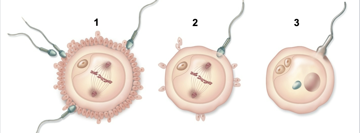
Capacitation and Fertilization
• Capacitation
– Period of sperm conditioning in female reproductive tract
– Prepares the acrosome for fertilization.
• Fertilization
– Occurs in the uterine tube
Three Phases of Fertilization
1. Corona radiata penetration
2. Acrosome reaction and zona pellucida penetration
3. Fusion of sperm and oocyte plasma membranes
and pronuclei
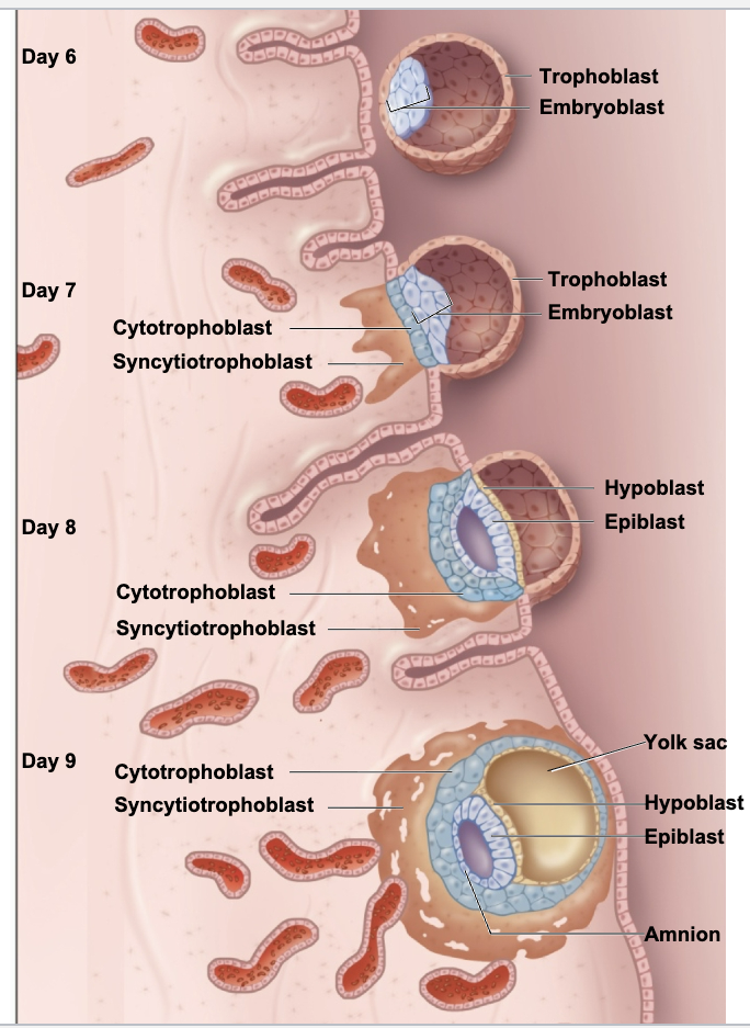
implantation
Usually begins ~7 days after fertilization
• Trophoblast subdivides
– Cytotrophoblast
– Syncytiotrophoblast
• Burrows into the stratum functionalis of the endometrium
• Produces human chorionic gonadotropin (hCG)
The Placenta
Anatomy
– Formed from maternal tissues (stratum functionalis) and fetal tissues (chorion)
– Chorionic villi immersed in a pool of maternal blood
– Mother and baby’s blood do not mix
FUNCTIONS
– Exchange of nutrients, respiratory gases, waste products and antibodies
– Production of estrogen and progesterone
Embryonic Period Overview (Weeks 3-8)
• Primitive streak forms
• Gastrulation
• Body folding
• Neurulation
• Limb buds form
• Organogenesis
sexual differentiation
• No difference in males and females before week 5
• Genital (gonadal) ridges become gonads starting in week 5
– Region on Y chromosome (SRY gene) causes testes to develop
– If no SRY gene is present, ovaries develop
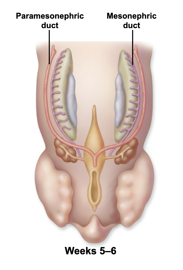
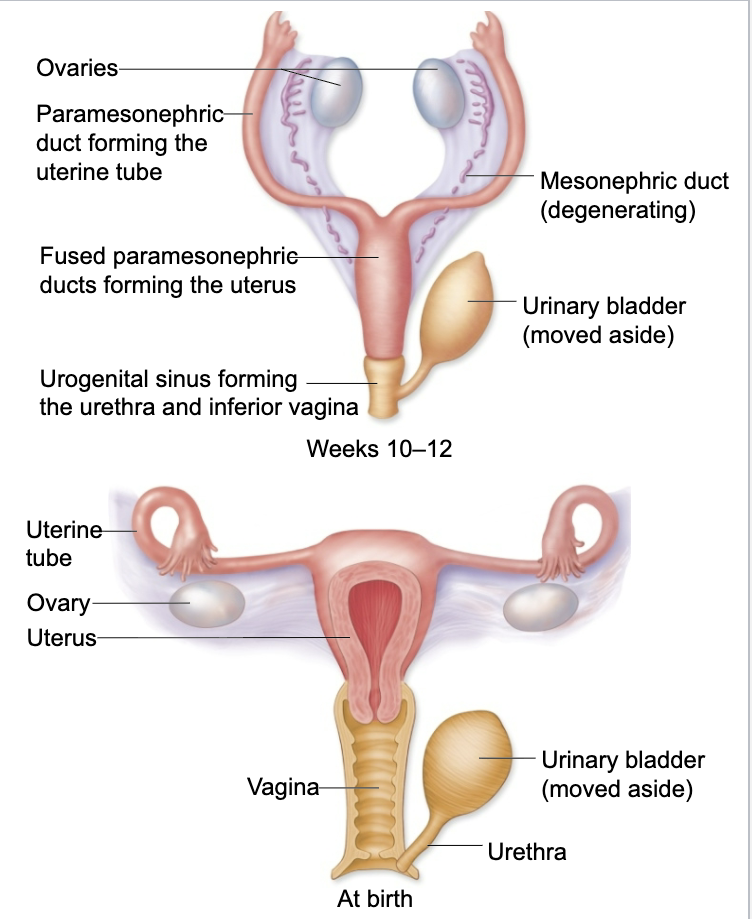
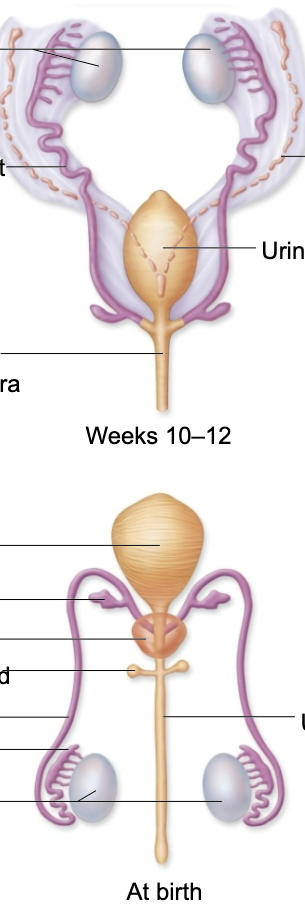
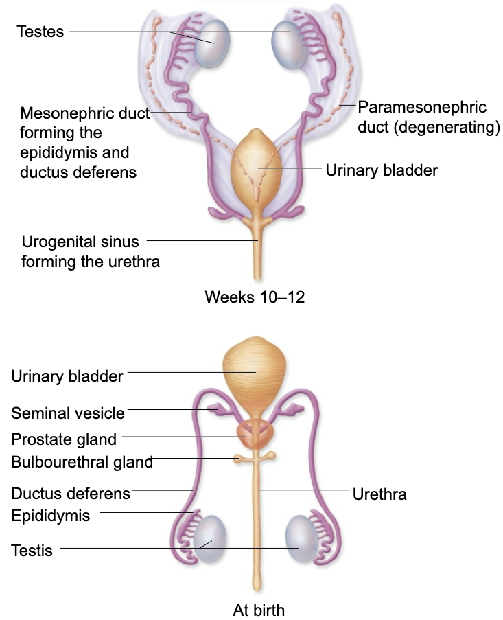
development of internal organs
• Embryos initially contain both male and female duct systems
– Paramesonephric ducts (female)
– Mesonephric ducts (male)
• Genes and hormones determine which duct system will degenerate
Internal Female Development
Internal Male Development
External Development
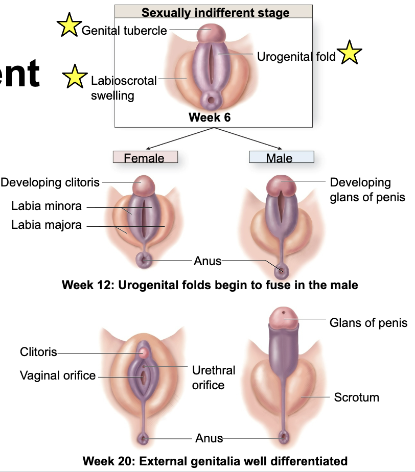
common function of ovaries and testes
Produce gametes and sex hormones
common function of Clitoris and Glans of penis
Contain autonomic nervous system axons that stimulate feelings of arousal and sexual climax
common function of Labia majora and Scrotum
Protect and cover some reproductive structures
common function for estibular glands and Bulbourethral glands
Secrete mucin for lubrication
late pregnancy
• Gestation lasts 38 weeks from conception (40 weeks from last menstrual period)
• Uterus expands ~20 times larger than normal
• Breasts enlarge and develop ability to produce milk
• Uterus myometrium becomes more active during last trimester. Mild contractions may occur.
• Hormones soften the cervix and loosen the pubic symphysis
• Baby “drops” and is normally oriented head down with face posterior
what happens during fetal period (weeks 9-38)
• Growth and maturation of existing organs
– Bones ossify
– Reproductive organs develop
– Brain enlarges
– Body elongates
– Limbs grow
– Organ systems become functional
– Baby movement begins
– Baby gains weight
Name the three phases of the ovarian cycle in order.
follicular, ovulation, luteal
What layer of the uterine wall is shed as menses?
stratum functionalis
Ovulation occurs after a surge in which hormone?
LH
Pick the correct order of female genitalia from posterior to anterior.
anus, vaginal orifice, urethral orifice, clitoris
True or false: Oogenesis and follicle development begin at the onset of puberty.
false
What two organs are reproductive homologues?
vestibular glands and bulbourethral glands
What happens first in fertilization?
corona radiata penetration
The ________ provides cooler temperature needed for sperm production.
scrotum
Erection is under ___ control; ejaculation is under ___ control.
Answer 1:
parasympathetic
Answer 2:
sympathetic