AUTOPSY
1/32
There's no tags or description
Looks like no tags are added yet.
Name | Mastery | Learn | Test | Matching | Spaced |
|---|
No study sessions yet.
33 Terms
Autopsy
The systematic examination of a cadaver for study or for determining the cause of death.
Auto - self
Opsy - look or appearance
Definition of “Autopsy” from Greek word
Determine the etiology or cause of death of a patient
Determine the pathogenesis
Preservation of tissues of the dead person for further examinations and for further research
Improvement of safety standards for the living.
An autopsy is performed as soon as possible to prevent post- mortem changes that can occur in the body.
Purpose of Autopsy?
Preparatory Measures
External Inspections
Internal Inspections
Dissection and Examination of organs
Analysis of Tissue, Fluids, and Other specimens
How is an Autopsy performed?
Preparatory Method
STEPS:
Consent is given by the nearest kin.
Documents such as the clinical abstract in coordination with the attending physician.
Medical records are provided
External Inspection
STEPS:
Scrutinize both anterior and posterior surfaces
Observe for signs and violence, lacerations, identifying marks, edema, distention, hemorrhages, and jaundice.
Internal Inspection
STEPS:
Primary incision of the abdomen and thorax
a “Y” incision is done for both males and females to expose the abdomen
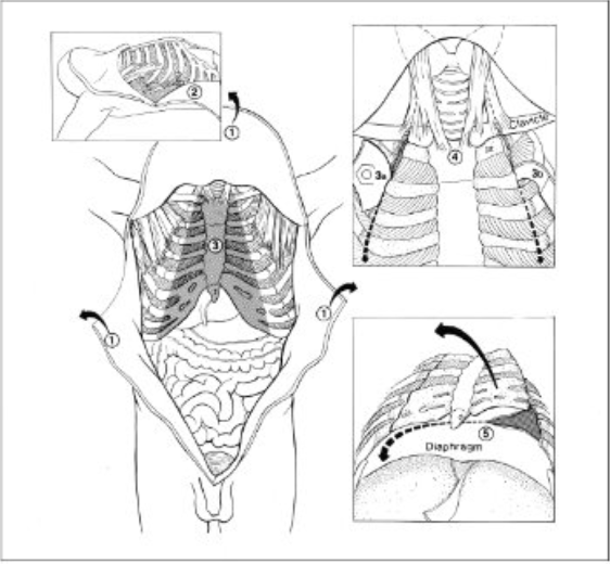
Reflect the skin flaps. Cut perpendicular to the ribcage. Make relaxing incisions in the peritoneum and musculature about 15cm above the symphysis
Check for tension pneumothorax. make a pool of water in the axilla. Push the closed clamp through an intercostal muscle beneath the water level.
Release the chest plate. Cut ribs medial to the costochondral junction and the clavicle lateral to the sternoclavicular joint with either:
-An oscillating saw; linoleum knife and bone shears; or pruning shears
Preserve the muscle attachments to the manubrium and head of the clavicle
Detach the diaphragm from the chest plate.
Inspect surfaces and contents of the pleural spaces.
INTERNAL INSPECTION:
Opening The Body
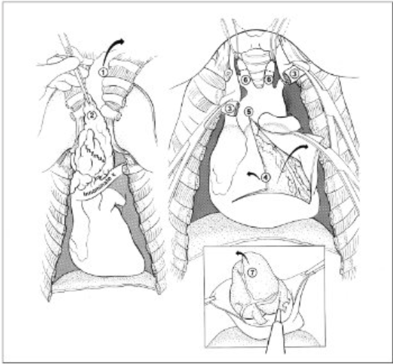
Reflect chest plate and strap muscles to expose the lower neck.
Blunt dissect thymic fat pad from the pericardium. Carry reflection upward to lower pole of thyroid. Cut the thymic vein where it enters the innominate vein.
Double-clamp, divide, and reflect the innominate vein.
Open pericardium and clamp edges. inspect pericardial surfaces and contents.
Extend the pericardial incision through the pericardial reflection.
Isolate and ligate the carotid arteries
Lift the heart cranially and draw blood samples from the left atrium.
INTERNAL INSPECTION:
Thorax
Examine the heart, Elevate, Palpate, and inspect the lungs.
Collect specimens for microbiology, toxicology, etc.
Take any cultures after searing the surface.
INTERNAL INSPECTION:
Thorax (In-Situ Examination)
Heart:
Dissect the heart starting from the part where blood first flows
Transverse
Start the incision from the inferior vena cava and cut through the superior vena cava
Kidneys:
Divide the kidney into anterior and posterior halves along the longitudinal axis of convexity
Lungs:
Sagittal section
Dissection and Examination of Organs:
Heart:
Kidneys:
Lungs:
Culture the blood from the heart aerobically and anaerobically
Inoculate on both liquid and solid media
Blood can be collected from other tissues
Analysis of Tissue, Fluids, and Other specimens:
What to culture?
Heat the metal spatula until glowing hot red, apply on the surface from which the culture is to be obtained.
Do not allow the area to be contaminated
Analysis of Tissue, Fluids, and Other specimens:
How to obtain the culture?
Heart’s Blood:
Collect using a sterile syringe with needle gauge (18-20) 20mL. on the ventricle or atrium by aspiration
Solid Organs:
Sterile by searing the surface and collect by plunging a sterile applicator stick on the organ
Analysis of Tissue, Fluids, and Other specimens:
How to obtain the culture: Heart’s Blood and Solid Organs?
Consent is given by the nearest kin.
Documents such as the clinical abstract in coordination with the attending physician.
Medical records are provided
How is an Autopsy performed?
Preparatory Measures
Scrutinize both anterior and posterior surfaces
Observe for signs and violence, lacerations, identifying marks, edema, distention, hemorrhages, and jaundice.
How is an Autopsy performed?
External Inspection
Primary incision of the abdomen and thorax
a “Y” incision is done for both males and females to expose the abdomen
How is an Autopsy performed?
Internal Inspection
Peritoneal Fluid:
Collect 50mL for culture and smear preparation
Pleural Fluid:
Collect 50mL for chemical analysis
Analysis of Tissue, Fluids, and Other specimens:
Analysis of Bodily Fluids: Peritoneal Fluid and Pleural Fluid
Lung Tissue for Culture:
Use of heated metal plate touched to the pleural surface
Localized cleansing using acetone or alcohol; slice the lung tissue and perform touch preparation
For Tuberculosis:
Lung should be fixed in 1:1 solution of 10% Formalin and 50% alcohol
Analysis of Tissue, Fluids, and Other specimens:
Lung Tissue for Culture?
For Tuberculosis?
Complete Autopsy
Types of Autopsy:
Examination of the organs of the 3 major cavities of the body (Abdomen, Chest and Head)
False.
Spinal cord is not removed and examined
False.
Blood vessels of the arms and legs aren’t examined.
Modified True or False.
The spinal cord is removed and examined
Blood vessels in the arms and legs are examined
Virchow’s Technique
Rokitansky’s Technique
Ghon’s Technique
Letullet’s Technique
4 Autopsy Techniques
Virchow’s Technique
Autopsy Techniques:
Organs are removed One by One
Widely used
Originally the first step was to expose the cranial cavity and from the back, the spinal cord, followed by the thoracic cavity, cervical cavity, and abdominal organs
Relationships between various organs may be hard to interpret.
Disadvantage of Virchow’s Technique
Rokitansky’s Technique
Autopsy Technique:
“In Situ” Dissection, in part combined with en-bloc removal
Ghon’s Technique
Autopsy Techniques:
Removal of the cervical, abdominal, and urogenital system organs as organ blocks / en-bloc
Letullet’s Technique
Autopsy Techniques:
Organs are removed en masse
Best for routine inspection and preservation of connections between organs and organ systems.
Best technique for preserving the vascular supply and relationships between organs
The body can be made available to the undertaker quickly, without having to rush the dissection and risk obscuring findings or destroying important specimens
Advantages of Letulle’s Technique
The organ mass is often awkward to handle, and the autopsy is difficult to perform without an assistant
Disadvantages of Letulle’s Technique
Prosecutor
Diener
Coroner
PERSONNEL IN AUTOPSY EXAMINATION
Prosecutor
PERSONNEL IN AUTOPSY EXAMINATION:
The individual that performs the dissection of the cadaver for anatomic demonstration and pathologic examination
Diener
PERSONNEL IN AUTOPSY EXAMINATION:
The laboratory worker who assists the performance of the autopsies and maintenance of morgues
Coroner
PERSONNEL IN AUTOPSY EXAMINATION:
An official whose duty is to investigate sudden, suspicious or violent death to determine its cause.