Module 2 Nervous System A&P for SLH
 Call Kai
 Learn
Learn Practice Test
 Spaced Repetition
 Match
Match Flashcards
 Knowt Play
1/62

There's no tags or description
Looks like no tags are added yet.
Name | Mastery | Learn | Test | Matching | Spaced | Call with Kai | Chat |
|---|
No analytics yet
Send a link to your students to track their progress
63 Terms
Central Nervous System
Brain & spinal cord
Command center that receives and processes info and control the response
Peripheral Nervous System
Cranial and Spinal nerves coming off brain stem and spinal cord throughout the body
Transmits sensory info to the CNS from the body and carries motor commands from the CNS to the body
Somatic nervous system
Carries sensory info and motor (voluntary) commands from CNS to skeletal muscles
Carries sensory information from peripheral organs to CNS
Autonomic Nervous System
Involuntary inner organs
Sympathetic: fight or flight
• Parasympathetic: rest and digest
• Enteric: functions of the GI tract
Efferent vs Afferent
When motor commands go from the brain to the muscles
Afferent neurons carry sensory information toward the central nervous system (brain) , while efferent neurons carry motor commands away from the central nervous system (to muscles).
Nerve impulses
electrical signals that travel through neurons to transmit information throughout
the nervous system, producing a specific response or function
Internruron
Connect similar neurons. Process info, not sending it
Motor Neuron
Transmits motor impulses from CNS to the body to be executed by muscles
Sensory Neuron
Transmits sensory information from sensory receptors to the CNS to be processed
Dendrite
Receives signals, send it to soma
Axons
Transmits signal through a neuron, covered in myelin (helps speed of signal sending)
Axon terminals
Where the axon comes to an end
Synapse
Specialized junction that allows a neuron to pass a signal to another cell
Glial Cells
Support & nourish neurons
Oligodendrocytes
lays down myelin on axons in the CNS
Schwann cells
lays down myelin on axons in the PNS
Microglia
immune defense and removal of cellular waste
Neurons
Signal sending/receiving cells in brain
Central sulcus
separates the hemisphere from front and back

Lateral sulcus/Sylvian fissure
separates temporal lobe from frontal and parietal lobes

Parieto-occipital sulcus
parietal and occipital lobes

Gray matter
information processing, decision- making, memory storage, muscle control
Made up of densely packed neuronal cell bodies/ neuron soma
White matter
Connects different gray matter areas and allows communication
between them through myelinated axons
Made up of axons, white because mylin
Inner part of cerebral hemisphere
Cerebral cortex
the visible surface of the brain (gray matter/outer layer)
-Outer portion of the cerebrum
-Most sophisticated part of brain
-Contains gyri and sulci
Laterilzation
When control of a function is dominated by 1 hemisphere relative to the other
Specialization
Control of a function is dominated by a particular region in the brain, regardless of hemisphere
Corpus Collosum
A white matter pathway that connects the 2 cerebral hemispheres
• Allows for communication between the left and right hemispheres
Frontal Lobe
Function: higher level cognitive skills, decision-making, problem solving, executive functioning, initiation of movement and inhibition of impulses
Linked to personality
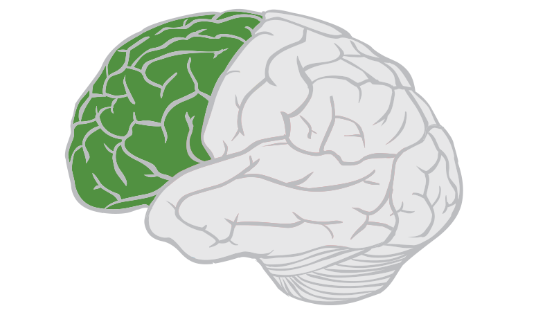
Pre-frontal cortex
Large anterior-most mass of frontal lobe
Primary motor cortex
Part of the frontal lobe responsible for voluntary movement
Generates the motor signal that is sent to the brainstem and spinal cord through neurons
Important regions control muscles involved in speech production and swallowing
Somatopic Organization of the Cortex
Representation of different areas of the brain's cortex correspond to different parts of your body.
Primary motor cortex → controls voluntary movement
Primary somatosensory cortex → processes sensations such as touch, pressure, pain, and temperature
Broca’s area
Left of frontal lobe
Planning and organizing motor behavior required for speech production
expressive language what we say vs receptive language- what we understand and take in

Temporal lobe
Conscious perception of auditory and smell info (hearing)
Located laterally/inferiorly of each cerebral hemisphere (below the lateral fissure)
• Contains Wernicke's area
• Contains hippocampus: memory

Wernicke’s Area
Understanding and processing language (receptive language)

Primary Auditory Cortex
Receives afferent signals of auditory stimuli (hearing)
Left hemisphere: receives auditory signals of speech sounds and transmits to Wernicke's Area to be processed for comprehension
Right hemisphere: receives auditory signals from the environment and transmits them to area just posterior for processing of non-speech sounds

Parietal Lobes
Conscious perception of touch, pressure, vibration, pain, temperature, and taste
Contains supramarginal gyrus and angular gyrus

Somatosensory Cortex
Receives and processes sensory information from the body
Touch, Pressure, Pain, Temperature, and proprioception (body position)
through the PNS to the primary sensory/ somatosensory cortex in the CNS
Basal ganglia
Group of neurons within cerebellum cortex part of the subcortical structures of the brain.
They help start, stop, and regulate movement. (DOES NOT CREATE)
Disorders: Parkinsons disease- tremors and slow movement
Supramarginal gyrus
Language/phonological processing, and sensory information
-in parietal lobe

Angular gyrus
Language comprehension, reading, writing, visual and language information
- located in parietal lobe

Occipital lobe
Visual stimuli & hearing

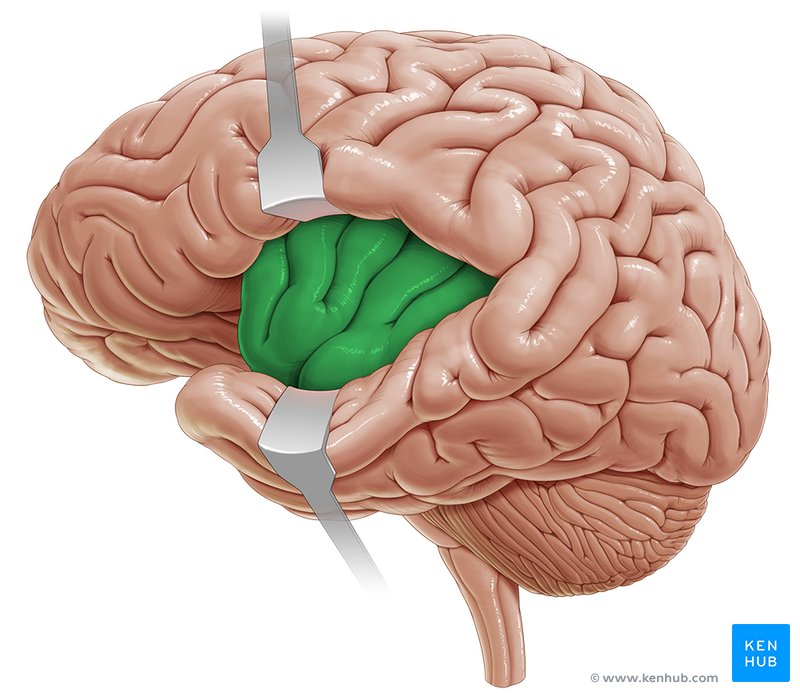
Insula
Involved in the control of speech, language, breathing, and swallowing
part of the limbic system (emotions, memory, etc.)
Under frontal and parietal lobes
Subcortical Nuclei
Deep gray matter structure that helps regulate movement sensation, emotion, memory, and body function.
Cranial nerves
- 12 pairs of nerves that control sensation and movement of head, neck, and face (sensory and motor)
10 of 12 in brainstem
Cranial Nerve V- Trigeminal
touch, pressure, and pain from mid and upper face and maxillary and motor
motor: muscles of jaw movements
sensory: lower face/teeth, outer ear and anterior 2/3 tongue (touch, temp, pain)
if damaged: Impaired mastication, Impaired oral sensation, Potential difficulties with feeding and swallowing
Cranial Nerve VII- Facial
Sensory: parts of external ear, taste for anterior 2/3rd of tongue
• Motor: muscles of facial expression
• Carries autonomic motor fibers to glands that secrete tears and saliva
• Aids in muscles of articulation
if damaged: bells palsy ex
Sensory Nerve VIII- Vestibulocochlear
Balance/coordination of head in space
Cochlear: Auditory/ hearing
Carries auditory and vestibular(coodination) info from the inner ear to the brainstem
Cranial Nerve IX- GlossoPharyngeal
(tongue)(throat)
Has voluntary and autonomic motor and sensory
components
• Motor: Innervates the stylopharyngeus muscle (in neck for swallowing)
• Autonomic motor: salivary gland secretion
• Sensory: parts of external ear, eardrum, upper part of pharynx
• Autonomic sensory: taste for posterior 1/3rd of tongue
Cranial Nerve X-Vagus
Motor: pharynx muscles, larynx, tongue
Autonomic motor: stimulation of mucous glands
Sensory: for pharynx, larynx, meninges, parts of the external ear and eardrum
Autonomic sensory: sensation from gut/bowels
Cranial Nerve XI- Spinal Accessory
a motor nerve that controls specific movements in the neck and shoulders
motor neurons are in the upper part of the spinal cord rather than the brainstem
• It innervates 2 muscles of the head and neck: Sternocleidomastoid muscle and Trapezius muscle
Cranial Nerve XII- Hypoglossal
Intrinsic and extrinsic muscles of tongue movement
Internal carotid arteries
travel upward through the neck and enter the skull.
They primarily supply the front and middle portions of the brain
Anterior cerebral artery (ACA)
Middle Cerebral Artery (MCA)
Middle Cerebral Artery
Blood vessel supplying blood to lateral frontal, parietal, and temporal lobes (needed for speech and language)
Part of internal carotid arteries

Vertebral arteries
travel upward through the vertebrae of the neck and enter the skull.
The two vertebral arteries join together to form Basilar artery

Basilar artery
Supplies blood to: Brainstem, Cerebellum, Inner ear
Eventually divides into the posterior cerebral arteries (PCAs).

Circle of Willis
a circular network of arteries located at the base of the brain.
provide collateral circulation, an alternate route for blood to reach brain tissue if one pathway becomes narrowed or blocked.
Like a roundabout connecting multiple roads:
Internal carotid system ↔ Circle of Willis ↔ Vertebrobasilar system

Spinal Nerves
Part of the PNS: 31 pair of nerves that act as communication lines from spinal cord and rest of the body
Motor: info from brain (CNS) to muscles & glands
Sensory (afferent) fibers: info from body to the brain (CNS)

Cortical Innervation Patterns
tells us whether a motor nucleus receives commands from one side or both sides of the brain
Describes how the motor cortex sends signals to motor
nuclei
Mostly bilateral
The two Cortical Innervation Patterns
Bilateral: Motor nucleus receives input from both sides of
the motor cortex
• Contralateral: Motor nucleus receives primarily opposite
side cortical input
Ventricles
four interconnected cavities (spaces) inside the brain that produces and circulates cerebrospinal fluid.
Cerebral spinal fluid
clear liquid that surrounds and protects the brain and spinal
cord, removes waste and provides nourishment
CSF Path
flows through the two lateral ventricles> third ventricle> fourth ventricle>
spinal cord and brain> reabsorbed into bloodstream
Cerebellum
Back part of the brain that helps make movements smooth, coordinated, and accurate.
Disorders: stroke- problems talking, walking and with coordination
