exam 4 new material
 Call Kai
 Learn
Learn Practice Test
 Spaced Repetition
 Match
Match Flashcards
 Knowt Play
1/46

There's no tags or description
Looks like no tags are added yet.
Name | Mastery | Learn | Test | Matching | Spaced | Call with Kai | Chat |
|---|
No analytics yet
Send a link to your students to track their progress
47 Terms

Carotid Endarterectomy
Invasive procedure that is performed in the OR. It is the removal of fatty plaque from the neck artery. A tube is inserted above & below the blockage to reroute blood flow.
Recommended if you have a severe narrowing in your CCA.


Angiogram
Imaging technique that uses xray imaging of vessels with an injectable die contrast (radio plaque) to visualize blocks
Vascular surgeon navigates through blockage with a wire with inflatable balloon

Angioplasty
Non invasive. Mechanical dilation of a blocked site in an artery.
After balloon device is positioned across blocked portion of the artery, the vascular surgeon inflates the balloon to expand the artery. The balloon is then deflated & removed.

Which vessels is angioplasty performed?
femoral artery in the groin
radial artery in wrist
reopening of a blocked artery
Which lab is angioplasty performed?
Cath lab with a balloon tipped catheter. Called non invasive because it is catheter based and is minimally invasive.

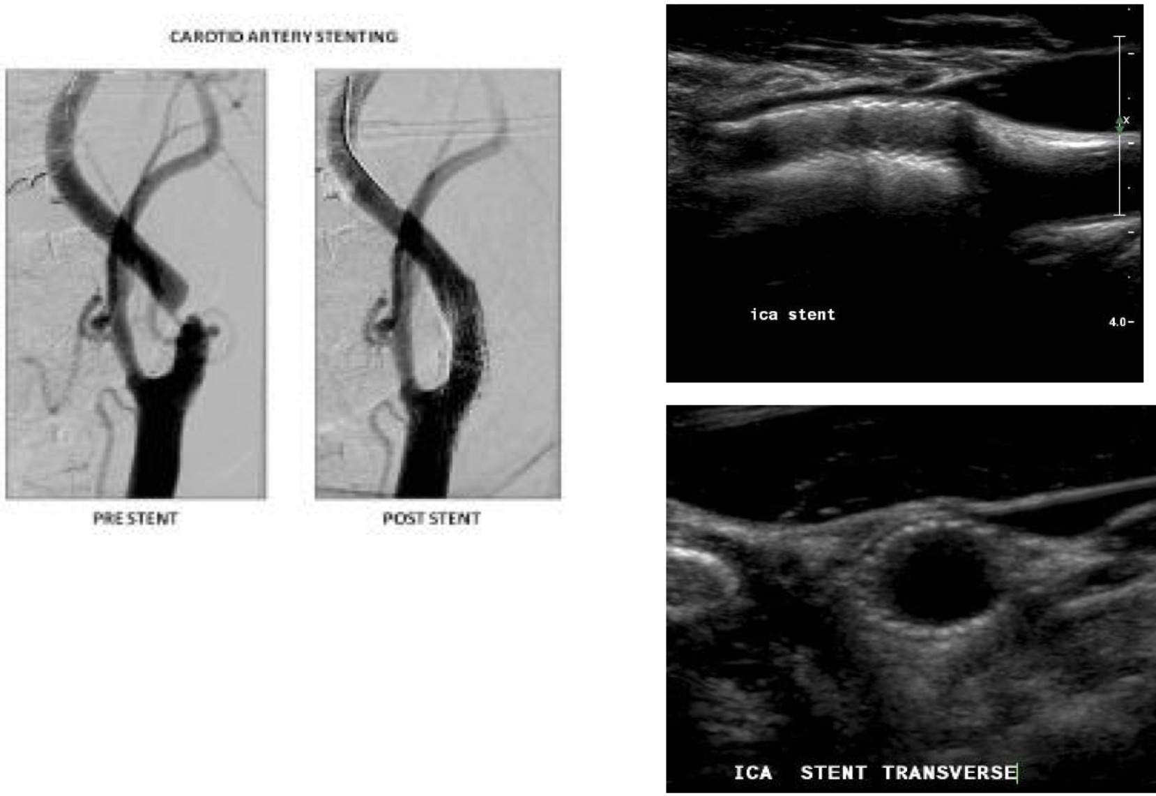
Carotid stents
Tube made of metal that is inserted into a vessel to keep it open. Stents are used during an angioplasty. Non invasive & performed in femoral or radial artery. Self expanding or balloon deployed.
Extracranial
Arteries that lay outside of the skull or cranium
vertebral
ICA
ECA
CCA
Intracranial
Arteries that lay inside the skull of cranium
ICA
vertebrals
Circle of Willis

Circle of Willis
Circle of vessels that lie at the base of the brain connecting the anterior & posterior circulatory systems. Provides important collateral pathway in cases of stenosis or occlusion. Developed through enlargement of secondary vessels after obstruction of a main channel.

% of normal circle of Willis
18%-20% of the time
Anterior circulatory system
Consists of the carotid arteries and their branches

Posterior circulatory system
Consists of the vertebral and basilar arteries & their branches

Posterior vessels of the circle of Willis
VA, basilar artery, posterior cerebral artery (PCA), posterior communicating artery (PCoA)
Vertebral Artery
1st branch off the SUB A
Enter the transverse process of the 6th cervical vertebra
Bend medically before entering cranial cavity through foramen magnum
R & L VA enter skull through foramen magnum and join to form a singular basilar artery
Forms the “Vertebrobasilar” circulation- supplies circle of willis
Supply the posterior portion of the brain with O2 rich blood
Basilar artery
forms the union of the two intracranial vertebral arteries
3cm long
BA extends anterior and superior from its origin
Terminates by diving into the posterior cerebral arteries
Posterior Cerebral Artery (PCA)
Perfuses the posterior hemisphere (occipital lobe) of the brain
Divided into 4 segments (P1-P4)
Only P1 & P2 are seen in transcranial Doppler
P1- origin to the PCoA
P2- distal to the PCoA
Posterior Communicating Artery (PCoA)
Courses posterior & medically from the posterior cerebral artery to join the ICA
2nd most common site for intracranial aneurysms
Most common collateral pathway, connecting the carotid and basilar
Anterior vessels of circle of Willis
ICA, OA, ACA, ACoA
ICA- anterior vessel of Circle of Willis
Supplies anterior portion of brain & eyes
4 segments
Cervical segment: ICA originates at the bifurcation & ends as the artery enters the base of the skull
Petrous segment: begins when ICA enters carotid canal
Cavernous segment: portion that forms the “S-shape” is termed the carotid siphone
Cerebral/Supraclinoid segment: termination into 4 branches
anterior cerebral
middle cerebral
posterior communicating
anterior choroidal
Ophthalmic artery- anterior vessel of Circle of Willis
1st branch off of ICA
Occurs at level of carotid siphon
Several branches- supraorbital, frontal, nasal
Anterior Cerebral Artery (ACA)
smaller of the terminal branches of the ICA
Carries 20%-30% of blood → brain
A1 connects the R and L sided ACAs
A2 joins branches of the PCA
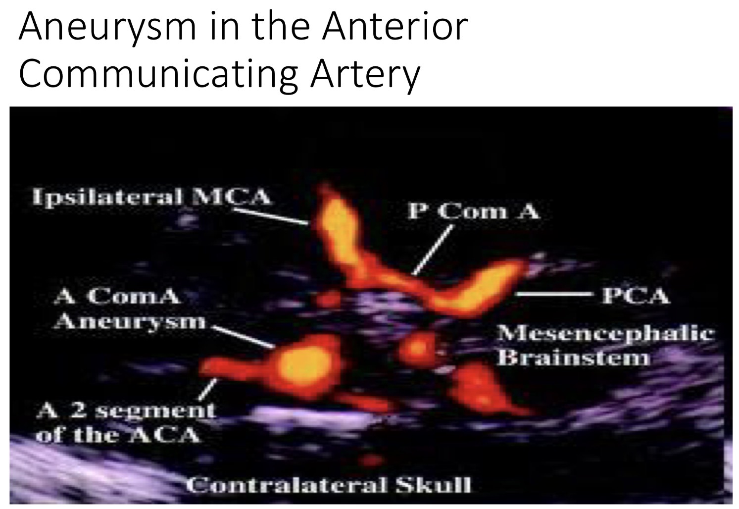
Anterior Communication Artery (ACoA)
Short vessel that connected the anterior cerebral arteries (A1-A2)
Most common site for intracranial aneurysms
Most common collateral pathway between R and L hemispheres
Middle Cerebral Artery (MCA)
Larger terminal branches of the ICA
Courses laterally towards the temporal bone
Carries 75%-80% of ICA flow
Larger than ACoAs
4 segments (M1-M4)
Only vessel that lays on the outside of Circle of Willis
Initial MCA bifurcation is the 3rd most common site for congenital aneurysms
The ACoA and PCoA are the 1st and 2nd most common sites for aneurysm formation

Transcranial Doppler (TCD)
Performed with a blind probe; will not see image (only waveform). 1-2MHz pulsed wave transducer

Transcranial Duplex Imaging (TCCD or TCDI)
Usually performed with phased array transducer 2-4MHz

Reasons for doing TCCD/TCDI
Stroke
Aneurysm
Cerebral microemboli
Stenosis
Sickle cell disease- disorders of hemoglobin; molecule in RBCs distort into sickle shape
TCD windows
Transtemporal
Transorbital
Suboccipital (transforiminal)
Submandibular

Transtemporal window
over the temporal bone
vessels identified:
MCA, ACA, PCA, TICA (terminal ICA), PCoA & ACoA
Transorbital window
Vessels identified:
ICA siphon, OA
Submandibular window
Vessel identified:
distal ICA
Sub-Occipital Window
Vessels identified:
VA, Basilar Artery (BA)

What is window A?
Transtemporal


What is window B?
Transorbital


What is window C?
Suboccipital (transforiminal)


What is window D?
Submandibular

Temporal approach: which vessels, directions, & depths? (MCA)
arterial segment: MCA (M1 & prox M2)
flow direction: towards transducer (M1), away from transducer (M2)
sample volume depth ranges: 30-60
Temporal approach: which vessels, directions, & depths? (ACA- A1)
arterial segment: ACA (A1)
flow direction: away from transducer
sample volume depth ranges: 60-75
Temporal approach: which vessels, directions, & depths? (PCA- P1)
arterial segment: PCA (P1)
flow direction: towards transducer
sample volume depth ranges: 60-75
Temporal approach: which vessels, directions, & depths? (PCA- P2)
arterial segment: PCA (P2)
flow direction: away from transducer
sample volume depth ranges: 60-65
Orbital approach: which vessels, directions, & depths?
arterial segment: OA
flow direction: toward transducer
sample volume depth ranges: 35-55
Foramen Magnum approach: which vessels, directions, & depths? (VA)
arterial segment: VA
flow direction: away from transducer
sample volume depth ranges: 60-90
Foramen Magnum approach: which vessels, directions, & depths? (BA)
arterial segment: BA
flow direction: away from transducer
sample volume depth ranges: 70-120

Anatomy 1-5
Brachiocephalic A
RCCA
R Sub A
R ECA
R Vert A

Anatomy 6-10
R ICA
R PCA
R PCoA
R MCA
R ACA

Anatomy 11-15
ACoA
L ACA
L MCA
L PCoA
L PCA

Anatomy 16-21
Basilar A
L ICA
L ECA
L Vert A
LCCA
L Sub A

What are vessels 1-10?
Anterior Communicating Artery
Anterior Cerebral Artery
Ophthalmic Artery
Internal Carotid Artery
Anterior Choroidal Artery
Posterior Cerebral Artery
Basilar Artery
Vertebral Artery
Posterior Communicating Artery
Middle Cerebral Artery
