NeuroDegen Diseases
 Call Kai
 Learn
Learn Practice Test
 Spaced Repetition
 Match
Match Flashcards
 Knowt Play
1/26

There's no tags or description
Looks like no tags are added yet.
Name | Mastery | Learn | Test | Matching | Spaced | Call with Kai | Chat |
|---|
No analytics yet
Send a link to your students to track their progress
27 Terms
Describe ALZHEIMER DISEASE (AD)
Stats
Genetic Stats
Clinical Presentation
Patho
STATS
MCC of dementia
onset > 50 yrs.
Prevalence increases with increasing age
Genetics Stats :
Majority = sporadic
5 - 10% = familial
Trisomy 21 -> develop manifestations of AD
Clinical Presentation:
impairment of higher intellectual function,
memory loss,
isorientation,
poor judgement,
Aphasia,
alterations in mood & behavior
Pathogenesis:
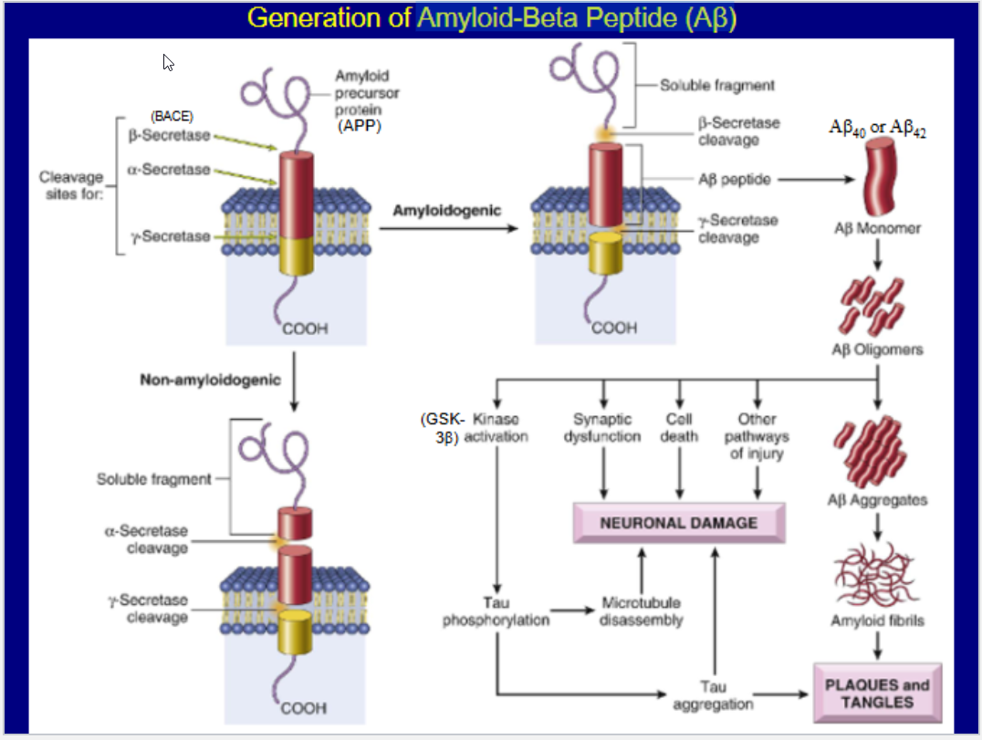
formation of amyloid beta peptide (AB)
an amyloidogenic peptide
NOTE:
Clinical assessment and imaging studies allow accurate Dx in 80% to 90% of cases
Describe the generation of Amyloid-Beta Peptide (Aβ)
List the Evidence implicating AB in the pathogenesis of AD
Which gene is largely responsible for AD
Alleles?
Fxn
How it increases risk
Describe the gross and Histological associations w/ AD
Evidence implicating AB in the pathogenesis of AD
found in brains of patients with AD
relatively specific for AD
AB = neurotoxic ( in vitro evidence)
some forms of familial AD have mutations in APP gene -> produces AB
APP gene= on chromosome 21 -> trisomy 21 -> gene dosage -> increases production of APP -> AB
mutations in genes encoding presenilin-1 & presenilin-2 -> familial AD -> increases y-secretase activity -> production of AB
ApoE Gene
Alleles:
E2, E3, E4 -> Isoforms ApoE2, ApoE3, & ApoE4
Fxn:
facilitate clearance of Aβ from the brain
ApoE4 = increases risk of AD
not as efficient as other isoforms in clearance of Aβ
increases production of Aβ42
exacerbates tau hyperphosphorylation
NOTE:
Mutations in tau gene -> Frontotemporal Lobar Degenerations, but not AD
Gross:
Cortical atrophy w/ secondary ventricular enlargement
Esp in frontal, temporal & parietal lobes,
medial temporal lobe are involved early -> atrophic in later stages
Histology:
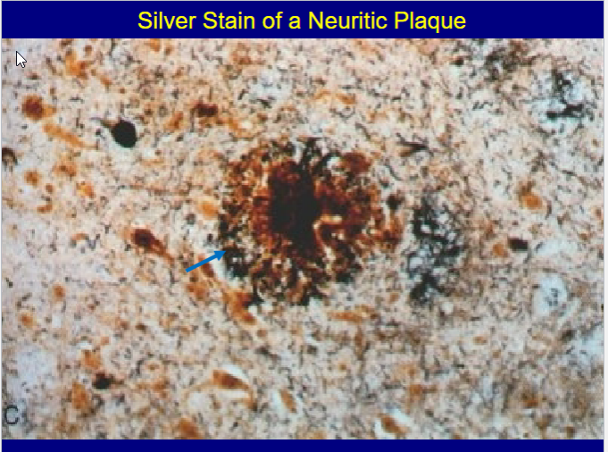
Neuritic Plaques:
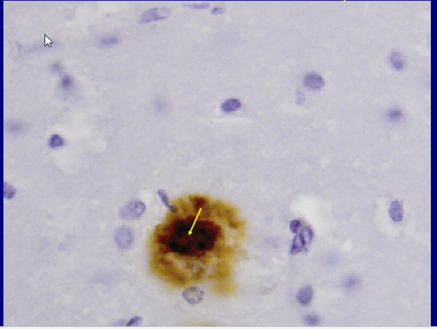
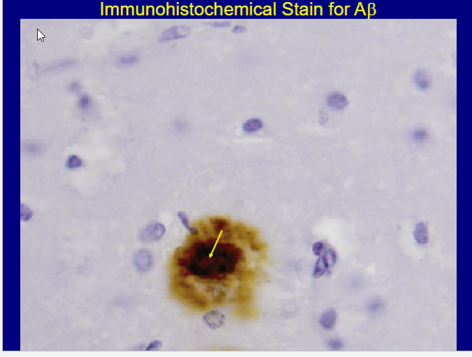
spherical collections of neuritic processes surrounding a central core of amyloid (AB)
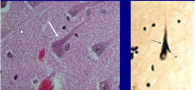
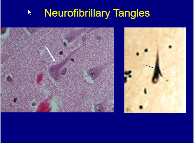
Neurofibrillary Tangles
cytoplasmic bundles of paired helical filaments composed of hyperphosphorylated tau
Cerebral Amyloid Angiopathy:
vascular deposits of amyloid (AB40)
What does a normal brain look like vs one w/ AD


Describe CTE
Pt Profile
Pathogenesis
Clinical Manifestations
Pathologic findings
Chronic Traumatic Encephalopathy (CTE)
Pts Profile:
individuals w/ history of mild repetitive TBI
Clinical onset of CTE can range from the 2nd to the 7th decades of life
Pathogenesis:
DAI
Diffuse Axonal Injury
Clinical manifestations
Behavioral & mood disturbances
Impulsive behavior
Mood swings with outbursts of rage
Substance abuse
Depression with suicidal behavior
Dysarthria
Dementia
Ataxia
Parkinsonism
Pathologic Findings
Cerebral atrophy w/ enlargement of ventricles
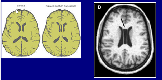
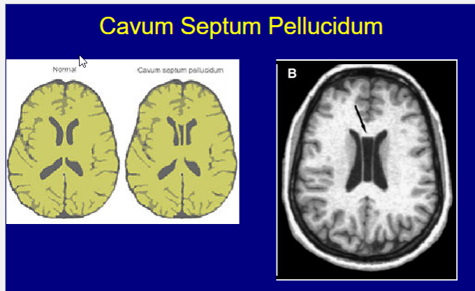
Fenestrated cavum septum pellucidum
NFTs around small blood vessels in cerebral cortex, particularly @ depths of the sulci
Severe: NFTs are present throughout the brain
TDP-43 inclusions
Describe Frontotemporal Lobar Degenerations (FTLDs)
What is it?
Clinical Manifestations
STATs
Pathos
Two types?
What is it?
Heterogeneous group of disorders associated w/ focal degeneration of frontal and/or temporal lobes
Clinical Manifestations:
Alterations in personality, behavior, and language
Precedes memory loss
Dementia (as it progresses)
STATs
same frequency as AD in those < 65
Patho:
Caused by aggregation of specific proteins
Two Types
FTLD-tau
FTLD-TDP
Describe Pick Disease
What is it?
Clinical Manifestations
Genetics
Patho
Morphology
What is it?
FTLD-tau
Clinical Manifestations:
Behavioral and personality changes
Language disturbances
Genetics:
Most cases are sporadic
Familial forms due to mutations in the MAPT gene
Patho:
Aggregation of tau w/in neurons
Morphology
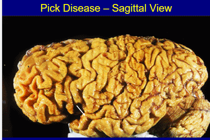
Atrophy of frontal & temporal lobes
“Knife-edge” atrophy
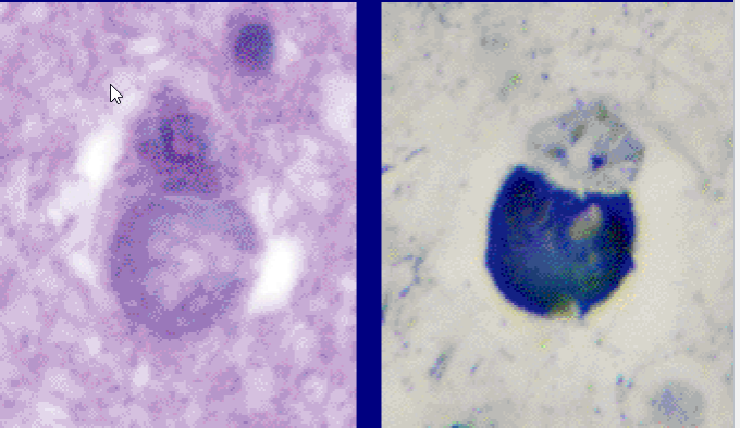
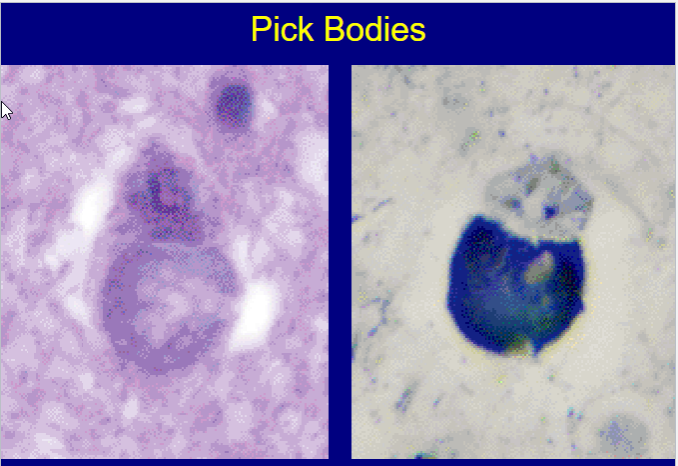
Marked neuronal loss with reactive gliosis
Many residual neurons exhibit “Pick bodies”

Describe PARKINSON DISEASE (PD)
Genetics
Clinical manifestations
Patho. (theories)
Histo
Neurochem
Rx
PARKINSON DISEASE (PD)
Genetics:
appears in the 6th - 8th decades
Most = sporadic, some autosomal dom./recessive
Clinical Manifestations:
Decreased Face. expression
Stooped posture,
festinating gait,
bradykinesia,
rigidity & pill rolling tremor,
± dementia
Pathogenesis (theories):
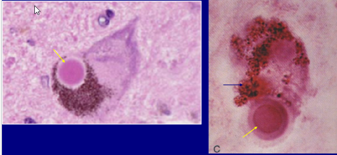
Aggregation of alpha-synuclein w/in neurons
major component of Lewy Bodies
autosomal dominant forms: mutations in the alpha-synuclein gene (SNCA)
mitochondrial dysfunction
Decreases ATP, Increases ROS + release of Cyt. C
PINK1 & parkin are involved in mitophagy
Mutation in genes = AR forms of PD
mutations in glucocerebrosidase gene
impairment of autophagy -> accumulation + aggregation a-synuclein
exposure to toxins
pesticides
Histology:
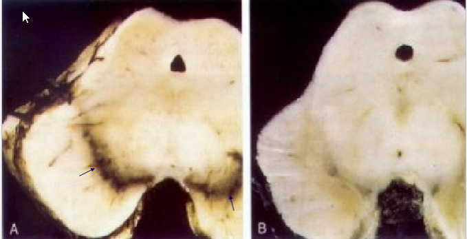
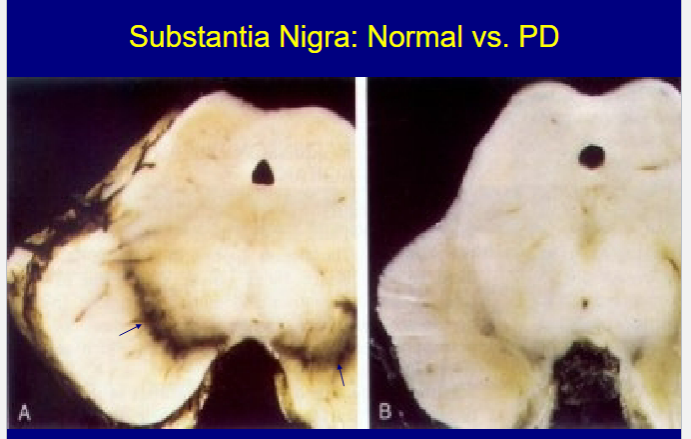
loss of pig. Dopa neurons in substantia nigra w/ gliosis,
intracytoplasmic Lewy bodies
Neurochemistry:
marked depletion of dopamine in the striatum
Rx:
L-DOPA

Describe the relationship between Parkinson Disease & Dementia
Relationship
Pt. Presentation
Characteristic Clinical features
Mech
Terminology
Parkinson Disease & Dementia
Relationship:
50 – 80% of patients with Parkinson disease will eventually develop dementia
Pt. Presentation:
Some have pathologic evidence of AD
Some have Lewy bodies in cortex + brainstem
Characteristic clinical features
fluctuating course
hallucinations
Mechanism:
neurons take up aggregates of α- synuclein -> PD spreads from lower brainstem up to cortex
Terminology:
"Dementia with Lewy Bodies"
If dementia develops before, concurrently with, or within 1 yr. of PD onset
“Parkinson Disease Dementia”
If PD has been present for at least 1 yr. before development of dementia
Describe HUNTINGTON DISEASE (HD)
Genetics
Anticipation
Patho
Clinical
Morph.
HUNTINGTON DISEASE (HD)
Genetics:
HTT gene on chromosome 4p encodes a protein “huntingtin”
encodes a polyglutamine tract in huntingtin
trinucleotide (CAG) repeat in coding region -> abnormal huntingtin
Anticipation:
The greater the # of trinucleotide repeats, the earlier the onset of disease
# of CAG repeats in a mutated HTT gene tends to increase during spermatogenesis
Pathogenesis:
Mutant huntingtin -> bind various trxn factors -> transcriptional dysregulation
proteasomal and autophagic degradation pathways Disruption
Clinical:
Choreoathetosis,
affective disorder
Dementia
Progress to death in ≈ 15 yrs
Morphology:
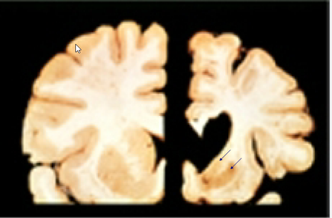
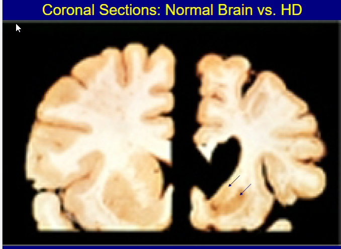
caudate nucleus & putamen
Atrophy
Loss of neurons with gliosis
Atrophy of frontal cortex w/ 20 dilatation of the ventricles
Intranuclear inclusions in surviving neurons
Cortical neurons are less severely depleted
NOTE:
Direct relationship between the degree of neuronal loss in the striatum & the severity of clinical Sx’s
Describe AMYOTROPHIC LATERAL SCLEROSIS (LOU GEHRIG’S DISEASE)
What is it?
STATs
Genetic Factors
Potential Proteins/Genes involved
ENV factors
Patho
Clinical Manifestations
What is it?
Worldwide degenerative disease of motor neurons progressive muscle weakness
STATs
incidence ≈2 per 100,000
Onset 5th decade or later, M/F 1.5-2 /1
Genetic Factors:
Familial ALS accounts for up to 20% of cases
two dozen genetic loci
Potential Proteins/Genes involved
Mutation in gene encoding SOD1: 20% of fam. Cases
SOD1 misfolds + aggregates -> death of motor neurons
Aggregation can occur w/o gene mutation
These Proteins -> both ALS & FTLD simultaneously
C9orf72
(GGGGCC) repeat in noncoding region
40% of familial ALS & 8-10% of sporadic ALS cases
TDP-43
FUS
Environmental Factors
Pesticides
Heavy metals
beta-N-methylamino-L-alanine (BMAA)
produced by cyanobacteria (blue-green algae)
can be incorporated into proteins -> misfolding
Pathology:
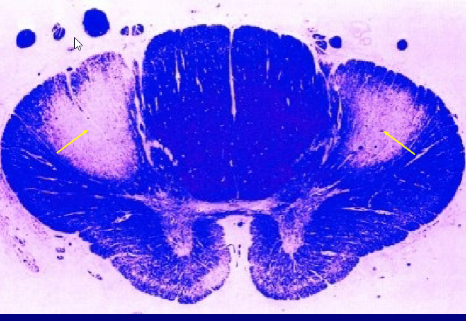
Loss of LMN in Ant. Horn of Spinal Cord
(Progressive Muscular Atrophy)
Loss of UMN in motor cortex w/ 2° loss of axons in the lateral columns
(Primary Lateral Sclerosis)
Involvement of CN motor nuclei
(Progressive Bulbar Palsy)
TDP-43 cytoplasmic inclusions
When C9orf72 & TDP-43 involved
Clinical:
Weakness of hands
often early symptom
spasticity of extremities with + Babinski sign
Progressive muscle weakness & wasting, fasciculations
Difficulty with speech & swallowing
Respiratory muscle weakness
NOTE:
course is ≈ 3-5 yrs.
Some ALS patients also have FTLD

Describe Spinocerebellar Degenerations
What is it?
MOA?
What is Spinocerebellar ataxias
What is it?
Group of degen diseases characterized by involvement of the:
Cerebellum
Spinal cord
Peripheral nerves
MOA:
Loss of neurons in affected areas -> secondary degeneration of white-matter tracts
Spinocerebellar ataxias
Group of AD inherited spinocerebellar degenerations
35 distinct subtypes
Many subtypes caused by trinucleotide repeat expansion mutations
Describe Friedreich Ataxia
STATs
Patho
Morphology
Clinical
Prognosis
STATs
Most common inherited ataxia
Patho:
GAA repeated in 1st intron of the frataxin gene -> defective Frataxin -> MIt. Dysfunction
Involved in assembly of iron-sulfur clusters
Present in mit. enzyme complexes I and II
Morphology:
Loss of dorsal root ganglion neurons -> 2degree degen of dorsal roots and dorsal columns
Loss of Betz cells -> 2degree degeneration of corticospinal tracts
Loss of neurons in dorsal nucleus of Clarke -> 2degree degeneration of spinocerebellar tracts
Loss of neurons in the cerebellum & some cranial nerve nuclei
Clinical
gait ataxia -> hand clumsiness + dysarthria
Onset; first decade of lifeP
Spastic weakness, + Babinski sign
Joint position + vibratory sense impaired
Decreased or absent DTRs
may have loss of sensation
pes cavus
Kyphoscoliosis
Cardiac Involvement
Prognosis
Most patients become wheelchair-bound within 5 years of onset
Life expectancy is 40 – 50 yrs.
Describe MS
What is it?
STATs
Clinical Progression
Symptoms
Gross features
Microscopic features
MULTIPLE SCLEROSIS (MS)
What is it?
Autoimmune demyelinating disorder
STATs
MC demyelinating disease
prevalence ≈ 3/1,000 in the US
Most commonly presents 20-40 yrs, mean ≈ 30 yrs
F/M ≈ 3:1
Clinical Progression:
Chronic relapsing & remitting course
episodes of neurologic deficits over weeks to months, followed by partial recovery
Symptoms:
Unilateral visual impairment (optic neuritis)
Internuclear ophthalmoplegia
Facial weakness,
vertigo
Ataxia
sensation & spastic weakness,
bladder dysfunction
Gross
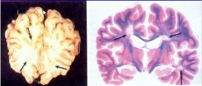
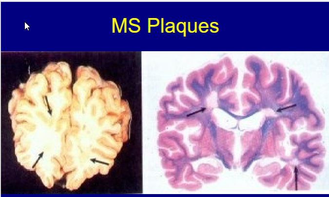
Hallmark = Plaque
relatively firm well- circumscribed area of dark discoloration in the white matter
involves
periventricular white matter (corona radiata),
optic nerves & optic chiasm,
spinal cord
others
Microscopic
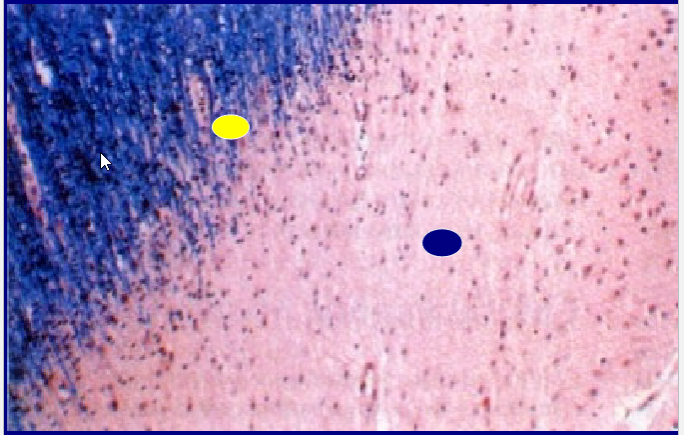
Active plaques
ongoing myelin breakdown w/ numerous foamy macrophages
(and some lymphocytes)
relative preservation of axons
Inactive
few inflammatory cells,
little to no myelin,
dense gliosis,
Decreased oligodendrocytes + markedly decreased axons

Describe MS
Patho
Diagnostic Feature
Genetic Factors
ENV factors
Prognosis
Pathogenesis
Autoimmune
Mediated by CD4+ T cells (TH1 & TH17) -> against CNS antigens
Act. Macrophages/Microglial -> demyelination
Diagnostic Feature:
Oligoclonal bands (OCBs)
Restricted bands on CSF electrophoresis composed of IgG
Imp in diagnosis/prognosis
Genetic factors
15× higher if a 1st degree relative has the disease
Multiple immune system genes are associated with MS
HLA-DR15 haplotype = strongest genetic association
Environmental factors
latitude gradient
higher prevalence at higher latitudes both north & south
(EBV)
May trigger via molecular mimicry
Prognosis
Significant variation in severity of relapses & rate of accumulation of neurologic deficits
average lifespan ≈ 7 years shorter than normal
individuals who don’t have MS