Internal Medicine EOR Topics
 Call Kai
 Learn
Learn Practice Test
 Spaced Repetition
 Match
Match Flashcards
 Knowt Play
1/86

There's no tags or description
Looks like no tags are added yet.
Name | Mastery | Learn | Test | Matching | Spaced | Call with Kai | Chat |
|---|
No analytics yet
Send a link to your students to track their progress
87 Terms

Premature atrial contractions (PACs)
Early beat originating from an ectopic pacemaker site in the atria, early beat interrupts the regularity of sinus rhythm, increase automacity of pacemaker cells
Same ectopic site= regular irregular rhythm
Different ectopic site= irregular, regular rhythm
Risks: caffeine, nicotine, cocaine, amphetamines, thyroid disease, post-MI, stress/anxiety, hypoxia, CHF, Pulm dz
Tx: Infrequent: no treatment, Frequent: underlying cause
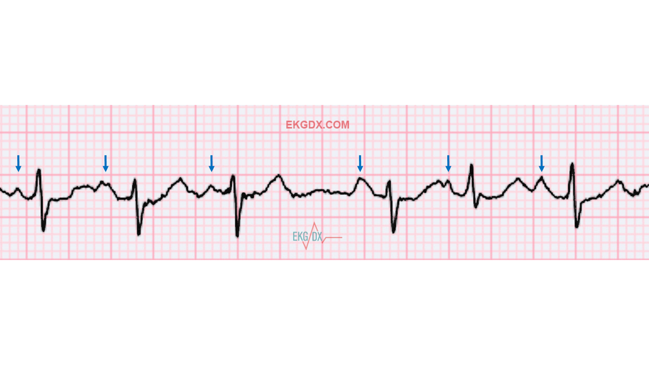
Multifocal atrial tachycardia and wandering atrial pacemaker (WAP)
Occurs when the pacemaker site shifts back and forth b/t sinus node and ectopic atrial sites; P-waves vary in size, shape or direction across the rhythm strip as the pacemaker “wanders” b/t multiple sites
3 different P-wave morphologies AND MAT: HR>100 OR WAP: HR<100
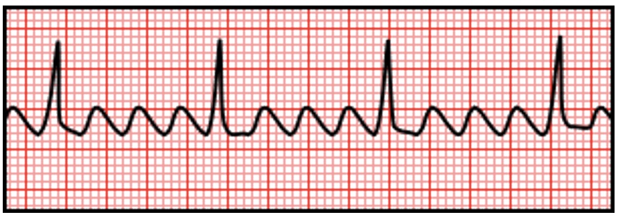
Atrial flutter
Originates in ectopic pacemaker site in the atria discharging atria impulses at a rate of 250-400 bpm; ventricular contraction rate typically regular and ranges from 75-150 bpm, blood pools and the atria cannot drain
rapid atrial impulses create a V-shaped waveform with a sawtooth pattern
TX:
Unstable: Cardioversion (synchronized electric shock)
Stable: Rate control (B-blocker or CCB) or Rhythm control (amiodarone or cardioversion with med failure), catheter ablation (PREFERRED LONG TERM MANAGEMENT)
Anticoagulation (decrease stroke incidence)
Onset <48 hrs can convert rhythm
Onset >48 hrs begin anticoag or schedule TEE for clot evaluation before cardiovert
Long term anticoag with a DOAC
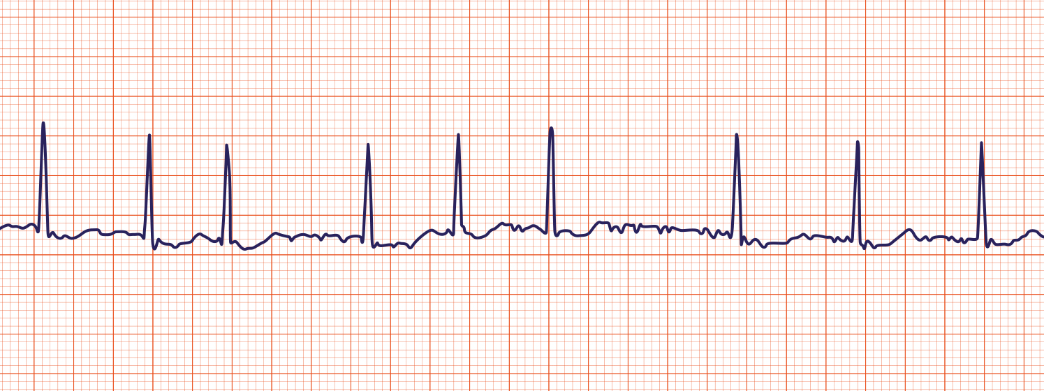
Atrial fibrillation (A-fib)
MC arrhythmia requiring treatment and can be secondary to many medical conditions; atria contracts 300-500 contractions in a minute
atrial pathology: increase atrial pressure, atrial dilation (disrupts underlying conduction path)
Atrial rate >300 bpm, irregular rhythm, variable ventricular rate, absence of P-waves, irregularly regular, varying R-R interval
Risks: enveloping blood clots, stroke, 60 and older
S/S: dizziness, palpitations, fatigue, mild dyspnea, chest pain
Tx: rate control, rhythm control, stroke prevention (anticoagulation)
Unstable: Cardioversion (synchronized cardioversion)→ TEE or anticoag first
DO a CHADS-VAS risk score (stroke risk >2 in men, >3 in women)
Stable: 1ST LINE- Rate control and anticoag for long term maintenance
Rate: Beta blocker (metoprolol, atenolol), CCBs (diltiazemn, verapamil), amiodarone
Anticoag: DOAC (apixaban, rivaroxaban)
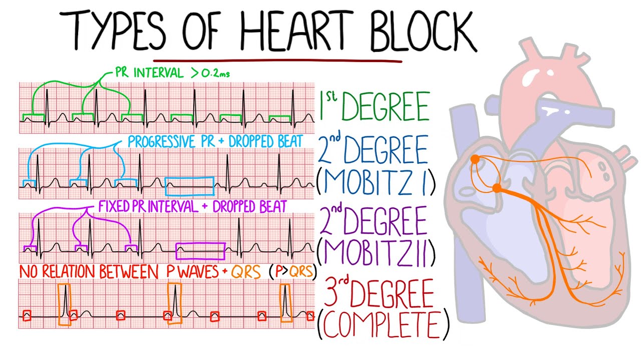
Heart block
Rhythms where there is a delay or block in the conduction of impulses from the atria to the ventricles, 3 classes
1. First degree AV block: all conducted just delayed
2. Second Degree AV block
A. Mobitz I: Progressive PR interval with a dropped beat
B: Mobitz II: Fixed PR interval with a dropped beat
3. Third degree AV block: non conducted, firing completely on their own
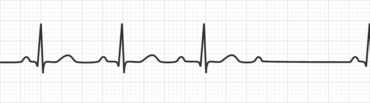
Second degree heart block Mobitz Type I (Wenckebach)
Initial impulse leaves the sinus node and travels through the AV node, is conducted through the ventricles but each successive impulse has increasing difficulty passing through the AV node until finally an impulse does conduct to the ventricles
PR intervals longer and longer until dropped QRS

Second Degree Heart block Mobitz Type II
More than one P-wave to each QRS complex
PR is normal or prolonged but will be consistent
Can progress suddenly to 3rd degree
TX: PERMANENT PACING if it does not resolve
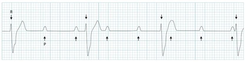
Third degree heart block
Impulses fail from atria to ventricles, complete heart block, beat totally separate of eachother
TX: permanent pacemaker
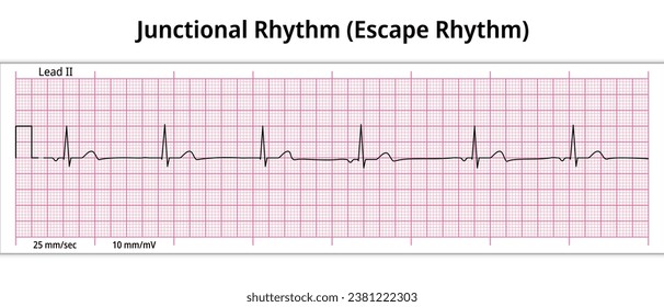
Junctional escape rhythm
Junctional arrhythmia the heart rate is low at 40-60 bpm, SA node fails to initiate impulse so a secondary pacemaker within AV junction takes over
P-waves typically absent/buried
Bradycardia

Premature ventricular contractions (PVCs)
Early ectopic impulse that originates in ventricular tissue either in the right or left ventricle, premature wide morphology
QRS complex is wide and morphology is different b/c of underlying rhythm
Pause follows the PVC, usually compensatory
Risks: MC in those w/ heart disease
Tx: not indicated if asymptomatic
Supraventricular arrhythmias
Originate ABOVE the cardiac ventricles
narrow QRS complex, less serious because ventricle activity is preserved
S/S
Fast heart rate: palpitations, chest pain, nausea, diaphoresis
Slow heart rate: dizziness, syncope

Supraventricular Tachycardia (SVT)
Often occurs in patients without structural heart disease, episodes start and end abruptly (seconds to hours)
Rate 140-240 bpm, regular rhythm (not sinus)
Loss of normal contour, P-wave differs in contour from sinus beats and is often buried in the T-wave (no sinus), Normal QRS complex
S/S: palpitations and anxiety
Tx: depends on pt stability
Unstable (hypotension, AMS, HF): cardioversion (synchronized electric shock)
Stable (least to most invasive)
Vagal maneuvers (carotid massage, Valsalva)
Bolus of adenosine IV →If that doesn’t work do another bolus of adenosine→ If ineffective do another bolus of adenosine
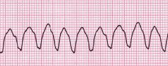
Ventricular Tachycardia (V-Tach)
Originates from an ectopic site in the ventricles discharging impulses at a rate of 140-250 beats per minute (complication of MI or cardiomyopathy)
3 or more premature beats
Tombstone appearance
Tx
Unstable: cardioversion (w/pulse)→ if recurs give amiodarone and shock again
Stable: amiodarone, if unsuccessful sedate patient and perform cardioversion as indicated
Long term: B-blocker or CCB
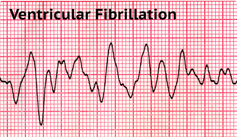
Ventricular fibrillation
No regularity, no complexes or waves present are able to be analyzed (no pulse), death is imminent unless rhythm is treated immediately
1. Defibrilate at highest energy level (unsuccessful do IV lines and intubate)→ Administer epinephrine and perform 5 cycles of CPR→ Continue drug therapy, CPR and defibrillation attempts
Once pt is revived, tx the underlying cause
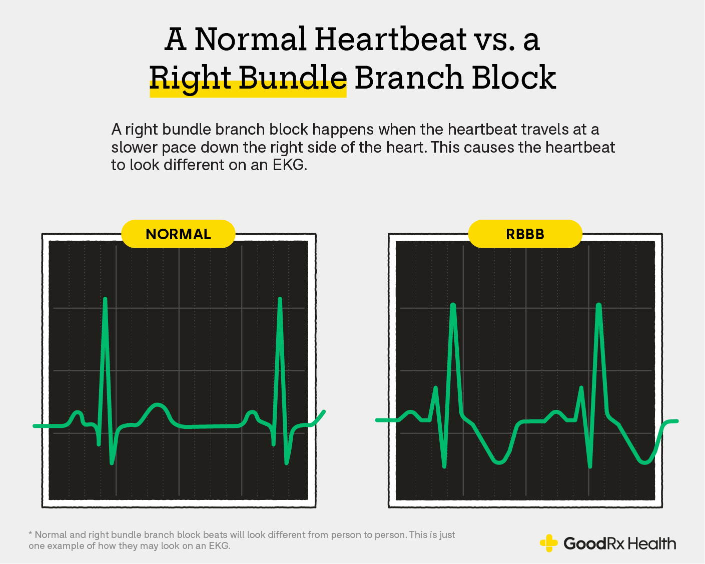
Right bundle branch block
QRS >120 ms elongated, in lead V1 or V2 an rSR or rSR (rabbit ears pattern is typical, wide or slurred S-waves in leads I and/or V6, T-wave inversion
Often incidental finding and benign in pts w/o heart disease
Dx: ECG, ECHO
TX: underlying cause, evaluate for pacemaker

Left bundle branch block
Prolonged QRS duration >120 ms with normal morphology; dominant S-wave in V1 and tall R waves in V6, often associated with underlying heart conditions like HTN or CAD
Risks: more significant cardiac issues (ischemic heart disease, cardiomyopathy, valvular heart disease)
S/S: fatigue, SOB, palpatations, chest pain/discomfort, syncope/presyncope
Dx: ECG, ECHO, evaluate for extensive heart disease
Tx: underlying cause, pacemaker, cardiac resynchronization therapy (CRT)

QT Prolongation
Delayed ventricular repolarization , longer reset of the heart after each beat
1. Congenital Long QT Syndrome: 1 in 2500 live births, long QT interval (usually >470 msec), ventricular arrhythmia, typically polymorphic ventricular tachycardia in presence of congenital deafness
2. Acquired long QT Syndrome: usually second to the use of antiarrythmic agents, certain abx, electrolyte abnormalities, myocardial ischemia, or bradycardia
S/S: asymptomatic; palpatations, syncope (can be misdiagnosed for seizures), cardiac arrest
DX: Personal and family history, ECG, exercise ECG for long QT, genetic testing (cardiac arrest)
Tx: Long QT- Beta blockers, Inherited Syndrome- cardiac arrest due to genetics, implantable cardioverter-defibrilator
refer to cardiology
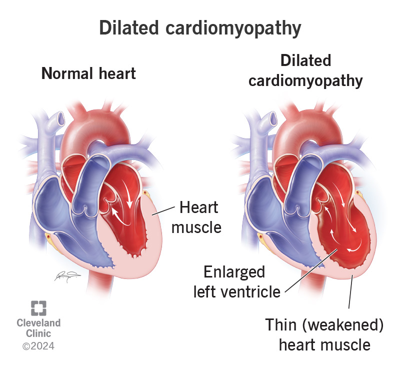
Dilated cardiomyopathy
LV or bi-ventricular chamber thinning and enlargement and systolic dysfunction, high end-diastolic and high end-systolic volumes; diminished cardiac output, interstitial and endocardial fibrosis, enlargement of the remaining heart chambers
Risks: viral, bacterial, chemo, ethanol, cocaine, uremia, thyroid hormone, pregnancy, HEAVY EXPOSURE, SMOKER
S/S: HF- progressive DOE, orthopnea, peripheral edema,JVD, PND, edema, abd pain, nausea, congestive cough, fatigue, weakness, rales
Acute presentation is often misdiagnosed as a viral URI in young adults
DX: Hx is important, ECG ± a Holter monitor, ECHO (LV dial, thin ventricular walls, decrease EF), Cardiac cath
TX: Underlying cause
ANRI or ACE/ARB
B- Blocker
Aldosterone agonist
SGLT2I w/ loop diuretic
Sx: LVAD, cardiac resynchronization (CRT), automatic implantable cardioverter-defibrillator, heart transplant
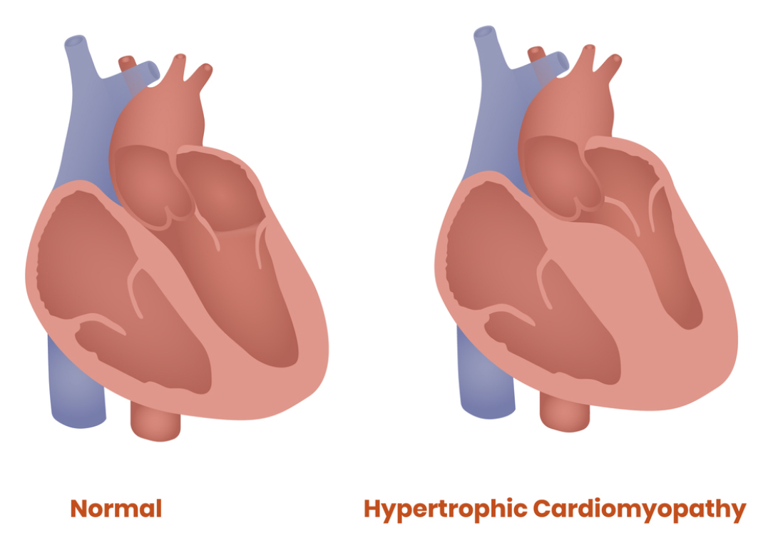
Hypertrophic cardiomyopathy
Genetic disease of heart muscle (myocardium)- frequently autosomal dominant, mutation in sarcomere (muscle gene), unexplained LV hypertrophy w/o dilation for the ventricles or a cardiovascular disease that could cause the degree of hypertrophy seen; LV is small and hypercontractile with increase wall stress
RISKS: STUDENT ATHLETES, males, 3rd decade, structural abnormalities of septum , subendocardial ischemia
1. Obstructive: midsystolic obstruction of flow through the LV outflow tract as a result of systolic anterior motion of the mitral valve toward the septum (SAM)
2. Non-obstructive
S/S: most patients asymptomatic until sudden death from V-tach or V-fib, fatigue, dyspnea, angina, palpatations, presyncope/syncope
PE: double apical impulse, S2 split, systolic ejection murmur
ECHO: LVH with no dilated LV
ECG: left axis deviation and abnormal Q waves
TX: BETA BLOCKER
Sx: surgical septal myectomy or alcohol septal ablation

Restrictive cardiomyopathy
Deposits in tissue causing stiffness (does not relax), non-dilated, non-hypertrophied ventricles w/ impaired LV filling; diastolic dysfunction, LV wall thickness may be increased by infiltrative disease, bi-atrial enlargement often severe
Risks: primary idiopathic (most cases), amyloidosis, sarcoidosis, hemosiderosis, genetics, Loeffler Eosinophilic Endocardial Disease
S/S: symptoms of HFpEF, orthostatic HoTN, syncope, orthopnea, fatigue
PE: R sided heart failure, cardiac cachexia, loud S3 murmur, systolic murmur, apical impulse, JVD
Amyloidosis: periorbital purpura, macroglossia, easy bruising
DX: ECHO (front line- biatrial enlargement and diastolic dysfunction), BX for amyloidosis, Lab (eosinophilia), ECG (90% abnormal- nonspecific ST-T wave changes, low voltage QRS)
Cardiac cath or Bx is the best
TX
Medication: Beta blocker or non-DHP CCB (verapamil, diltiazem)
Tx underlying disorder ex. Chemo for amyloidosis
Pacemaker implantation
Coronary artery disease (CAD)
Blood vessels become narrowed or blocked by a buildup of fatty plaque
Risks: atherosclerosis, smoking, poor diet, high cholesterol, diabetes, HTN, family history
S/S: angina, SOB, fatigue, “silent”
Tx: lifestyle changes, treat underlying
Acute coronary syndrome
Spectrum of clinical symptoms that include
Unstable angina, acute myocardial infarction (N-STEMI, STEMI)
Acute myocardial injury and evidence of ischemia and elevated troponin with one of the following
Ischemic symptoms (chest pain, dyspnea)
Development of pathological Q waves on ECG
ECG changes indicative of ischemia (ST segment elevation or depression)
Imaging evidence of new loss of viable myocardium
Identification of coronary thrombus by angiography or autopsy
STEMI
Occlusive coronary thrombus at site of pre-existing atherosclerotic plaque; ST elevation denotes an acute coronary occlusion and warrants immediate reperfusion
COCAINE considered in young individuals w/o risk factors
Stress cardiomyopathy can mimic a STEMI
S/S: worsening angina, occur at rest, MC in early morning, more severe than angina and builds up to maximum intensity, nitroglycerin has little to no affect on the pain, cold sweats, diaphoresis, weakness, apprehension, pre/syncope, N/V, dyspnea, cough, wheezing, can be Brady or tachy, decreased cardiac output, RA hypertension, Kussmaul sign, soft heart (LV dys), atrial gallop (S4), mitral regurg, pericardial friction rub, cyanosis/decreased body temp
Dx: cardiac enzymes, ECG, CXR
Management of a STEMI
Medication
Aspirin immediately through the door
Anticoag- heparin, use bivalrudin if undergoing PCI
Nitro/morphine for s/s
ACE
Beta-blocker
DAPT for reperfusion (Aspirin, prasugrel/ticagrelor)
All pts on antithrombitic tx need prophylactic GI tx with PPI (rabeprazole, esomeprazole)
Reperfusion Tx (PCI-preffered, or fibrinolytic)
Primary PCI: usually a stent (drug-eluding, bare metal if unable)- give glycoprotein with heparin for pts undergoing PCI
After PCI stenting DAPT w/ ASA and P2Y12 inhibitor for 1 yr
Fibrinolysis: recombinant DNA products (tPA, reteplase), require heparin
Be careful of previous hemorrhagic stroke, ischemic stroke, intracranial neoplasms, head trauma, internal bleeding
DAPT with ASA and clopidogrel
Killip Classification in pts that experience MI to evaluate for heart failure
Class I: No evidence of HF
Class II: mild-moderate HF (S3 gallop, rales, increased JVP)
Class III: pulmonary edema
Class IV: cardiogenic shock BP <90 mmHg and hypoperfusion
N-STEMI
S/S of ACS with positive biomarkers and ECG without ST elevation
TX: Medium/high risk hospitalize the pt with limited activity
Medication
Aspirin upon arrival and then lifelong
Add ticagrelor or prasugrel to aspirin
Anticoagulation: Heparin
Nitrates PRN for chest pain
Beta-blocker (CCB 3rd line)
Statins
Home meds: Aspirin, Statin (atorvastatin), Beta blocker (metoprolol), Nitrate PRN, (DAPT only if stent)
Evaluate with GRACE and TIMI scores to determine need for PCI (most will not need)
Bivalrudin (anticoagulation) for pts going to PCI
Glycoprotein IIB/IIIA for pts undergoing PCI- tirofiban
Unstable angina
Angina pectoris or equivalent ischemic discomfort with at least one of three features:
1. Occurs at rest or minimal exertion and prolonged
2. Severe and new onset
3. Recent acceleration- crescendo pattern
Signal rapidly worsening heart disease and high risk for MI
On ACS spectrum work up as MI, admit to chest pain observation unit
Management is per non-ST elevation MI (STEMI)
Stable angina
Manifestation of stable CAD due to atherosclerotic heart disease, coronary vasospasm may occur on lesion site or “normal” vessel; can also be caused by congenital abnormalities, embolism, arteritis, dissection, severe hypertrophy, severe aortic stenosis/regurgitation, in response to increased demands (anemia or hyperthyoidism)
Risks: CAD, stress, exertion, meals, excitement, morning hours, cold exposure
S/S: Helps to remain upright (preload pain), tight squeeze, pressing burn, Levine sign, substernal pain L of mid sternum, radiates to L shoulder and upper arm
PE: significant increase in BP/or hypotension, arrhythmia, gallop rhythm, diaphoresis/pallor
DX: supported if nitroglycerin permits increased exertion tolerance or prevents angina entirely, Labs to evaluate for ACS
ECG: usually normal, angina episode can show down sloping of ST-segment, may have occasional T wave flattening or inversion
Tx: Identify trigger, Nitro before activity, long—acting nitrate if that doesn’t work, Beta-blocker (1st line) + Aspirin if not CI
Vasoplastic angina (Prinzmetal (variant) angina)
Ischemia from vasoconstriction, chest pain w/o normal precipitating factors
ST elevation rather than depression
Risks: women under 50 yo, early morning, AWAKENS pt, arrhythmia, conduction deficit
DX: vasoconstrictor challenge (NOT a good idea)
TX
ST-segment elevation= coronary arteriography- CCB and nitrates for prophylaxis
Lesion= med therapy or revascularization
No lesion= endothelial disruption
Heart failure
Heart cannot pump blood to the body at a rate commensurate with its needs, or can only do so at the cost off high filling pressure
Systolic HF (HFrEF): heart failure with reduced ejection fracture ,<40%, loss of contractility, excess preload or afterload (pump failure), HR too slow or too rapid (MCC OF ISCHEMIC CARDIOMYOPATHY)
Diastolic HF (HFpEF): LV or RV filling is abnormal, impaired myocardial relaxation or chamber noncompliance/stiffness (EF IS preserved)→ HFpEF>50%
R Sided HF: fluid retention predominant, congestion of the peripheral tissues (MC due to LHF)
L sided HF: dyspnea predominant, impaired gas exchange and pulmonary edema
RISKS: ischemic cardiomyopathy, HTN, valvular disease, pericardial disease, chronic arrhythmias and cardiomyopathies
Heart failure signs and symptoms
Left HF: exertional dyspnea, orthopedic, PND, Pulm edema, chronic productive cough (transudate), tachycardia, fatigue, exercise intolerance
Right HF: fluid distention (JVD, edema, hepatic congestion, ascites), loss of appetite, nausea, gut edema, impaired GI perfusion, abdominal discomfort (ascites)
Acute: SOB at rest or w/ exertion, orthopnea, PND, RUQ discomfort
Chronic: fatigue, peripheral edema, anorexia, abdominal distention
Primary hypertension
HTN without an identifiable secondary cause
RF: genetics, age, diet, sedentary lifestyle, obesity, stress
Tx: Stage 1 HTN with CVD, CKD, diabetes, Prevent risk >7.5%
BP >130/80 mmHg after 3-6 mo of lifestyle change
Stage 2 HTN (>140/90 mmHg): start 2 drugs (single pill combo preferred)
ACE/ARB+CCB (MC)
Secondary Hypertension
Appears suddenly and caused by an underlying medical condition; kidney disease, thyroid problems, adrenal gland tumors, me
Tx: Stage 1 HTN with CVD, CKD, diabetes, Prevent risk >7.5%
BP >130/80 mmHg after 3-6 mo of lifestyle change
Stage 2 HTN (>140/90 mmHg): start 2 drugs (single pill combo preferred)
ACE/ARB+CCB (MC)
Hypertensive urgency (severe asymptomatic hypertension)
Chronic uncontrolled HTN or medication nonadherence, slow vascular adaptation, NO ORGAN DAMAGE
>180 or >120 without organ damage
Risk: non-compliance with meds, abrupt withdrawal of B-blocker or clonidine, pain/anxiety/stress
Dx: BP measurement and evaluation for organ damage
Tx: Outpatient or observation unit
Usually always a PO med- ACE/ARB, CCB (DHP), BB
Med adherence, diet, and at home BP monitoring
Hypertensive emergency
Sudden rise in systemic vascular resistance, endothelial injury, fibrinoid necrosis of arterioles, ischemia, organ dysfunction
usually >180, >120 WITH ORGAN DAMAGE
Risks: med non-adherence, renal disease, drugs, post-op, aortic dissection
S/S: organ damage, headaches, confusion, HF, aortic dissection, acute kidney injury, retinal hemorrhages, stroke symptoms
DX: confirm BP, Labs, Imaging (for signs of organ damage)
TX: Need ICU and continuous monitoring
IV Meds (sodium nitroprusside, B-Blocker, CCB), NO ACE
Orthostatic hypotension
Exaggerated increase in blood pressure upon standing, reflects autonomic dysregulation or abnormal vascular response
Risks: impaired baroreflex buffering, arterial stiffness, increase norepinephrine release, older adults w/ HTN, DM
S/S: may be asymptomatic, light-headedness, headache, palpatations, chest discomfort
Dx: measure orthostatic BP, 24 ambulatory BP, R/O other causes
TX: Identify and tx underlying cause, BP control, lifestyle control
Vasovagal hypotension
Vasovagal reflex is due to a nervous system-cause vasodilation, bradycardia, dilation of arteries, decrease in blood pressure due to stress, a sudden unexpected pain, fear, blood drawing, unpleasant sight, sounds or smells or Valsalva maneuvers (prolonged standing, bearing down)
S/S: Prodrome (hallmark)- nausea, diaphoresis, tachycardia, pallor, better with lying down or removing trigger
Dx: Hx, ECG (r/o cardiac causes), Tilt table test (+ in Vasovagal)
Tilt Test: exaggerated drop in BP, light headed/dizzy, presyncope/syncope
Tx: First line: avoid stimuli , avoid injury during episode, counter pressure and education
Second line: Meds- Minodrine (1st line), B-blockers or SSRIs
Dyslipidemia
Abnormal levels of lipids (fats) in the blood that are divided on the basis of density
Risks: Primary (genetic)- familial hypercholesterolemia, Secondary- sedentary lifestyle, T2DM, excessive alcohol, poor diet, CKD, B-blockers, atypical antipsychotics
S/S: >1000 triglycerides- eruptive xanthomas, High LDL- tendinous xanthomas, lipidemia retinalis
Dx: Labs (lipid panel/profile)→ start screening at 20 and do once a year
Tx: Lifestyle changes, statins if lifestyle measure don’t improve
Ezetimbe (Zetia), PCSK9-I (Repatha, Praluent), Omega 3 fatty acids, Bempedoic acid (nexletol)
Cardiogenic shock
Heart is not filling enough, blood pump problems decrease in CO leads to oliguria, hepatic failure, anaerobic metabolism, lactic acidosis, hypoxia→ further impairs cardiac function
RF: age, female, large anterior wall MI, previous MI, previous CHF, DM
S/S: HoTN, tachy/bradycardia, cool/clammy skin, oliguric, AMS, LV failure (tachypnea, rales, frothy sputum), valvular dysfunction and septal defect, JVD and abdominal jugular reflex
Dx: Hx, ECG(consistent with MI), CHR- CHF, 2D TEE, cardiac enzymes, coag parameters, serum lactate, BNP to predict HF after MI
Tx: medical therapy (Dobutamine OR Dopamine + Norepi) until permanent coronary patency (reperfusion modalities)
stabilize pt- ET tube, IV, O2, pulse ox
consult cardio or thoracic surgery and admit to ICU
Hypovolemic shock
Loss of circulating volume (external- dehydration, blood loss: internal- loss of vascular integrity)
15%: well compensated, little to no change
15-30%: low BP, oliguria, hypoxemia
30-40%: impaired compensation, severe shock
40-50%: refractory, death or severe organ damage
Tx: restore volume- judicious fluid replacement, optimize O2, vasoconstrictor is BP is still low with fluids
Obstructive shock
Mechanical or physical obstruction to cardiac flow; decreased CO→ body initially compensate with vasoconstriction to increase BP→ pressure will cause back flow/obstruction
RF: cardiac tamponade, large PE, tension pneumothorax, air embolism
Tx: urgent eval and management of underlying cause
Acute rheumatic carditis
Inflammation of heart tissues that occurs as a serious complication of acute rheumatic fever after an untreated strep infection
S/S: new heart murmur (MC mitral), SOB, chest discomfort, tachycardia, fatigue
Tx: Abx to tx strep infection, anti-inflammatory drugs
Infective endocarditis
Microbial infection of valvular or endocardial surface of the heart (bacterial or fungal); commonly involves heart valves, produces lesion-vegetation
Risks: Males, age >60 yo, IV drug use (R sided tricuspid), poor dentition, 20% on prosthetic valves, underlying heart condition, impaired immune system, HIV, hx of bacteremia, indwelling intravascular device, chronic hemodialysis (indwelling catheter)
Staph aureus (MC, severe s/s), strep viridians, enterococcus
S/S: Fever, chills, sweats, anorexia, weight loss, malaise, cough
PE: murmur, temp, emboli, splenomegaly, clubbing
Petechiae: microbleeding in peripheral capillaries
Splinter Hemorrhages: bleeding/lines under the nails
OSLER NODES: “OUCH”- painful violaceous nodules on finger, toes, and feet
JANEWAY LESIONS: painless, erythematous macules on palms or soles
ROTH SPOTS: retinal hemorrhages w/ pale center
Dx: TEE, blood cultures (prior to abx)
Tx: Empiric Vanc+Cef
Myocarditis
Inflammatory disease of the cardiac muscle; infectious, cardio toxins, hypersensitivity reactions, systemic disorders
1. Primary: acute viral or post-viral immune response
2. Secondary: non-viral pathogens, medications (cocaine), chemicals, physical agents; injury can be fulminant, subclinical (subacute), or chronic
*Chronic injury subgroups can benefit from immunosuppression
COVID19: viral infection, possibly after the vaccination, younger males (CORTICOSTEROIDS)
Viruses: Coxsackie C, cytomegalovirus, Hep C, Echovirus, Herpes 6, EBV, Flu, COVID, some childhood
S/S: Days→ weeks; acute febrile illness or respiratory infection
Dyspnea, pleural/pericardial chest pain, arrhythmia
PE: Tachycardia, gallop rhythm, heart failure, pericardial friction rub
*CAN MIMIC AN MI W/ ST CHANGES AND BIOMARKERS
Dx: Bx dependent
Tx: Abx, colchicine, NSAIDs→ may need cardiac transplant or long term LV assist device
Pericardial effusion
Accumulation of fluid in the pericardial sac→ gradual= stretch and accommodate OR quickly= cardiac tamponade (1st affected= right atrium and ventricle)
Transudate= L heart failure, overhydration, hypoproteinemia
Exudate= renal failure, malignancies, infection
S/S: underlying cause, dull chest pain, dyspnea, muffled heart sounds, poorly palpated apical pulse, pulsus paradoxus
Dx: ECHO (fluid around the heart), CXR (enlarged flask shaped cardiac silhouette)
Tx: Underlying, NSAIDs, Large/cardiac tamponade= pericardiocentesis
Pericarditis
Less than 2 weeks, inflammation of the pericardium
Risks: idiopathic, infection, surgery, CT disease, radiation, pericardial injury. myxedema, Infectious→ Viral: coxsackie, echo, influenza, EBV, varicella, hepatitis, mumps, HIV, COVID→ Bacterial: rare (TB), Males <50 yo, Uremic, neoplastic, post MI (Dressler syndrome), radiation, CT disease, drug induced
S/S: chest pain (significant, continuous, sharp), dyspnea, fever, pericardial friction rub, TB: TB symptoms, Bacteria: inflammatory, toxic, critically ill (septic), Uremic: w/ or w/o symptoms, absent fever, Neoplastic: Often painless, increase risk of pericardial effusion
Dx: Clinical based on criteria
Tx: Tx underlying, NSAIDs, colchicine
Aortic stenosis
Mid-systolic (ejection) murmur due to stenosis of the aortic semilunar valve; degenerative/calcific or congenital uni/bicuspid valve
Risks: age, males, smoker, Hx of HTN, aortic SCLEROSIS, concentric LV hypertrophy (pressure overload)
S/S: associated with severe AS (non-severe is asymptomatic), dyspnea on exertion, presyncope/syncope, exertional angina- SAD (syncope, angina, dyspnea, w/ diminished carotid pulse)
PE: systolic murmur, ± an ejection click, palpable LV thrill/heave, delayed/diminished carotid pulse
DX: TTE (ECHO)
ECG: LVH
CXR: cardiomegaly, calcification of the aortic valve
TX: Sx in pts with symptomatic severe ALWAYS, Sx in asymptomatic severe as indicated
SAVR (open heart): bio prosthetic or mechanical
Mechanical valve= permanent Warfarin tx
On-X= Warfarin+ ASA
INR target 2-3, average 2.5
TAVR (MC transfemoral): always a bio prosthetic valve
*Ross procedure: younger pt with bicuspid valve
Aortic regurgitation
Early diastolic murmur due to insufficiency of the aortic valve
Risks: bicuspid valves, ineffective endocarditis, HTN, secondary to aortic root disease
Chronic AR: eccentric hypertrophy (dilation, grow outward for more space)
Acute AR: most likely from aortic dissection or endocarditis
S/S
Chronic: asymptomatic, exertional dyspnea, fatigue, PND, pulmonary edema
Acute: LV failure→ pulmonary edema, develops rapidly
PE: (widened pulse pressure and other weird stuff)
Widened pulse pressure, Watter/Hammer Corigan pulse, Quincke pulse (nail bed), Duroziez sign (sys and dia built over femoral artery), de Musset (head bob), Hill sign (BP in leg >40 mmHg higher than in arm), prominent and laterally displaces apical impulse
DX: TTE confirmatory, EKG= mod/severe LVH, CXR= cardiomegaly ± dilated aorta
Tx: Afterload reduction if hypertensive (ACE/ARB)
Acute AR: Sx replacement even if infection (SAVR tx of choice, repair possibly for younger pts)
Chronic AR: prognosis is poor when s/s start
Coarctation of the aorta
Localized narrowing of the aortic arch just distal to the origin of the left subclavian artery; thought to be related to accessory ductal material that contracts soon after birth
Risks: secondary HTN, cerebral berry aneurysms, age
S/S: if no cardiac failure in infancy, usually no s/s until LV failure, strong arterial pulses in the neck, HTN in arms, femoral pulses are weak
Associated w/ Turner Syndrome (X-linked)- may have webbed neck
DX: ECHO/Doppler, cardiac cath done if stent is needed
TX: peak gradient >20 mmHg or collateral blood vessels consider endovascular stent (if correct anatomy) or resection of coarctation site, balloon dilation
Long term f/u with cardiologist
Rheumatic heart disease
Permanent, chronic heart condition caused by the scarring and deformation of one or more heart valves→ typically the Mitral or the aortic valve
S/S: “Silent”→ may cause severe damage 10-30 years after the infection, SOB, fatigue and muscle weakness, chest pain or discomfort, heart palpitations, swelling (edema)
Dx: Hx, auscultation, ECHO, strep titers
Tx: secondary abx prophylaxis, symptom management, surgical intervention if necessary
Mitral regurgitation
Insufficiency of the mitral valve and leaking during systolic
1. Primary MR: abnormality of 1 or more components of the valve
Mitral valve prolapse (MC), rheumatic heart disease, endocarditis, congenital
2. Secondary MR: alterations in the left heart geometry and function
Ventricular (CAD< cardiomyopathy), atrial (A-fib, HFpEF)
S/S: (SEVERE if ERO>40 cm²)
Chronic MR: asymptomatic, exertional dyspnea, fatigue, A-fib (dilation of aorta)
Acute MR: pulmonary edema
DX: ECHO
TX: Repair (preferred)- primary wait until indicated, secondary wait until severe s/s, acute emergency Sx
Mitral valve prolapse
Valve leaflets balloon upward as the ventricle contracts (regurg); midsystolic clicks ± regurgitant systolic murmur
Risks: Pectus excavatum, scoliosis, connective tissue disease, 10% OF HEALTHY WOMEN
S/S: usually asymptomatic, chest pain, dyspnea, fatigue, palpatations, hyperandrenergic syndrome (controversial)
Dx: ECHO
TX: Mitral valve repair, refer to cardiology
Mitral stenosis
Calcification, congenital, iatrogenic, rheumatic heart disease; A-fib is common, LA enlargement due to increased LA pressure
S/S: A-fib, chest pain, pregnancy worsens s/s, Pulm HTN, diastolic
Mild/moderate: >1.5 cm, asymptomatic, symptomatic with extreme exertion
Severe: <1.5cm, decrease exercise tolerance and increase exertional dyspnea
Dx: ECHO
Tx: A-fib (control heart rate), valvular A-fib tx with WARFARIN (NO DOAC W/ VALVULAR A-FIB)
Tx of choice- percutaneous balloon valvuloplasty (repair)→ replacement if stenosis and regurg at the same time
MAZE at same time for A-fib
INR target 2.5-3.5
Pulmonic regurgitation
RV tolerated volume load better than pressure load, tends to tolerate low pressure PR for longer
High pressure causes: Pulm HTN
Low pressure causes: dilated Pulm, annulus, congenital bicuspid/dysplastic valve, plaque from carcinoid dz
S/S: most asymptomatic, if marked S/S of RH volume overload
PE: palpable RV lift possible, Graham Steel murmur (high pressure PR), low pressure PR can be absent
Tx: Treatment of the primary cause
HTN: tx HTN
Carcinoid heart dz: PVR with bioprosthetic
SX: if valve is damaged (usually bioprosthetic)
Pulmonic stenosis
Most often congenital and associated other cardiac lesions
1. Domed valve (MC)
fused or absent commissures with thickened PV leaflet
2. Dysplastic valve (Noonan Syndrome)
irregular with prominent leaflet thickening→ small valve annulus and decreased mobility
*MC is valvular stenosis of the pulmonic valve itself
*Severe PS→ RV hypertrophy
S/S (Mild <36mmHg, Mod 36-64 mmHg, Severe >64 mmHg)
Mild: asymptmatic
Mod/Severe: dyspnea on exertion, syncope, chest pain, RV failure
PE: palpable parasite renal lift due to RVH, cyanosis
DX: ECHO
TX: Intervention in symptomatic pts and severe PS
Percutaneous balloon valvuloplasty= TX of choice in domed valves
Pulmonary valve replacement= dysplastic valve or Noonan syndrome (bioprosthetic)
Tricuspid regurgitation
Secondary/functional TR (MC): often whenever RV dilation
Pulm HTN, pulmonary valve stenosis or regurgitation, cardiomyopathy, MI
Primary TR (least common cause)
Valve injury from pacemaker, endocarditis, congenital
S/S: identical to RV failure; fatigue, weakness, SOB, increased JVP, peripheral edema, hepatomegaly, ascites, S3 common
PE: ± RV heave on palpation, uni or bilateral pleural effusions common in Pulm HTN
DX: ECHO
TX: Mild- diuretics, fluid overload
SX- annual dilation at the end of diastole exceeds 4.0 cm and patient is symptomatic- Repair over replacement if possible
Tricuspid stenosis
MCC worldwide- rheumatic fever
MCC US-carcinoid syndrome and stenosis of prior TV replacement or repair
*Frequently accompanies by tricuspid regurgitation
S/S:fatigue, dyspnea, JVD, hepatomegaly, ascites, peripheral edema
SEVERE: pressure ½ time is >190 ms and valve area is <1 cm
Tx: Diuretics, and a valve replacement (MC)
Percutaneous balloon valvotomy if severe
Aortic aneurysm
Localized dilation or out pouching of abdominal aorta, can be apart of aging, 90% are below renal artery (healthy is 2 cm)
RF: Aneurysm present when diameter exceeds 3 cm, Rarely rupture until >5 cm, Male to female ration 4:1, occurs in 2% of men over 55 yo, Involve the aortic bifurcation and often the common iliac arteries
Dx: US (screen), CT for emergent surgical planning
Tx: Emergent surgical consult
Aortic dissection
Spontaneous initimal tear develops, blood dissects into the media of the aorta and blood flows into the lining and not the lumen, HTN is a key finding, often confused for an MI
If there is an absence of HTN, abnormalities of smooth muscle, elastic tissue, or collagen are present
Type A: arch proximal to the left subclavian artery
Type B: proximal descending thoracic aorta beyond the left subclavian artery
S/S: severe sudden onset of chest pain, radiates down back-anterior chest-and neck, HTN, syncope or paralysis of lower extremities, new murmur, peripheral pulses diminished
Dx: EKG (to eliminate MI confusion)- can show LVH might be clear, CT Chest and Abdomen (TEST OF CHOICE): line (dissection)
Tx: Bring down BP: Beta blockers (labetelol or Esmolol), CCB if doesn’t work (nicardipine or nitroprusside IV)
Sx
Type A: URGENT Sx repair
Type B: early thoracic stent repair if signs of malperfusion
Uncomplicated: BP control, urgent Sx not required, monitor with yearly CT
Acute arterial insufficiency
Usually occurs acutely as a result of thrombosis or embolism (MCC- emboli from heart)
emboli from A-fib/flutter, mitral stenosis, transmural infarction
10x more likely in lower extremity than upper (most common superficial femoral artery)
Acute occlusion= severe ischemia (no collateral circulation)
Must catch within 4-6 hours to minimize the risks of limb loss
AV malformation
Abnormal tangled web of blood vessels where high-pressure arteries that connect to low pressure veins that completely skip the capillary bed→ this increased the risk of a rupture and life threatening internal bleeding and steals oxygenated blood from healthy tissues
Dx: CT (emergency), MRI (non-emergency), digital subtraction angiography
Tx: endovascular embolization, microsurgical resection, stereotactic radio surgery
Atherosclerosis
1. Stage 1: endothelial dysfunction leads to formation of a fatty streak
2. Stage 2: LDL enters the intima and becomes oxidized, attracts macrophages
3. Stage 3: plaque matures to fibrous cap, vessel of lumen narrows
Plaques can remain stable or progressive or narrowing of arteries or can rupture
Risks for rupture: high lipid content, high concentration of macropahage, thin fibrous cap (signs of young, immature plaques)
Can be asymptomatic; angina, arrhythmia, MI, possible sudden death
Coronary Heart Disease
Risks:
Nonmodifiable: age (Men >45, women >55), family hx, Males, Race (Hispanic, south asain, Indian)
Modifiable: Hyperlipidemia (hypercholesterolemia), HTN, smoking, DM, obesity, sedentary, diet/alc, psychosocial, METABOLIC SYNDROME
S/S: Always in a state of inflammation (high-sensitivity CRP inflammatory marker)
Atherosclerosis: CHD results from endothelial cell dysfunction, dyslipidemia and inflammation
Deep vein thrombosis
Blood clot (thrombus) that forms in venous system, deep veins, or R side of the heart
Can lead to PE or underlying cardiac dysfunction of stasis
Virchow’s Triad: venous stasis, endothelial injury, hypercoagulability
Risks: atrial dilations, catheters, pacemaker, ICD
DX: Venous Doppler, ECHO (atrial/vent thrombi), CT Pulm angio (PE), D-dimer
Tx: Anticoag (heparin then DOAC/Warfarin, IVC if no meds), thrombolysis or surgical thrombectomy (massive unstable)
Peripheral artery disease (PAD)
Most commonly atherosclerotic disease of the arteries that perfuse the limbs an occlusion can reduce or completely obstruct blood flow to distal tissues (arterial insufficiency) it’s chronic or acute→ occlusion of 50% of arterial lumen (75% of vessel diameter), produces significant reduction in blood flow→ Decreases blood flow causes tissue hypoxia and ischemia (severe: tissue necrosis w/ gangrene)
Dx: First- Ankle-Brachial Ratio/Index (ABI/AAI)- normal is 1.0-1.1, <0.9 is PAD, <0.5 is critical PAD, Second- USN: arterial duplex scanning, Third- CTA/MRA: precise location and extent of vascular occlusion with contrast (Gold standard test)
Tx: Reduce cardiovascular risk (smoking cessation, lipid/BP control, weight loss, antiplatelet), Exercise program (daily walking and tracking point of claudication)
Meds (after initial lifestyle changes)
Aspirin
High dose statin (atorvastatin)
± Rivaroxaban (DOAC)
± Cliostazol (symptom relief of claudication)
SEVERE: vascular reconstruction or angioplasty and stenting, arterial grafts, thromboendarectomy (repairing diseased arteries), amputation in the case of gangrene
F/U 3-6 months with stable disease and consult vascular surgery
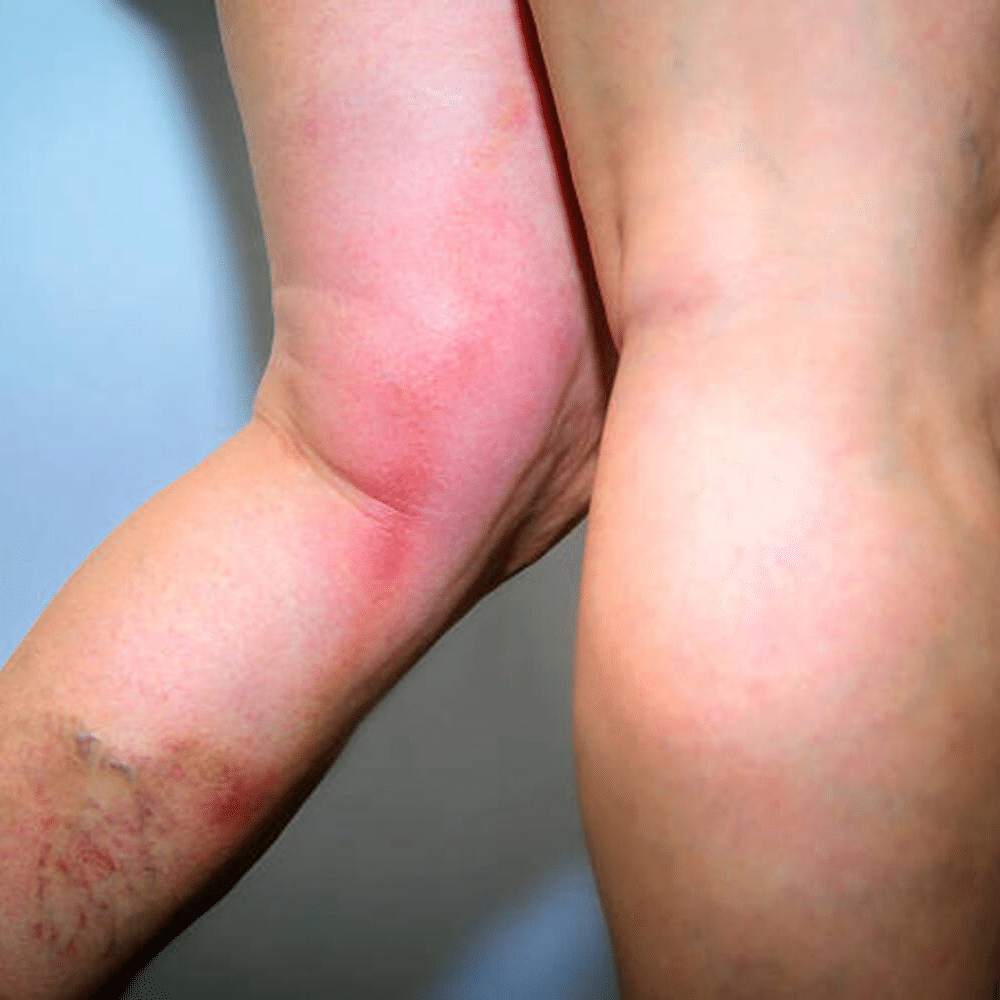
Superficial venous thrombophlebitis
Red, painful in duration along a superficial veins, MC @ the site of an IV Line
Risks: Pregnancy, trauma, varicose veins, systemic hyper-coagulable states, IV lines
Monitor site for SEPTIC phlebitis (erythema, fever, fatigue, leukocytosis)
Dx: Hx and PE
Tx: Symptomatic: analgesics (Tylenol) and warm compresses
Septic thrombophlebitis: remove lines/catheters, IV abx (VANC and CEFTRIAXONE)
Varicose veins
Dilated tortuous superficial veins in the legs due to high venous pressure
Risks: prolonged standing, pregnancy, heavy lifting, hereditary
S/S: dull aching heaviness of legs, worse w/ standing, itching over the ankles or large varicosities
older patients bleed with minimal trauma
Dx: Hx and PE
Tx:
Non-surgical: medical grade compression stockings
Scleortherapy: sclerosing agents to veins
Surgical: thermal ablation or vein stripping
Giant cell arteritis (GCA)/ Temporal arteritis
Chronic-inflammatory disease involving large and medium sized arteries, can be widespread causing stenosis and aneurysm of affected vessels
Risks: Age (almost never before 50, usually 70-79), MC in women, caucasians (Scandinavian), Polymyalgia Rheumatica
S/S: fever, fatigue, weight-loss, headache (scalp tenderness), jaw claudication, ocular involvement (amaruosis fugax)
Dx: Labs- increased ESR, CRP, TEMPORAL ARTERY BIOPSY
Tx: 1 mo prednisone (do steroid taper)
Granulomatosis with polyangitis (Wegner’s granulomatosis)
Rare, chronic autoimmune disease that cause inflammation of small to medium blood vessels (vasculitis)→ causes clusters of inflammatory cells called granulomas
S/S: Upper respiratory (sinus, saddle nose deformity), lungs, kidneys, fevers/night sweats/joint pain
Dx: ANCA blood test, X-ray/CT for noduels, bx
Tx: Prednisone +Rituximab
Polyarteritis Nodosa
Rare, systemic AI disease with necrotizing inflammation of medium and small muscular arteries that causes microaneurysms
S/S: typically spares the lungs, painful red subcutaneous nodules, deep skin ulcers, livedo reticularis (purple lace like web discoloration), damage to peripheral nerves, difficult to control high blood pressure
Dx: tissue bx, angiography
Tx: IV glucocorticoids, Aziathoprine (maintenance)
IgA Vasculitis (Henoch-Schonlein Purpura)
Immune complex mediated small vessel vasculitis; IgA deposits on the inside walls of capillaries, venules and arterioles
S/S: palpable purpura, arthralgia, GI distress, Kidney involvement/nephritis
Dx: skin biopsy, urine analysis and serum creatine
Tx: self limiting
Chronic venous insufficiency
Inadequate venous return over a long period of time
Risks: prior DVT, leg trauma/surgery, obesity
S/S: progressive pitting edema, itching-dull-aching after standing, taut shiny skin at ankle, brownish discoloration of the skin, ulcerations above the ankles, frequent cellulitis
Dx: Hx and PE
Tx: prevention of primary causes, graduated compression stockings, meticulous wound care in cellulitis
Acute respiratory distress syndrome
onset of respiratory distress, progressive respiratory failure within 7 days of known clinical insult, new bilateral radiographic pulmonary opacities not explained by pleural effusion, atelectasis, or nodules/heart failure or volume overload→ alveoli cannot expand or exchange oxygen
impaired oxygenation of paO2/fiO2 <300 mmHg w/ peep 5 cm of H2O or more
Risks: ICU patients, age, organ dysfunction, chronic medical illness, lung injury, pneumonia (MC), severe sepsis, aspiration
S/S: profound dyspnea, labored breathing, tachypnea, intercostal retractions, crackles, marked hypoxemia that is refractory to supplemental O2
Dx: Chest x-ray (new bilateral radiographic opacities), diffuse or patchy bilateral infiltrates, air bronchograms, possible PE
Tx: treat underlying cause, decrease tidal volume ventilation, use low setting PEEP vent, prone position, monitor airway pressures, conservative fluid management (dieresis), HOB at 30 degrees, pressure ulcer prevention
Acute respiratory failure
respiratory dysfunction causing impairment of ventilation severe enough to threaten vital organs (consequence of a disease)
ABGs criteria: PO2 <60 mmHg, PCO2 >55 mmHg
Risks: lung parenchyma infections, lung parenchyma disorders, pleural/chest wall/airway disorders
S/S: of the underlying cause, hypoxemia (cyanosis, confusion, tachypnea, lethargy), hypercapnia (dyspnea, headache, hyperemia, asterixis)
Dx: ABG (use criteria)
Tx: adequate oxygenation, possible mechanical ventilation, general supportive care
Asthma
airway inflammation resulting in reversible bronchoconstriction (obstruction); airway edema and inflammation, airway hyperactivity and remodeling→ IgE mast cell response
Risks: Black pts, low socioeconomic status, obesity, genetic (atopic patients), childhood males
Triggers: allergens, exercise, URIs, rhinosinusitis, PND, aspiration, GERD, change in weather
S/S: can be asymptomatic b/t exacerbations→ dyspnea, wheezing, coughing, severe disease can be nocturnal, hyperresonance to percussion (excess air), possible accessory muscle use
Dx: Hx and PE confirmed with PFTs, bronchodilator challenge, bronchial provocation testing
Tx
Intermittent- low dose ICS-formoterol PRN
Mild persistent- low dose ICS-formoterol PRN (can add SABA)
Moderate persistent- low dose ICS-formoterol maintenance with reliever therapy
Severe persistent- medium dose ICS-formoterol maintenance with reliever therapy
Bronchiectasis
manifestation of disease→ Obstructive Pulmonary disease; manifestation of dilation and destruction of larger bronchi caused by chronic inflammation or infection (diffuse or focal)
Risks: CF, immune defects, recurrent infection, TB and HIV, sometimes idiopathic
S/S: chronic cough with copious purulent sputum, hemoptysis, Pleuritic chest pain, dyspnea (75%), Persistent crackles at lung base, clubbing
Dx: Hx, Chest X-Ray (dilated airways), High Resolution CT (gold standard), Culture (P. aeruginosa, S. Pneumonia and S. aureus, H. flu (CF))
Tx: Abx, inhaled bronchodilators
Complications: hemoptysis, cor pulmonale, secondary visceral abscess at distant sites
COPD
small airway disease with parenchymal obstruction; obstructive pulmonary disease that is non/partially reversible
Emphysema: “pink puffer”, permanent abnormal distention of air spaces distal to the terminal bronchiole with destruction of alveolar septa and attachments to bronchial walls, lung hyperinflammation, air flow limit and trapping, barrel chest, absent breath sounds
Chronic bronchitis: “blue bloater”, chronic productive cough for at least 3 months for 2 years, cyanotic, crackles/rhonchi/wheezing
Can be mixed
Risks: smokers (MC), low birth weight, low socioeconomic status, air pollution, family hx, allergies
Dx: based on pt history
Tx: smoking cessation, pulmonary rehab, vaccinations, bronchodilators, ICS, supplemental O2 if needed
Idiopathic pulmonary fibrosis
Chronic, progressive, and irreversible lung disease characterized by the scarring (fibrosis) of lung tissue without an identifiable cause→ over time the normal lung tissue will be replace by a thick, stiff scare tissue that prevents alveoli from transferring O2
S/S: progressive dyspnea, persistent dry cough, digital clubbing, constitutional symptoms
Dx: CT, PFTs
Tx: antifibrotic drugs, supportive care, lung transplant
Acute bronchitis
infectious disorder; inflammation (self-limited) of the large airways of the lung that extends to the tertiary bronchi; leads to inflammation and thickening of bronchial and tracheal mucosa; can be obstructive with bronchial hyperresponsiveness
MC from a viral infection (No abx needed), Can be bacterial
Influenza A and B, parainfluenza, RSV, COVID, adenovirus, rhinovirus
Risks: adults, winter season, exposure to virus/bacteria
S/S: cough of recent onset, extended period of cough >7 days, coughs that start nonproductive and progress to productive coughs, URI, wheezing, dyspnea
PE: cough, wheeze, rhonchi, URI symptoms, can vary at time of exam
Dx: Clinical and r/o pneumonia
Tx: supportive care
Acute epiglottis
life-threatening airway emergency caused by inflammation and swelling of the epiglottis and surrounding supra epiglottic tissues- rapidly obstruct airway
Risks: Children 2-6, unvaccinated/immunocompromised pts
Etiology: H Fly type B, Strep, Staph including MRSA
S/S: (4D,S: Dysphagia, drooling, dysphonia, dyspnea, stridor)
Dx: SECURE AIRWAY, Lateral neck X-Ray (thumbprint sign)
Tx: Intubation, IV antibiotics (ceftriaxone), IV corticosteroids
Hib vaccine for prevention
Mnemonic: Tripod child with thumbprint sign
Empyema
Collection of pus in the pleural cavity
Risks: bacterial pneumonia, thoracic sx or trauma, aspiration, esophageal perforation
S/S: high fever, chills/nightsweats, pleuritic pain, progressive SOB,
Dx: CT with contrast, US, thoracentesis
Tx: medical drainage vs sx options
Influenza
nfectious disorder, viral respiratory infection caused by the influenza viruses and other viral respiratory pathogens; trick immune system through antigenic drift and antigenic shifts
Risks: fall/winter, young children, old age, pregnancy, impaired respiratory function
S/S: Rhinitis, cough, sore throat, increase fever with chills, severe myalgia, anorexia/malaise, ocular symptoms
Dx: Rapid influenza diagnostic tests→ can also use PCR
Tx: Supportive care, antivirals if critically ill
Bacterial pneumonia
Risks: Strep pneumoniae, H flu, klebsiella pneumoniae, staph aureus, psuedomonas, Atypical- Mycoplasma, legionella, chlamydia
S/S: acute onset high fever, productive cough with purulent sputum, pleuritic chest pain
Dx: CXR- lobar consolidation in typical, interstitial infiltrate in atypical, blood cultures and sputum gram stain
Tx
Outpatient: Amoxicillin OR Doxy OR Azithromycin
Inpatient: Respiratory fluoroquinolone OR Ceftriaxone+Macrolide
Viral pneumonia
Influenza is the MCC of viral CAP; others include RSV, parainfluenza, adenovirus, COVID
S/S: acute/subacute onset of fever, cough, dyspnea, ± sputum production, sweats, chills, rigors, chest discomfort, pleurisy, myalgias, fatigue
Elderly patients: present atypically (falls, AMS, lethargy)
PE: increase temp, tachypnea (besides Legionarre’s), O2 desat, inspiratory disease, inspiratory crackles, (+) special tests, dullness to percussion
Dx: Chest XRAY, isolate with sputum culture, Pneumonia severity index (PSI)
90 or less= tx at home
90 more more= hospital
Tx: supportive, can give antivirals for influenza within 48 hrs of symptom onset
HIV-related Pneumonia
Directly related to CD4 T-lymphocyte count; Pneumocystis jirovecii pneumonia (PCP) under 200 cells/microliter (if under 200 can do prophylaxis with Bactrim)
S/S: subacute dyspnea on exertion, non-productive cough, fevers, exertion hypoxemia
Dx: markedly elevated LDH, CXR- bilateral diffuse ground glass perihilar infiltrates
Tx: Bactrim and can add systemic corticosteroids
Histoplasma capsulatum
Risks: Ohio and Mississippi River valleys, bat dropping or bird roosts, caves
S/S: palatal/oral ulcers, hepatosplenomegaly
Dx: urinary antigen , CXR- hilar lymphadenopathy and granulomas
Tx: Itraconazole/Amphotericin B
Blastomyces dermatitidis
Risks: Great Lakes and ohio river valleys, decaying wood, organic soil
S/S: verrucous skin lesions and osteomyelitis
Dx: smashed broad based budding yeast on smear