Histology Pictures
 Call Kai
 Learn
Learn Practice Test
 Spaced Repetition
 Match
Match Flashcards
 Knowt Play
1/60

There's no tags or description
Looks like no tags are added yet.
Name | Mastery | Learn | Test | Matching | Spaced | Call with Kai | Chat |
|---|
No analytics yet
Send a link to your students to track their progress
61 Terms

Simple squamous epithelium
Description: single layer or flattened cells with disc-shaped central nuclei and sparse cytoplasm.Simple of the epithelia.
Functions: allows materials to pass by diffusion and filtration in sites where protection is not imp.
Location: kidney, air sacs lungs, lining of heart
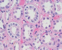
Simple cuboidal epithelium
Description: single layer of cubelike cells with large, spherical central nuclei
Function: secretion and absorption
Location: kidney tubules, ducts and secretory portions of small glands, ovary surface

simple columnar epithelium
Description: single layer of talls cells with round oval nuclei
Function: Absorption; secretion of mucus enzymes and other substances
Location: Digestive tract

Pseudostratified columnar epithelium
Description: single layer of cells of differing heights, some not reaching the free surface.
Function: secrete substances, particularly mucus
Location: upper respiratory tracts, lines th trachea, sperm-carrying ducts.

Stratified squamous epithelium
Description: thick membrane composed of several cell layers
Function: protects underlying tissues in areas subjected to abrasion
Location: esophagus, mouth and vagina.

Transitional epithelium
Description: basal cells cuboidal and columnar
Function: stretches readily,permits stored urine to distend urinary organ
Location: lines the ureters, bladder and part of the urethra

Connective tissue proper: loose connective tissue, areolar
Description: gel-like matrix with all three fiber types; fibroblast, macrophages,mast cells and some white cells.
Function: wraps and cushions organs
Location: under epithelia of body.

Connective tissue proper: loose connective tissue, adipose
Description: matrix as in areolar, but very sparse
Function: provides reserve food fuel
Location: under skin, around kidneys and eyeball, abdomen and breast
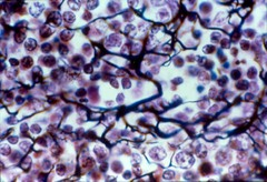
reticular
Description: loose network of reticular fibers in a gel-like ground substance
Function: fibers form a soft internal skeleton that supports other cells types including white blood cells, mast cells, and macrophages.
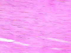
dense regular
Description: few elastic fibers
Function: attaches muscles to bones or to muscles
Location: tendons, most ligaments, aponeuroses

dense irregular
description: some elastic fibers
Function: withstands tension exerted in many directions
Location; organs and joints, dermis of skin, submucosa of digestive tract.
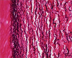
elastic
description: containing high portion of elastic fibers
Functions: allows tissue to recoil after stretching maintaining pulsatile flow of blood through arteries
Location: walls of large arteries.

Cartilage: hyaline
Function: supports and reinforces
Location: embryonic skeleton, costal cartilages of the ribs, trachea and larynx.
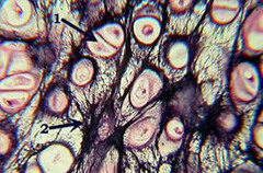
Cartilage: elastic
Function: Maintains the shape of a structure while allowing great flexibility
Location: support external ear

Cartilage: fibrocartiage
Function: tensile strength allows it to absorb compressive shock
Location: intervertebral discs, knee joint
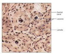
other: bone (osseous tissue)
Function: supports and protects
Locations: bones

Connective tissue: Blood
function; transport respiratory gases, nutrients, wastes, and other substances.
Location: contained within blood vessels

skeletal muscle
Function: voluntary movement
Location: in skeletal muscles attached to bones or occasionally to skin
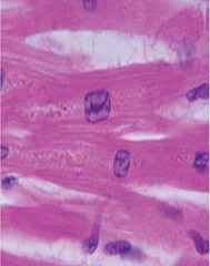
cardiac muscle
Function: as it contracts it propels blood into the circulation.
Location: the walls of the heart.
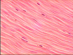
Smooth muscle
Function: propels substances or objects along internal passageways. (foodstuffs, urine, baby)
Location: mostly in the walls of hollow organs

Nervous tissue
Function: Neurons transmit electrical signals form sensory receptors and to effectors
Location: brain, spinal cord and nerves

elastic

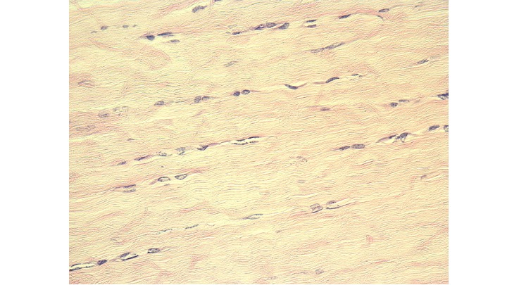
Identify the connective tissue below.
fibrocartilage
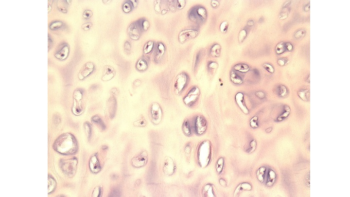
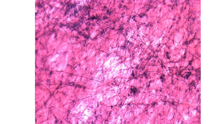

Identify the type of cell highlighted in blue.
red blood cell

Identify the connective tissue below.
elastic

Identify the type of cell highlighted below.
osteocyte

Identify the type of fiber indicated below (they are stained blue).
reticular
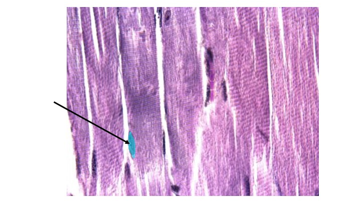
Identify the highlighted structure.
nucleus

Identify the structure highlighted below.
neuroglia cell

Identify the highlighted structure.
nucleus
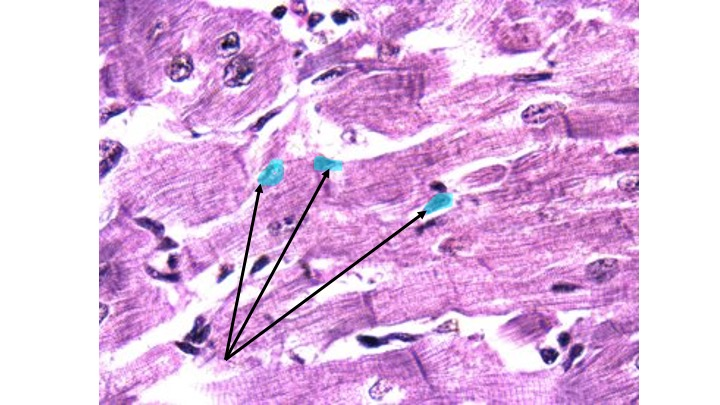
Identify the type of muscle tissue.
skeletal muscle

Identify the epithelial tissue.
nonkeratinized stratified squamous epithelium
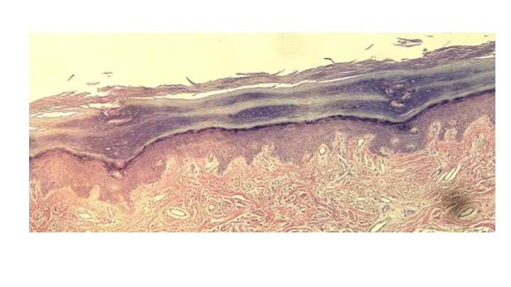
Identify the epithelial tissue.
keratinized stratified squamous epithelium

Identify the cellular projection indicated by the arrow.
microvilli

Identify the epithelial tissue.

Identify surface indicated by the arrow as either apical or basal.
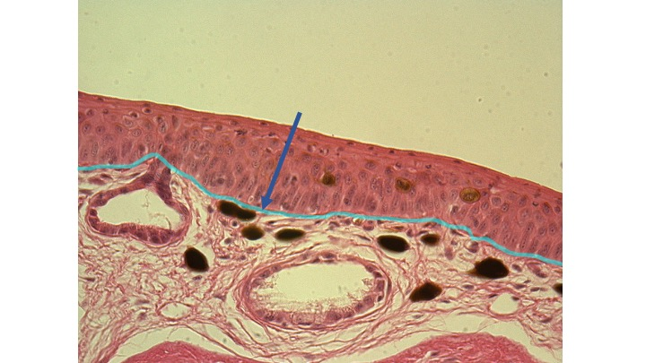
Identify the structure highlighted on the image.
basement membrane

Identify the cellular projection indicated by the arrow.
cilia

Identify the connective tissue below.
bone

Identify the type of connective tissue.
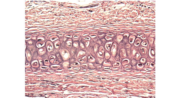
Identify the connective tissue below.
elastic cartilage

Identify the type of cell highlighted below.

Identify the connective tissue below.
blood

Identify the connective tissue below.

Identify the type of muscle tissue.
smooth

Identify the structure highlighted below.
nerve cell
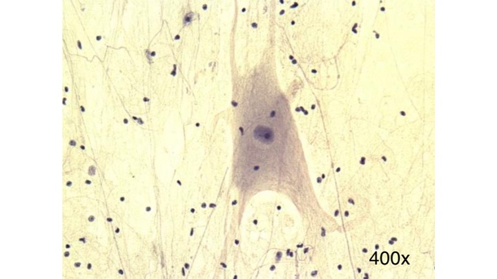
Identify the type of tissue.
nervous tissue

Identify the type of muscle tissue.
cardiac