Microbio - Module 2: Bacterial Cell Structure
 Call Kai
 Learn
Learn Practice Test
 Spaced Repetition
 Match
Match Flashcards
 Knowt Play
1/75

There's no tags or description
Looks like no tags are added yet.
Name | Mastery | Learn | Test | Matching | Spaced | Call with Kai | Chat |
|---|
No analytics yet
Send a link to your students to track their progress
76 Terms
How was the term "prokaryote" originally defined in 1962?
By the features organisms lack compared to eukaryotes (specifically lacking a membrane-bound nucleus, a cytoskeleton, membrane-bound organelles, and internal membranous structures).
Why is defining "prokaryotes" based on absent features scientifically inaccurate?
Modern biochemical, genetic, and genomic analyses show that Bacteria and Archaea are fundamentally distinct domains of life, not just two forms of a single structural group.
Who proposed abandoning the term "prokaryote" in 2006, and why?
Microbiologist Norman Pace, because the term fails to reflect the evolutionary differences and unique traits of Bacteria and Archaea.
Which two domains of life were historically grouped together under the term "prokaryote"? (originally no longer now)
Bacteria and Archaea.
Since the microbial world has a variety of morphologies, what are the three groups? explain for ea
shape
cocci—spherical cells
rods—oblong cells
arrangement
determined by plane of division
determined by separation or not
size—varies
whats the general shape of cocci (s., coccus)?
spherical
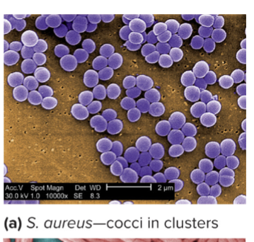
match ea cocci arrangment to its description:
Diplococci
Streptococci
Staphylococci
Tetrads
Sarcina
bacilli
vibrios
how do spirilla and spirochetes differ in their structure?
mycelium
pleomorphic
Diplococci: Divide and remain in pairs.
Streptococci: Divide on 1 plane to form chains.
Staphylococci: Divide in random planes to form grape-like clusters.
tetrads: Divide in 2 planes to form a square of 4.
Sarcina: Divide in 3 planes to form a cubic packet of 8.
rod shaped
comma shaped
spirilla—rigid, spiral-shaped. spirochetes—flexible, spiral-shaped
network of long filaments
bacteria that r variable in shape
bacterial cell size
whats considered small, avg, very large?
small = 0.3 um (mycoplasma)
avg = 1.1-1.5 x 2-6 um long (e. coli)
very large = 600 × 80 um
size-shape relationship
do cells want a low or high surface area-to-volume ratio (S/V ratio)? And why?
high surface area to vol ratio
inc efficiency of nutrient uptake and diffusion of
molecules within a cell.
what are good protective mechansism from predation for cells?
Large size and odd shape
what are the common bacterial structures?
whats a cell envelope and what does it consist of?
what are the external structures?
—layers that surround the cell
plasma membrane
cell wall
cytoplasm
nucleoid
external structures
pili
flagella
Bacterial Cell Envelope
Besides the plasma membrane and cell wall, the cell envelope includes AT LEAST ONE additional layer, what could they be?
capsule
slime layer
whats the most important membrane that required for all living organisms?
plasma membrane
plasma membrane functions
Innermost membrane that encompasses the cytoplasm.
Selectively permeable barrier that acquires nutrients and
eliminates waste.
Interacts with external environment.
• Detects and responds to surrounding chemicals.
• Transport systems used for nutrient uptake.
• Metabolic processes (that is, respiration &
photosynthesis
Plasma Membrane Structure is Dynamic
explain the structure
what is it composed of? which part is hydrophilic, hydrophobic?
Plasma membrane is a thin structure (7 to 8 nm) composed
of 2 lipid sheets.
Amphipathic lipids
• Hydrophilic—Polar ends that interact with water.
• Hydrophobic—Non-polar tails that are insoluble in water
and interact with each other.
Bilayer forms spontaneously
in aqueous environment.
• Hydrophilic on surface.
• Hydrophobic ends buried.
what proteins are on the plasma membrane? What is the function of the second type of protein?
peripheral membrane proteins—Loosely connected to
the membrane and easily removed.
• 20 to 30% of the total membrane proteins.
Integral membrane proteins—Amphipathic proteins that
are embedded within membrane and not easily removed.
• Hydrophobic region buried in membrane lipids.
• Hydrophilic regions project from the surface.
• Carry out important functions (that is, transport).
whats the plasma membrane mainly composed of what kind of lipids? what are the other smaller lipids?
mainly phospholipids
SMALLER LIPIDS:
hopanoids—Hydrophobic molecule similar to cholesterol.
• Distort the bilayer, which impacts the fluidity and shape in membrane region.
• Form functional membrane microdomains that are platforms for protein complex assembly.
what do bacterial cells take for nutrients? where are they found?
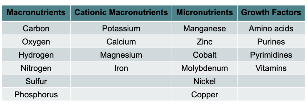
Macronutrients—required in large amounts.
• Found in organic molecules (that is, proteins, lipids,
nucleic acids, and carbohydrates).
• Cations contribute to activity and stability of molecules and
cell structures.
• Important in cellular processes (that is, protein synthesis).
Micronutrients—required in small amounts.
• Ubiquitous in nature and usually present in adequate
amounts to support microbial growth.
• Work to assist enzyme catalysis and maintain protein
structure.
what are growth factors? can u name four of them?
Organic compounds required for survival.
• Essential cell components (or their precursors) that the
cell cannot synthesize and must be supplied by
environment.
microbes can only take in nutrients in what way? What mechanisms do they use?
Microbes can only take in dissolved particles across a
selectively permeable membrane.
microorganisms use transport mechanisms
Passive diffusion
Facilitated diffusion
Primary and secondary active transport
Group translocation
Passive diffusion
mlcls move fr region of highr concentration to one of lowr concentration
Requires a large concentration gradient for adequate nutrient uptake.
The rate of diffusion decreases as more nutrients accumulate in the cell.
• H2O, O2, and CO2 easily cross the plasma membrane via passive diffusion.
facilitated diffusion
what is it? done with the help of what and name them? is it energy dependent? what is direction of movement? what impacts the rate of uptake? when would transport stop?
Movement across the plasma membrane with the help of
transport proteins.
• Channels—proteins that form pores for substances to
pass through.
• Carriers—proteins that have high substrate specificity in
transport.
Truly diffusion because it is not energy dependent.
• Direction of movement is from high to low concentration.
• Size of concentration gradient impacts rate of uptake.
• Rate increases with the concentration gradient.
• If the gradient is lost, transport stops.
active transport
Transport of molecules against the concentration gradient.
Energy-dependent process.
• ATP or proton motive force used.
Involves carrier proteins that control the rate of transport.
• When the solute concentration is high, carrier saturation
effect is observed
Primary Active Transport
does it use energy?
Use energy from ATP hydrolysis to move substances against concentration gradient without modifying them.
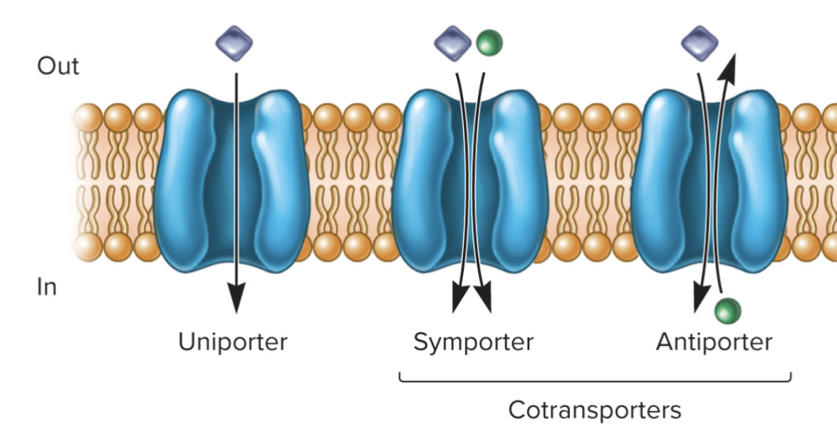
Uniporters—single molecule transported across membrane.
ATP-binding cassette (ABC) transporters
Consist of:
• 2 hydrophobic membrane
spanning domains.
• 2 cytoplasmic associated ATP-
binding domains
Secondary Active Transport
Use potential energy of ion gradients to cotransport
substances without modifying them.
• Move both the ion and the substance across the membrane.
• Symport—2 substances both move in the same direction.
• Antiport—2 substances move in opposite directions.
Group Translocation
Energy dependent transport that chemically modifies the molecule as it is brought into cell.
Best known translocation system is phosphoenolpyruvate:
sugar phosphotransferase system (PTS).
• Imports sugars while
phosphorylating them

wht do microorganisms require when building molecules? why is it important?
Microorganisms require iron for building molecules important in
energy-conserving processes.
• Ferric iron is very insoluble so uptake is difficult.
Siderophores—secreted by bacteria and complex with ferric ion
for transport into cell
bacterial cell wall
functions?
Cell wall functions:
• Maintains shape of the bacterium.
• Helps protect cell from osmotic lysis and toxic materials.
• May contribute to pathogenicity.
Peptidoglycan (murein)—Rigid structure lying outside the plasma membrane.
Two types of bacteria based on Gram stain.
• Gram-positive: stain purple; thick peptidoglycan
• Monoderm—single membrane
• Gram-negative: stain pink or red; thin peptidoglycan and outer membrane.
• Diderm—plasma membrane and an outer membrane
Peptidoglycan Structure
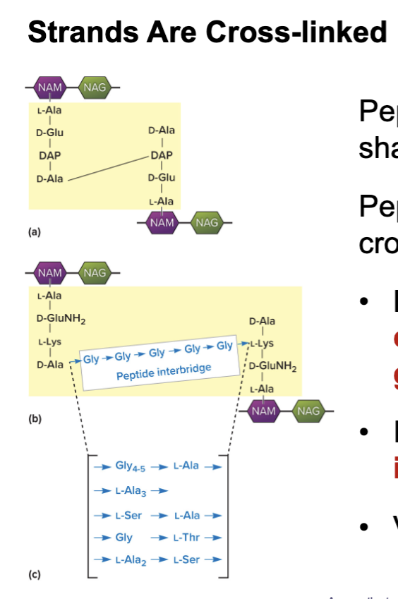
Meshlike polymer of identical subunits forming long strands.
Two alternating sugars:
• N-acetylglucosamine (NAG).
• N- acetylmuramic acid (NAM).
Alternating D- and L- amino acids.
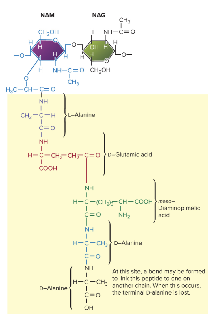
peptidoglycan chains are crosslinked by ____ for strength. Between ea amino acid theres a direct cross-link of what? Theres also an indirect link of what?
peptides for strength
carboxyl group and amino groups
peptide interbridge (may form)
gram-positive cell walls
whats it mainly composed of?
what kind of acid may it contain?
Composed primarily of peptidoglycan.
May also contain teichoic acids (negatively charged).
• Polymers of glycerol.
• Help maintain cell envelope.
• Protect from environmental substances.
• May bind to host cells to initiate infection.

Periplasmic Space of Gram-Positive Bacteria
which is more complex, gram neg or pos?
it contains few what?
why is it clinically important? what does it contain?
less complex than Gram-neg
btwn plasma membrane and cell wall
periplasm has relatively few proteins
the periplasmic space is of particular clinical importance in that it is the site, in some
species, that contains beta-lactamase, an enzyme responsible for degrading the
penicillin group of antibiotic drugs, leading to penicillin resistance.
Gram-Negative Cell Wall Basic Structure
which is more complex, gram neg or pos?
describe structure of peptidogylcan and locaation?
whats outer membrane composed of?
What does it NOT have that gram positive does have?
• More complex than Gram-positive.
• Consist of a thin layer of peptidoglycan surrounded by an outer membrane.
• Outer membrane composed of lipids, lipoproteins, and lipopolysaccharides.
• No teichoic acids.

Gram-Negative Cell Walls
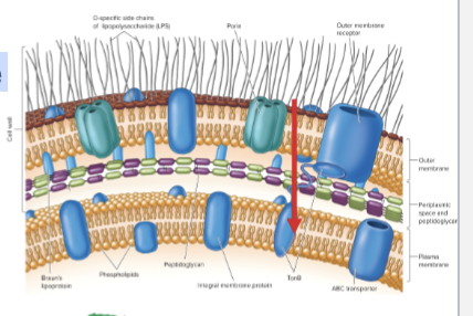
Outer membrane (OM) outside thin peptidoglycan layer. Braun’s lipoproteins connect OM to peptidoglycan. Peptidoglycan is approximately 5 to 10% of cell wall weight.
Periplasmic space differs from that in Gram-positive cells.
• May constitute 20 to 40% of cell volume.
• Many enzymes present in periplasm.
• Hydrolytic enzymes, transport proteins and other proteins.

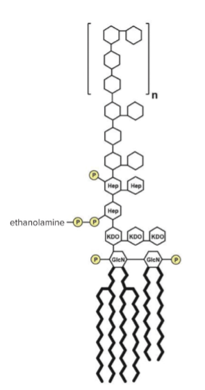
LPS—Lipopolysaccharide
wht are the three parts
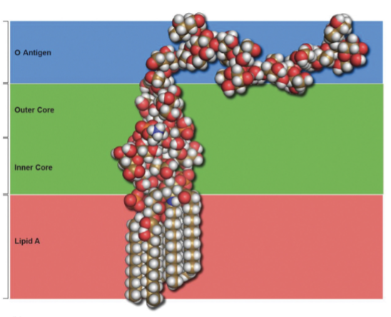
Consists of three parts:
• Lipid A—buried in outer membrane.
• Core polysaccharide—10 sugar structure joined to Lipid A.
• O side chain (O antigen)—polysaccharide that extends outward from the core
Importance of LPS
• Contributes to negative charge on cell surface.
• Helps stabilize outer membrane structure.
• Creates a permeability barrier.
• Host defense protection.
• Acts as an endotoxin.
Gram-Negative Membrane Transport
Two-step process:
• First the solute crosses the outer membrane
into the periplasm.
• Then crosses the plasma membrane.
Facilitated transport by porins.
Channels to let small molecules (600 daltons) pass

comparison gram +ve vs -ve
explain this diagram
The diagram of the gram-positive cell wall shows alternative
NAG(N-acetylglucosamine) and NAM (N-acetylmuramic acid) in a chain; these are shown as alternative orange and blue spheres. The chains or orange and blue spheres are connected to other chains with smaller yellow spheres in a chain labeled pentapeptide and smaller green spheres labeled tetrapeptide. Each NAG in the chain is connected to the NAG in the chains next to it by both a tetrapeptide (green) connected to a pentapeptide (yellow circles). The diagram of the gram-negative cell wall has the same NAG and NAM chains. But this time they are linked with a direct line to the chains next to them
cell walls and osmotic protection
hypotonic environments
hypertonic environments
• Solute concentration outside cell less than inside cell.
• Water moves into cell and cell swells.
• Cell wall protects from lysis.
• Solute concentration outside cell is greater than inside.
• Water leaves cell and cytoplasm shrivels up.
• Plasmolysis
evidence for protection of cell wall
remember that bacterial cell wall is largely made of peptidoglycan, mesh net that surrounds cell thats made fr repeating sugars—NAG and NAM
lysozyme breaks bond btwn NAG and NAM
penicilin inhibits (prevents) bacteria fr making strong peptidoglycan → since cell wall is weak → cell undergoes lysis(bursts cuz cell wall prevents cell fr bursting) if in hypotonic solution (less solute outside than inside)

what type of environment can cells tht lose a cell wall is survival possible in?
isotonic environments
protoplast—gram positive bacterial cell tht has lost its cell wall
spheroplast—gram negative cell that lose cell wall ONLY PART OF CELL WALL IS REMOVED (outer membrane remains) in isotonic environments
mycoplasma—bacteria that naturally dont have cell wall, BUT plasma membrane more resistant to osmotic pressure
gram-positive organisms appear what color? gram neg are what color?
pos = purple/blue
neg = pink/red
Extracellular Vesicles (EVs)
• Small membrane-bound particles (20 to 400 nm in size).
• Develop when a membrane buds out, pinches off, and is released from the cell.
• Gram-Positive EV—Made of the plasma membrane surrounding a small amount of cytoplasm.
• Gram-Negative EV—Made of LPS- containing OM surrounding a sample of periplasm
are extracellular vesicles cells? explain
NOOOOO
they do not reproduce
Can carry some ATP but do not have the ability to conserve
energy.
They play role in cell-cell interactions.
• Transfer genetic information between cells.
• Transfer toxin molecules.
Components Outside of the Cell Wall
Outermost layer in the cell envelope.
• Capsules—well-organized layers made of polysaccharides
that are covalently bonded and difficult to wash away.
• Slime layers—polysaccharide layers that are unorganized
and easily washed away.
• Glycocalyx—polysaccharide extension that aids in
attachment to solid surfaces.
• S-layers—geometric pattern made of protein that aid in
protecting from ion and pH fluctuations.
Components Outside of Cell Wall—Capsules
Well organized and not easily removed from cell.
Usually composed of polysaccharides.
Visible in light microscope.
Protective advantages.
• Resistant to phagocytosis.
• Protect from desiccation.
• Exclude viruses and detergents.
components Outside of Cell Wall—Slime Layers
• Similar to capsules except diffuse, unorganized, and
easily removed.
• Slime may facilitate motility
Components Outside of Cell Wall—S Layers
Regularly structured self-assembling layers of protein or glycoprotein.
In Gram-negative bacteria, S layer adheres to outer membrane.
In Gram-positive bacteria, associated with peptidoglycan.
S Layer functions:
Protect from ion and pH fluctuations, osmotic stress, enzymes, and predation.
Maintains shape and rigidity.
Promotes adhesion to surfaces.
Protects from host defenses.
Potential use in nanotechnology.
name 6 Bacterial Cytoplasmic Structures
• Cytoskeleton
• Intracytoplasmic membranes
• Inclusions
• Ribosomes
• Nucleoid
• Plasmids
define Protoplast and Cytoplasm
Protoplast—plasma membrane and everything within.
Cytoplasm—material bounded by the plasma membrane
Bacterial Cytoskeleton
Cytoskeleton—Protein filaments that polymerize to form
functional filaments that extend to full inner dimensions of the
cell.
Homologs(similar) of eukaryotic cytoskeletal elements have been identified in bacteria.
• Actin filaments, microtubules, and intermediate filaments.
Functions are similar as in eukaryotes:
• Participate in cell division.
• Localize proteins.
• Maintain cell shape.
Best Studied Examples of Bacterial Cytoskeleton
Molecules
FtsZ—many bacteria
• Forms ring at center of a dividing cell that constricts
as daughter separates.
MreB—many rods
• Maintains shape by positioning peptidoglycan synthesis machinery.
CreS—maintains curve shape
Intracytoplasmic Membranes
(folds or bumps in bacterial plasma membrane)
Plasma membrane infoldings.
• Observed in many photosynthetic bacteria.
• Observed in many bacteria with high respiratory
activity.
• May be aggregates of spherical vesicles.
Inclusions
Formed by aggregation of organic or inorganic substances.
Primary function of inclusions is to segregate cellular
components so they do not diffuse freely in the cytoplasm.
Granules, crystals, or globules of organic or inorganic
material that are stockpiled by the cell for future use.
Some are enclosed by a single-layered protein or lipid shell.
• May be referred to as microcompartments.
• Often used to sequester enzymes that produce toxic
intermediates.
📦 Store materials for later
🚧 Keep substances separated
🧪 Can contain enzymes that make toxic substances
Easy memory:
Inclusions = "inside storage containers." 🦠📦
Microcompartments
Not bound by membranes but compartments for specific
functions.
Carboxysomes—CO2 fixing bacteria.
• Contain the enzyme carbonic anhydrase that
release CO2 into a shell so it accumulates to high
concentration.
• Then RuBisCO makes sugar.
Gas Vacuoles
• Involved in bacterial movement.
• Provide buoyancy to aquatic bacteria.
tiny air-filled floatation devices in bactereia (bacterial floaties) help them float/move in water
• Made of aggregates of hollow, cylindrical gas vesicles.
Magnetosomes
Found in aquatic bacteria.
Magnetite particles for orientation in Earth’s magnetic field.
like tiny compass inside water bacteria, helps direct bacteria where to move
• Cytoskeletal protein MamK helps form magnetosome chain
Ribosomes
Complex protein/RNA structures
• Sites of protein synthesis.
• Bacterial and archaea ribosome = 70S (← what they’re called)
Bacterial ribosomal RNA (rRNA) has TWO parts:
• small subunit = 30s
16S rRNA in small subunit
• large subunit = 50s
23S and 5S rRNA in large subunit
The Nucleoid
• Usually not membrane bound (few exceptions).
• Location of chromosome and associated proteins.
• Usually 1 closed circular, double-stranded DNA molecule.
• Supercoiling and nucleoid proteins aid in folding and structure
Plasmids
Extrachromosomal DNA that is usually small, closed circular
DNA molecules.
Exist and replicate independently of chromosome.
• Episomes—are plasmids that able to integrate into bacterial chromosome.
• Inherited during cell division.
Benefit the survival of the organism by
1) providing antibiotic resistance to naturally occurring antibiotics in a
competitive environmental niche
2) producing toxins under similar circumstances, or
3) allowing the organism to utilize particular organic compounds that
would be advantageous when nutrients are scarce
Plasmid Classification
plasmids can be classified based on:
WHAT THEY DO
HOW THEY MOVE BTWN BACTERIA
1. Functional Classification = WHAT THEY DO
Plasmids are classified into five primary functional classes:
Fertility (F) Plasmids: Contain tra genes used for conjugation, allowing them to transfer DNA between bacteria via a sex pilus (←tiny connection/bridge)
Resistance (R) Plasmids: Carry genes that provide protection against antibiotics or poisons, such as the widely studied pBR322.
Col Plasmids: Encode bacteriocins (like colicin), which are proteins that kill other competing bacteria.
Degradative Plasmids: Enable the host to digest unusual substances like toluene, camphor, or salicylic acid.
Virulence Plasmids: Turn a bacterium into a pathogen by carrying toxin or infection-related genes (e.g., the Ti plasmid in Agrobacterium).
Transferability & Mobility (MOB)
Plasmids are classified by how they move between host cells:
•Conjugative: Self-transmissible plasmids that carry all necessary genes for conjugation.
•Mobilizable: Lack the full machinery but can "hitchhike" if a conjugative plasmid is present.
•MOB Typing: A sequence-based system that groups plasmids based on the amino acid
sequence of their relaxase proteins (e.g., MOBP, MOBQ)
External Structures
Extend beyond the cell envelope in bacteria.
Function in protection, attachment to surfaces, horizontal
gene transfer, cell movement.
• Pili and fimbriae
• Flagella
Pili and Fimbriae
Fimbriae (s., fimbria); pili (s., pilus).
• Short, thin, hairlike, protein appendages (1,000/cell).
• Can mediate attachment to surfaces, motility, and DNA uptake.
Sex pili (s., pilus).
• Longer, thicker, less numerous (10/cell).
• Genetically encoded on plasmids.
• Required for conjugation.
Flagella
Threadlike, locomotor appendages extending outward from plasma
membrane and cell wall.
Functions:
• Motility
• Attachment to surfaces
• Virulence factors
Patterns of flagella distribution:
• Monotrichous—one flagellum.
• Polar flagellum—flagellum at end of cell.
• Amphitrichous—one flagellum at each end of cell.
• Lophotrichous—cluster of flagella at one or both ends.
• Peritrichous—spread over entire surface of cell
Bacterial Flagella
Thin, rigid protein structures that cannot be observed with bright-field
microscope unless specially stained.
Ultrastructure composed of 3 parts:
• Filament—extends from cell surface to the tip.
• Basal body—embedded in cell envelope.
• Hook—short curved segment
Motility
Flagellar movement
• Swimming
• Swarming
• Spirochete motility
Twitching and gliding motility
Chemotaxis
Swimming
Flagellum rotates like a propeller.
• Very rapid rotation up to 1100 revolutions/sec.
• In general, counterclockwise (CCW) rotation causes forward motion (run).
• In general, clockwise rotation (CW) disrupts run causing cell to stop and tumble.
Mechanism of Flagellar Movement
Flagellum is a two-part motor producing torque:
Rotor—moving parts
• C (FliG protein) ring turn and
interact with stator.
Stator—stationary parts
• Form channel through plasma membrane.
• Protons move through Mot A and Mot B channels using
energy of proton motive force.
• Torque powers rotation of the basal body and filament
Swarming
Occurs on when cells move in unison across a moist surfaces.
• Most swarmers have peritrichous flagella.
• Commonly, the cell produces a molecule that lowers surface tension.
Twitching and Gliding Motility
Twitching motility
Gliding
- Occurs on solid surface.
Does not involve flagella.
May involve Type IV pili and slime.
• Pili at ends of cell.
• Short, intermittent, jerky motions.
• Cells are in contact with each other and surface
Smooth movements that do not require appendages.
Chemotaxis
• Movement toward a chemical attractant or away from a chemical repellent.
• Chemical attractants and repellents bind chemoreceptors that transmit signals throughout the chemosensing system.
• In presence of attractant/repellant, tumbling frequency is reduced; runs toward/away from compound are longer.
• Behavior of bacterium altered by temporal concentration of chemical
The Bacterial Endospore
Complex, dormant structure formed by some bacteria.
Form in response to nutrient depletion.
Resistant to numerous environmental conditions:
• Heat, UV radiation, gamma radiation, chemical disinfectants, and desiccation.
Endospore Structure
• Spore surrounded by thin covering called exosporium.
• Thick layers of protein form the spore coat.
• Cortex, beneath the coat, thick peptidoglycan.
• Core has nucleoid and ribosomes
Sporulation
Process of endospore formation.
Organized process that occurs over several hours.
Normally starts when growth slows due to lack of nutrients.
• Produces a dormant cell that can persist until nutrients are
available and growth resumes.
Endospore Formation
Steps of Endospore Formation
DNA Replication (Axial Filament Formation):
The bacterium copies its DNA and stretches the genetic material into a long, thread-like structure across the cell.
Asymmetric Cell Division (Septum Formation):
A membrane wall (septum) divides the cell unequally into a small forespore (the future
spore) and a larger mother cell.
Engulfment:
The mother cell membrane grows around and completely engulfs the forespore, placing a double membrane around it.
Cortex Formation:
A thick layer of peptidoglycan called the cortex, builds up betweentthe wo membranes.
Spore Coat Formation: A tough, protein-based outer coat forms around the cortex, giving the spore high resistance to chemicals and heat.
Maturation and Dehydration:
Dipicolinic acid and calcium are added to the core while water is pumped out, making the spore metabolically dormant and heat-resistant.
Release:
The mother cell lyses and releases the mature endospore into the environment.
Formation of Vegetative Cell
Three Stages:
• Activation
• Prepares endospores for germination.
• Germination
• Starts when germinant receptors detect small molecules (that is,
sugars and amino acids).
• Outgrowth
• Emergence of vegetative cell