LIVER
 Call Kai
 Learn
Learn Practice Test
 Spaced Repetition
 Match
Match Flashcards
 Knowt Play
1/56

Earn XP
Description and Tags
school slides
Name | Mastery | Learn | Test | Matching | Spaced | Call with Kai | Chat |
|---|
No analytics yet
Send a link to your students to track their progress
57 Terms
Fatty liver can lead to
steatohepatitis
what can steatohepatitis lead to
cirrhosis
viral hepatitiis signs
jaundice, dark urine, clay-coloured stool
viral hepatitis labs
increased AFT, ALT, bilirubin
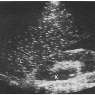
acute hepatitis sono
“starry sky” , hepatomegaly, splenomegaly, thick gb walls
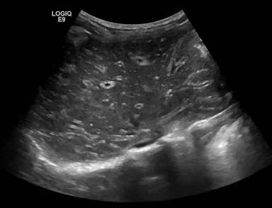
chronic hepatitis may lead to
cirrhosis, HCC, liver failure
what are the causes of cirrhosis
alcoholism, viral hepatitis (B+C), biliary obstruction, wilson disease, budd-chiari, hemochromatosis
what are the s/s of cirrhosis
jaundice, weight loss/fatigue, hepatomegaly, ascites
cirrhosis can lead to
portal hypertension, varicosities, PV thrombosis, splenomegaly, HCC
what are the sono of cirrhosis
lobe volume redistribution
coarse echotexture, nodular surface, hepatomegaly, splenomegaly, ascites
corkscrew appearance of hepatic artery
monophasic HV flow, heptofugal PV flow
collaterals
recanalization of umbilical vein off of LPV
esophageal varices
what is glycogen storage disease
lack of enzyme that breaks down glycogen into glucose (hypoglycemia- chronic hunger) therefore abnormal accumulation of glycogen in the tissues
von Gierke is the m/c
can be caused by oral contraceptives
what is the sono of glycogen storage disease
increase echogenicity and attenuation
hepatomegaly
solid liver masses (adenomas)
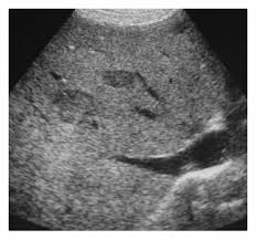
what is hemochromatosis
excess iron deposits in tissue
what is wilson disease
excess copper accumulation in tissues
hemochromatosis and wilson disease can lead to
chronic hepatitis, cirrhosis, HCC
what is the most common cause of portal hypertension
cirrhosis
what is the sono of portal hypertension
dilated PV veins ( > 13mm) , splenic veins, mesenteric vv
heptofugal PV flow
hepatosplenomegaly
Portal hypertension can be relieved by
TIPS
commonly between right PV and right HV
what can portal vein thrombosis lead to
cavernous transformation of portal vein (worm-like vessels)
what is budd-chiari syndrome
hepatic veins are occluded
possibly IVC too
what are the s/s of budd-chiari
ascites, RUQ pai, hepatosplenomegaly, increased LFT, leg edema with IVC occlusion
what is the sono of budd chiari
enlarged caudate
rt lobe atrophy
low vis of HVs
clot in HV or IVC
no flow in HVs
ascites
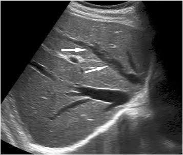
what is the most common source of infection for pyogenic abscess
cholangitis
what are the s/s of pyogenic abscess
fever and chills
increased WBC
RUQ pain
N&V
increased LFT
hepatomegaly
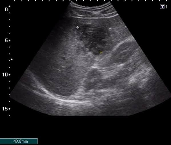
what is the sono of pyogenic abscess
variable appearance, usually singular lesion
hypoechoic with TT
complex with debris and irregular walls
fluid level
dirty shadow (with gas forming bacteria)
hypervascularity around the lesion
what is candidiasis (fungal abscess) caused by
a species of candida
occurs in immunocompromised hosts
chemo, organ transplant, HIV
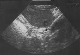
what is the sono of candidiasis (fungal abscess)
multiple small hypo lesions with echogenic center
bulls eye, target lesions, wheel within wheel
what is amebic abscess caused by
protozoan parasite (entamoeba histolytica)
from contaminated water
primary site is colon
what are s/s of amebic abscess
can be asymptomatic
abdo pain, diarrhea, leukocytosis, low fever
what is the sono of amebic abscess
round or oval
hypoechoic
debris
TT
what is hydatid echinococcal cyst caused by
parasitic tapeworm (echinococcus granulosum)
exposure to livestock, dog feces
what is the s/s of hydatid echinococcal cyst
urticaria (rash/itchy skin)
ruq pain, hepatomegaly
leukocytosis
obstructuve jaundice (increased ALP and direct bilirubin)
cysts can get large and rupture
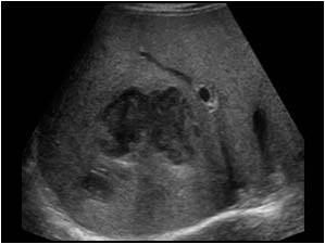
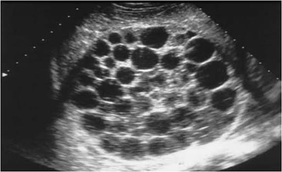
what is the sono of hydatid echinococcal cyst
simple cyst
complex cyst
daughter cysts
honeycomb sign (septa)
mobile debris
water lilly sign
what is schistosomiasis caused by
common parasitic infection in africa, asia, south america
worms in contaminated water penetrate skin and travel
causes itchy skin/rash
schistosomiasis clinic
portal hypertension
splenomegaly
ascites
gastroesophageal varices
what is the sono for schistosomiasis
irregular hepatic surface
hyperechoic thick PV walls
“clay-pipe stem” patterm
portal hypertension signs
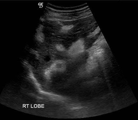
what is the m/c benign liver mass
cavernous hemangioma
what is the second m/c benign liver mass
FNH
influenced by hormones (OC may cause increase in size)
F > 40 yrs
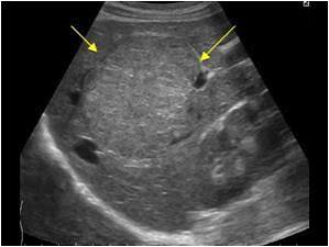
what is the sono of FNH
isoechoic / hyper
central scar
well defined
peripheral and central colour (spoke-wheel pattern)
what is liver adenoma associated with
long hx of oral contraceptive use
GSD type 1 (von Gierke)
F > M
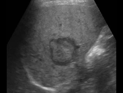
what is the sono for adenoma
m/c hypoechoic (can be hyper/iso)
hypo with hypo ring
complex - hemorrhage/necrosis
what is the m/c primary liver cancer
HCC (hepatoma)
male prevalence
HCC can….
invade PV and HV
can lead to budd-chiari
what can cause HCC
cirrhosis
chronic hep B, C
exposure to carcinogens
hemochromatosis
von gierke
wilson disease
what is the s/s for HCC
increase AFP
increase LFT
signs of cirrrhosis
weight loss
fever/night sweats
palpable mass
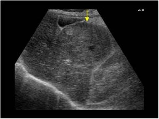
what is the sono of HCC
variable
solitary hypo mass
multiple heterogeneous masses
hypo halo around mass (bulls eye or target sign)
signs of cirrhosis or chr hepatitis
what is the most common cancer of liver
liver metastatic disease
what are the s/s of liver mets
hepatomegaly
weight loss
RUQ pain
jaundice
ascites
what is the sono of liver mets
well defined hypo
well defined hyper
diffuse inhomogeneous exchotexture with no focal mass
bulls eye/target sing
hypo halo, hypo center
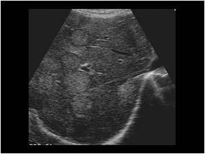
what is liver lymphoma
malignant (hodgkin/non-hodgkin)
lymphocyte proliferation in LNs
usually liver is metastatic site for lymphoma (non-hodgkin)
what are s/s for liver lymphoma
increase LNs
fever, fatigue, weight loss, night sweats, bone pain, splenomegaly
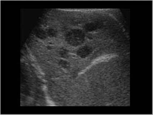
what is sono for liver lymphoma
diffuse or parenchymal change
hypo masses with target sign
what is passive hepatic congestion
develops secondary to congestive heart failure
dilated IVC, HV, SMV, PV, splenic V
what is pneumocystis carinii
m/c oportunistic infection in pt with AIDS
also affects organ transplantation, chemo pt
what is the sono of pneumocystis carinii
diffuse tiny non-shadowing echogenic foci
replacement of liver parenchyma by echogenic clumps of Ca +2