CLIN MED: GI (small intest, colon, rectum, misc)
 Call Kai
 Learn
Learn Practice Test
 Spaced Repetition
 Match
Match Flashcards
 Knowt Play
1/149

There's no tags or description
Looks like no tags are added yet.
Name | Mastery | Learn | Test | Matching | Spaced | Call with Kai | Chat |
|---|
No analytics yet
Send a link to your students to track their progress
150 Terms
Ileus (intestinal)
-pathology
-seen where
Lack of motility in the intestines, = buildup and blockage of food material, gas, and fluids.
-No mechanical obstruction present.
-Usually seen in hospitalized patients.

Ileus types
Paralytic (or adynamic) ileus: most common type
-paralysis of the intestinal muscles, leading to a loss of peristalsis.
Postoperative ileus:
-after surgery, particularly abdominal surgery, due to the effects of anesthesia, manipulation of the bowel, or inflammation.
Pseudobstruction (Ogilvie's syndrome):
-symptoms similar to mechanical obstruction but without any physical blockage, often seen in critically ill patients.
Ileus - causes
Surgery: Abdominal/Pelvic surgeries
Electrolyte imbalances: Hypokalemia, hyponatremia, or hypocalcemia can impair muscle contractions.
Medications: Opioids, anticholinergics, and certain anesthetics
Infections or inflammation: Peritonitis, appendicitis, or pancreatitis can lead to ileus.
Systemic illnesses: Sepsis, pneumonia, or severe metabolic disorders.
Neurological conditions: Parkinson's or spinal cord injuries can disrupt normal bowel motility.
Ileus- SXS
Abdominal distention-can be profound.
Abdominal pain or discomfort: often mild, diffuse, and continuous.
Nausea and vomiting
Bowel sounds: Diminished or absent.
Constipation or inability to pass gas: Due to the lack of bowel motility.
Inability to tolerate an oral diet
No peritoneal irritation
Ileus - labs/imaging
-Check serum electrolytes
-CBC, U/A -> r/o infection
X-ray: (test of choice) (acute abdominal series)
-Shows dilated loops of bowel and air-fluid levels, with no definitive site of obstruction.
CT scan:
•Provides more detailed imaging to rule out mechanical obstruction or other causes, such as tumors or hernias.
Ultrasound:
May be used in children, to differentiate between ileus and mechanical obstruction
Ileus - treatment
-NPO
-NG tube: decompress stomach and relieve distention and vomiting.
-IVF: prevent dehydration and maintain electrolyte balance while the bowel is at rest.
•Correction of underlying causes:
--> Electrolyte imbalances
--> Infections
--> Medications
Diet can be enhanced as bowel function returns.
Small bowel obstruction
-pathology
-leads to
-untreated complications
Blockage (completely or partially) of intestinal content flow by a mechanical cause such as adhesions, hernias, or tumors.
-accumulation of food, fluids, and gas
-untreated complications include --> Bowel ischemia, perforation, and peritonitis

classification of SBO causes
Mechanical obstruction - A physical barrier is blocking the intestinal lumen.
--> Complete obstruction: No passage of gas or stool beyond the obstruction.
--> Partial obstruction: Some contents can still pass through
--> Strangulated obstruction: surgical emergency
-Results in vascular compromise leading to bowel ischemia, gangrene, and bowel necrosis.
-If not treated will lead to septic shock and death
Small bowel obstruction - etiologies
#1 Adhesions: Most common cause
-Scar tissue that forms after previous abdominal surgeries.
-cause loops of the small bowel to stick together, leading to narrowing or blockage.
-appendectomy, colorectal surgery, and gynecologic and upper gastrointestinal (GI) procedures
#2 Tumors: 2nd most common cause
-Benign and malignant growths in the small bowel or from nearby structures can cause obstruction
Hernias:
-A loop of the small intestine becomes trapped in a hernia, cutting off the flow of intestinal contents.
Intussusception:
-Part of the bowel telescopes into an adjacent part, leading to obstruction
Volvulus:
-Intestine twists on itself, leading to blockage
Inflammatory bowel disease: (Inflammation or strictures = narrowing and obstruction)
Foreign bodies (blockage from objects or masses of undigested food)

Small bowel obstruction - sxs
-Abdominal pain, colicky.
-Proximal obstructions:
--> Less distension, shorter delay to presentation, early nausea and vomiting.
-Distal obstructions:
-Significant distension,
-Nausea and vomiting
-Dehydration
-Cramping, intermittent pain is typical, especially around the periumbilical area.
-If colicky pain becomes constant, ischemia, and/or infarction should be considered
-Obstipation seen only with complete obstruction.
Abdominal exam:
•Abdominal scars, tenderness, distension, and high-pitched tinkling bowel sounds, followed by decreased sounds if the bowel becomes paralyzed.
Small bowel obstruction
-imaging --> shows
X-RAY: Plain Films
•Show dilated loops of small bowel and air-fluid levels, suggesting obstruction.
•Will show no gas in the colon if the obstruction is complete.
•May be impossible to distinguish adynamic ileus from obstruction on x-ray, although entire bowel usually involved with an ileus.
•Typically will see distended loops of small bowel proximal to the point of obstruction with air/fluid levels and collapsed bowel distal to the obstruction.
CT SCAN:
Most useful if plain x-rays non-diagnostic, will yield the diagnosis in 80% of cases.
Can be used to demonstrate cause of the obstruction.
Small bowel obstruction - treatment (non surgical)
1. Admit for observation
2. Aggressive fluid resuscitation.
3. Bowel decompression usually with NG tube.
4. Broad-spectrum antibiotics -if strangulated.
5. Monitoring:
-If there are no signs of bowel ischemia or perforation
-Some obstructions (especially partial obstructions) resolve with conservative management.
Small bowel obstruction - treatment (surgical)
Surgical intervention:
-Required if conservative measures fail
Surgery may involve:
-Lysis of adhesions:
-Resection of the bowel
-Repair of hernias
Small bowel obstruction - immediate surgery indications
-Free air on plain xrays or CT = bowel perforation
-Signs of ischemia
-Complete or closed loop obstruction

Ogilvie Syndrome (Acute Colonic Pseudo-obstruction)
-pathology
-risk factors
-contributing factor
-epidemiology
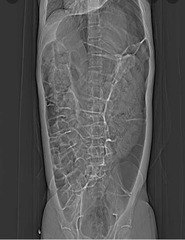
Spontaneous massive colonic dilation without mechanical obstruction, occurring in postoperative or critically ill patients.
Dilation predominantly affects the cecum and ascending colon, and if left untreated, excessive distension can compromise the blood supply to the colon, leading to ischemia or perforation.
Risk factors: hospitalization, post-surgical (3-5 days), trauma, metabolic imbalance, malignancy, MI, CHF, pancreatitis, or recent neurologic event
-Opioid pain medications may be a contributing factor
-usually elderly age group
Ogilvie Syndrome (Acute Colonic Pseudo-obstruction)
-sxs
-abdominal distension, often significant.
-Diminished bowel sounds
-Patients have minimal to no abdominal pain
-Signs of peritonitis absent unless perforation has occurred—No rebound, guarding or rigidity
-Nausea and vomiting may be present
Ogilvie Syndrome (Acute Colonic Pseudo-obstruction)
-labs/imaging
-treatment
IMAGING
X-rays demonstrate dilation of the cecum and proximal colon
TREATMENT
-NG or rectal tube can be placed for decompression
-Increase patient mobility to promote colonic movement.
-Neostigmine: a cholinesterase inhibitor- stimulates colonic motility by increasing acetylcholine levels.
Colonoscopy- decompress the colon
Surgical intervention: Last resort
-Reserved for cases where there is evidence of ischemia, perforation, or failure of conservative and endoscopic measures.
-Surgical options involve the resection of the necrotic bowel.
Avoid opioids and anticholinergics
Ogilvie Syndrome (Acute Colonic Pseudo-obstruction)
-complications
Colon ischemia
-Excessive distention can compromise blood flow to the bowel.
Colon perforation
-Increases significantly when the cecum becomes distended over 12 cm.
-Perforation can lead to peritonitis and sepsis, which are life-threatening
Sepsis
-If ischemia or perforation occurs, bacteria from the gut can enter the bloodstream, leading to widespread infection.
Appendicitis
-epidemiology
-pathology
-causes
-if untreated
Most common surgical emergency.
-Ages 10-30 years
Occurs when the lumen of the appendix becomes blocked, leading to infection and inflammation.
Causes of obstruction include:
•Fecalith (obstruct the appendix duct partially or completely.)
•Infection
•Foreign body
•Cancer
If untreated, the inflamed appendix can rupture, leading to peritonitis.

APPENDICITIS - signs and symptoms
Pain is colicky, starting in periumbilical area!
•Within 12 hours, pain localizes to RLQ!!!
-McBurney's point (1/3 the distance from the anterior superior iliac spine to the navel).
-nauseous, may have vomiting
•Vomiting starts after onset of pain
•If vomiting starts BEFORE pain, it is probably something else!
•High fevers/chills: something else, or the appendix has ruptured.
APPENDICITIS: physical exam
PROM
+ Psoas sign (pain on passive extension of right hip)
+ Rovsing sign,Rebound tenderness
+ Obturator: (pain w/ passive flexion and internal rotation of R hip)
McBurney's point: Localized tenderness with guarding in RLQ
APPENDICITIS
-labs
-imaging
LABS
-Moderate leukocytosis (10,000-20,000 WBCs)
-Elevated CRP
IMAGING
CT scan: Most accurate test, providing detailed images of the appendix and helping to rule out other conditions like diverticulitis or gynecological issues
Ultrasound: a swollen, inflamed appendix and is often used in children and pregnant women to avoid radiation exposure
APPENDICITIS treatment
Laparoscopic appendectomy-
-Early, uncomplicated appendicitis
-Associated with shorter recovery times and less postoperative pain.
Open appendectomy: Performed if the appendix has ruptured or if the patient have peritonitis
Antibiotics: Given preoperatively to reduce the risk of infection
Drainage of abscess: need to be drained before or after surgery.
Hernia
-types
-fears
-treatment
TYPES:
•Incisional
•Femoral
•Indirect
•Direct
•Umbilical
Fears: Incarcerated or not
Laparoscopic hernia repair!
Incisional Hernia
-patho
-epidemiology
-risk
The intestine pushes through the abdominal wall at the site of previous abdominal surgery.
-MC in elderly or obese who are inactive after abdominal surgery.
-Infections at the incision site, poor wound healing, and multiple surgeries can increase the risk.

Femoral Hernia
-patho
-epidemiology
-how common
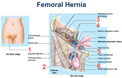
A femoral hernia occurs when the intestine enters the canal carrying the femoral artery into the upper thigh.
-most common in women, especially those who are pregnant or obese.
-Less common than inguinal hernias but have a higher risk of strangulation.
Direct Inguinal Hernia
-patho
-epidemiology
Arises from protrusion of abdominal viscera through Hesselbach's triangle.
-Hernia enters through a weak point in the fascia of the abdominal wall.
-Located medial to the inferior epigastric vessels
epidemiology
-elderly males due to pressure and tension on the muscles and fascia.
-Males are 10x more likely
-Lower risk of strangulation

Indirect Inguinal Hernia
-how common
-epidemiology
-pathology
-located
-risk of _____ and ______
Most common type of hernia
epidemiology
-younger males (newborns, children, and young adults)
occur due to a failure of the processus vaginalis to close during fetal development. (congenital defect).
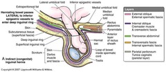
Located lateral and superior to the inferior epigastric vessels and protrude through the deep inguinal ring into the inguinal canal into the scrotum.
Higher risk of incarceration or strangulation
Umbilical Hernia
-patho
-epidemiology
Part of the small intestine passes through the abdominal wall near the navel.
-Common in newborns, obese women, or those who have had many children.

Incarcerated/strangulated Hernia
-patho
-strangulation leads to
If the contents of the hernia become trapped in the weak point in the abdominal wall, it can obstruct the bowel, leading to severe pain, nausea, vomiting, and the inability to have a bowel movement or pass gas
Strangulation: An incarcerated hernia can cut off blood flow to part of your intestine
Strangulation
-patho
-occurs when
-leads to
-treatment
An incarcerated hernia is one that becomes trapped outside the abdominal wall and cannot be manually or spontaneously reduced back into its normal position.
-blood supply to the incarcerated tissue is cut off.
--> lead to necrosis, gangrene, and perforation of the bowel. (Potentially fatal)
-urgent surgical intervention, especially if there are signs of strangulation or bowel obstruction.
Meckels Diverticulum
-location
-describe diverticulum
-epidemiology
Most common congenital abnormality of the small intestine.
Located in the distal terminal ileum, about 2 feet from the ileocecal valve.
-true diverticulum, involving all layers of the intestinal wall.
--> 2 in long
**occurs in 2% of the population, and only 2% of those who have it develop symptoms.

Meckels Diverticulum
-rule of 2's
-sxs
RULE OF 2's
-2-4% incidence
-2:1 ratio-M:F
-2 feet from ileocecal valve
-Up to 2 inches
SXS
-Causes painless rectal GI bleeding but can be massive
-Can lead to obstruction due to intussusception or volvulus
-Perforation
-Infection
-Prognosis is excellent after surgical removal of a symptomatic Meckel's diverticulum
Intusussepction
-patho
-untreated
-epidemiology
One part of the intestine telescopes into an adjacent part.
-Often starts near the junction of the small and large intestine, where the ileum invaginates into the colon.
-Blocks the passage of intestinal contents and compresses the blood vessels in the intestinal wall.
UNTREATED --> ischemia, necrosis, and perforation of the bowel.
common in children, particularly between the ages of 6 months and 3 years.

INTUSUSSEPCTION
-can be caused from
-sxs
-imaging
-treatment
Meckels can be lead point
SXS
-Intermittent abdominal pain,
-"Currant colored" jelly stools
-Sausage-shaped mass in the right upper quadrant
TESTING
Barium or air enema: diagnostic and therapeutic.
-The enema shows the intussusception as a blockage and can often push the telescoped section of the bowel back into place, reducing the intussusception.
**Surgery if intussusception not able to be reduced by the barium enema.
Duodenal intestinal atresia
-patho
-epidemiology
-sxs
-imaging
portion of the duodenum is completely or partially absent or closed, preventing the normal passage of stomach contents into the small intestine.
-Obstruction causes a blockage that can lead to vomiting shortly after birth
-Occurs in 1:10K live births
-30% associated with congenital chromosomal disorder-- Downs common
SXS
-bilious or non-bilious vomiting,
-abdominal distentio
-inability to tolerate food.
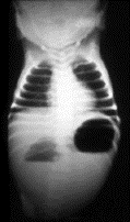
Radiology finding: "double bubble sign"
--> Represents a distended stomach and duodenum
Ischemic bowel disease
-patho
-types
Occurs when there is reduced blood flow to the intestines, leading to tissue damage or death due to lack of oxygen and nutrients.
Acute and chronic mesenteric ischemia
--> Small intestine
Ischemic colitis-
--> Colon
Mesenteric Ischemia
-patho
Poor perfusion into the intestines, resulting from oxygen compromise.
-The superior mesenteric artery (SMA) supplies all the small intestines and the proximal two-thirds of the transverse colon.
-The inferior mesenteric artery (IMA) supplies the remaining colon and the superior portion of the rectum.

ACUTE Mesenteric Ischemia
-pathology
-risk
Abdominal pain occurs because the bowel is starved for oxygen.
-Without oxygen, the bowel becomes ischemic, loses its normal functions, leading to ileus or bowel paralysis.
Over time, the ischemic mucosal barrier will perforate, allowing microbes to enter the normally sterile peritoneum.
-Sepsis is a major complication of untreated ischemic bowel disease.
ACUTE Mesenteric Ischemia
-results from
-causes (4)
Results from an acute interruption of blood flow.
-Usually caused by a blockage in the blood supply the intestines.
Causes include:
1. Arterial embolism: A CLOT that travels from the heart and lodges in the mesenteric arteries, cutting off blood supply
2. Arterial thrombosis: A blood clot forms directly within the mesenteric arteries, often due to atherosclerosis.
3. Venous thrombosis: A blood clot forms in the veins leading to impaired blood outflow and subsequent ischemia.
4. Non-occlusive ischemia: Caused by a low blood flow state such severe hypotension, rather than an actual blockage.
ACUTE Mesenteric Ischemia
-sxs
-labs
Classically present as sudden severe, periumbilical abdominal pain out of proportion
•Nausea, vomiting
•Bloody stools if bowel injury is severe
•Abdominal distension
Labs may show:
•Leukocytosis and a metabolic acidosis
ACUTE Mesenteric Ischemia
-imaging
CT angiography: imaging test of choice
•Shows reduced blood flow in the mesenteric vessels.
X-ray reveals:
•Ileus with distended bowel loops, air-fluid levels, or wall thickening.
•Free air on X-ray indicates perforation
Magnetic resonance angiography (MRA)
•If patient can not tolerate CTA
ACUTE Mesenteric Ischemia
-treatment (more severe cases & mild)
-mortality
SEVERE: (infarction, ischemia, peritonitis, perforation, or sepsis)
-laparotomy with surgical embolectomy. --> Remove the clot and restore blood flow
--> Remove damaged intestine if the bowel has become necrotic to prevent sepsis
MILD (or cannot tolerate surgery)
-Embolic AMI - thrombolytics to break up the clot.
•Thrombotic AMI - intra-arterial thrombolytics with endovascular angioplasty and stent placement
***Nonocclusive AMI (poor perfusion)
•Fluid, blood products, and vasopressors can be used based on the etiology.
CHRONIC Mesenteric Ischemia
-patho
A long-standing reduction in blood flow to the intestines, typically due to atherosclerosis affecting the mesenteric arteries.
Chronic narrowing of the arteries supplying the intestines, often due to atherosclerosis, which restricts blood flow during periods of high demand, such as after meals.
--> Atherosclerotic plaques in the mesenteric vessels, limit but do not completely occlude normal blood flow.
CHRONIC Mesenteric Ischemia
-risk factors
-Cardiovascular disease
-Atherosclerosis
-Advanced age (>45 years in men, >55 years in women)
-History of smoking
-Hypertension
-Hyperlipidemia
-Diabetes
-CKD
-Obesity
CHRONIC Mesenteric Ischemia
-sxs
Postprandial (after a meal), dull, crampy, vague abdominal pain.
-occurs because food shunts blood away from the intestines or increases the demands of the stenotic segments.
-Does not progress to infarction because, when the food is gone, blood flow returns.
-Because of the postprandial pain, patients start to limit food intake
Chronic weight loss with the abdominal pain.
Diarrhea or malabsorption: Due to impaired intestinal function from reduce blood flow
CHRONIC Mesenteric Ischemia
-testing
1. begin with a CT angiogram
--> If intervention is being planned, an MRA should be done.
Asymptomatic patients may be treated with medical management and lifestyle modification.
--> decrease smoking weight loss, blood pressure, and diabetes and lipid control.
Symptomatic revascularization is indicated.
-Revascularization can be done by open surgery
--> Thrombectomy or arterial reimplantation with bowel resection
--> Endovascular angioplasty and stenting.
Ischemic Colitis
-patho
-causes
Intestinal ischemia, affecting the colon.
•Usually occurs when blood flow to the colon is reduced.
Causes include:
•Hypotension-
•Arthrosclerosis
•Medications: vasoconstrictors
•Surgery: Abdominal or cardiovascular surgery may temporarily reduces blood perfusion
Ischemic Colitis
-sxs
-Abdominal pain associated with hematochezia within 24 hours after onset of pain.
--> Hematochezia is a sign of ischemic injury to the bowel lining
-May be associated with the need to defecate.
-Bowel infarcted is associated with gangrene and tissue necrosis, leading to perforation and sepsis
Ischemic Colitis (Colonic Ischemia)
-testing/diagnosis
CT scan.
--> Results will show wall thickening or pneumatosis coli—air in the colon wall
-->If the CT is negative, a colonoscopy can be done to visualize the ischemic tissue.
Ischemic Colitis (Colonic Ischemia)
-treatments
-treatment with IVFs and antibiotics.
untreated
-colonic ischemia may progress to gangrene and bowel perforation.
**Signs suggestive of perforation, such as abdominal guarding, hypotension, or peritoneal symptoms, indicate a need for surgical exploration.
Surviving patients are at risk of developing a stricture at the affected site, increasing risk of bowel obstruction
IBS
-patho
-epidemiology
-tests
Functional GI disorder defined by chronic abdominal pain with altered bowel habits
Women in 20s-30s more often affected than men.
no specific diagnostic tests, and it is a diagnosis of exclusion in association with a complex of symptoms.
IBS - classification
IBS-D (Diarrhea-predominant IBS):
-Frequent, loose stools, often accompanied by urgency
IBS-C (Constipation-predominant IBS):
-Infrequent, hard stools that are difficult to pass
BS-M (Mixed IBS):
-Alternating between diarrhea and constipation
IBS-U (Unsubtyped IBS):
-Patients whose symptoms do not clearly fit into the other categories.
IBS - SXS
-bloating
-diarrhea (predominant diarrhea (almost never nocturnal)
-constipation
-abdominal pain
-cramps
-distention
•Some people may experience nausea, lethargy, and back pain (defecation relieves pain)
•Lack of warning symptoms
RED FLAG
•Unintentional weight loss
•Rectal bleeding
•New-onset IBS-like symptoms in an elderly patient.
IBS - risk factors
-Mental health illnesses (depression, anxiety) have higher risk of IBS.
•Childhood abuse
•Physical illnesses such as fibromyalgia and migraines are also associated with IBS.
IBS - diagnosis criteria
The Rome IV criteria is used to make a diagnosis of IBS.
--> defined as: recurrent abdominal pain averaging at least 1 day a week for 3 months that is associated with at least two of the following:
-Pain is relieved by a bowel movement
-pain is related to a change in frequency of stool
-pain is related to a change in the appearance of stool.
IBS - pathology-dysmotility
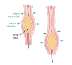
Abnormal gastrointestinal motility: Increased or slowed contractions of the intestinal muscles leading to diarrhea or constipation.
Visceral hypersensitivity: Heightened sensitivity to pain or discomfort from normal gut activity.
Gut-brain interaction: dysfunction in the communication between the CNS and the gut
Stress: emotional stress, anxiety, and depression are strongly linked to IBS and worsens symptoms.
Infections: history of GI or SB bacterial overgrowth may trigger IBS symptoms.
Dietary factors: high-fat foods, caffeine, alcohol, can worsen IBS symptoms.
IBS - diagnosis/workout
IBS is often a diagnosis of exclusion.
--> Exclusion of alternative diagnoses that can cause IBS symptoms may be needed.
NO definitive tests for IBS (diagnostic work-up is done to rule out lactose intolerance, celiac disease, IBD and cancer)
-colonoscopy
-WBC count
-Hgb normal C-reactive protein
-Fecal calprotectin levels.
-Stool ova and parasite testing rule out infectious causes
IBS - general treatment
Lifestyle changes, dietary modifications, medications, and psychological therapies.
Dietary modifications include:
--> Reducing foods high in fermentable carbohydrates to relieve bloating and gas.
--> Increasing dietary fiber
--> Soluble fiber is recommended.
--> Avoid dairy, caffeine, alcohol, or spicy foods.
**Probiotics may help improve gut health and relieve IBS symptoms in some individuals
IBS - treatment for mild-intermittent sxs
Mild-Intermittent Symptoms
-Physical exercise - regular physical activity can promote healthy digestion.
-Avoid gas-producing foods
-Stress management: techniques like mindfulness, yoga, or relaxation exercises can reduce stress and improve gut health
IBS-C treatment
Gut motility agents:
·Lubiprostone (Amitiza)
·Linaclotide (Linzess)
·Plecanatide (Trulance)
Osmotic laxatives - polyethylene glycol may be used to soften stools.
--> Can stimulate intestinal fluid secretion, transit, and relieve constipation.
IBS-D treatment
-Alosetron (Lotronex)- a 5HT-3 serotonin-receptor antagonist
-Antidiarrheal agents: (loperamide, Lomotil)
-Antispasmodics: (Dicyclomine, Hyoscyamine)- reduce spasms
-Eluxadoline (Viberzi)-mu-opioid receptor agonist; relieves pain/slows bowel.
-Rifaximin- non-absorbed antibiotic- small intestinal bacterial overgrowth
-Tricyclic antidepressants- helpful for reducing hypersensitivity
CLOSTRIDIUM DIFFICILE COLITIS
-patho
-cause
-causes
Infection of the colon caused by the bacteria Clostridium difficile.
CAUSE
After antibiotic use, which disrupts the normal gut flora, allowing C. difficile to overgrow and produce toxins that damage the colon.
Causes inflammation of the colon, leading to diarrhea and, in severe cases, the formation of pseudomembranes, or toxic megacolon
CLOSTRIDIUM DIFFICILE COLITIS
-risk factors
-Age >70 years
-Hospitalization or long-term health care facility
-Enteral feeding
-Previous C. difficile infection
-Chronic acid suppression (PPI)- reduce stomach acid, which alters the gut environment and promote C. difficile growth.
Exposure to antibiotics (including perioperative prophylaxis)
•Ampicillin, amoxicillin, clindamycin (most common), cephalosporins, and fluoroquinolones.
CLOSTRIDIUM DIFFICILE COLITIS
-sxs
•Watery diarrhea and abdominal cramping/pain
•Recent antibiotic use (risk persist up to 3 months after antibiotic is discontinued.
•Foul-smelling diarrhea and sometimes bloody.
•Fever (
CLOSTRIDIUM DIFFICILE COLITIS
-labs
The best initial test is a stool C. diff toxin test or PCR.
-C-difficile toxin test: Detects toxins A and B produced by the bacterium in the stool.
--> Most commonly used test but may miss some cases.
-Nucleic acid amplification test (NAAT) or PCR test:
--> Detects the genes responsible for toxin production and is more sensitive than toxin test
CLOSTRIDIUM DIFFICILE COLITIS
-diagnostic test
CT scan shows mucosal thickening, colonic wall thickening, pericolonic inflammation, or signs of complicated infection in severe cases.
Endoscopy-colonoscopy or sigmoidoscopy
•Evaluates for the presence of pseudomembranes and exclude other conditions.
•Flexible sigmoidoscopy may miss 15-20% of pseudomembranes (from the proximal colon).
CLOSTRIDIUM DIFFICILE COLITIS
-treatment
-discontinue
PHARM:
First-line: Oral vancomycin for 10 days is the most effective initial therapy.
-not effective --> fidaxomicin.
Metronidazole: mild cases or in combination with vancomycin for severe cases.
After multiple recurrences, the choice is fecal transplantation.
DISCONTINUE/AVOID:
-offending antibiotic, whenever possible.
-Antimotility agents
-indiscriminate use of antibiotics.
IBD (inflammatory bowel disease)
-patho
-encompasses two different dieases
-epidemiology
inflammation of the digestive tract.
1. Crohn disease (female)
2. Ulcerative colitis (UC) (male)
Age of onset for IBD is 15-30 years, with a second peak at ages 50-80 years.
IBD
-UC vs Crohn's causes and prevention
BOTH - genetic component identified
-Infections and imbalances in the gut microbiome are implicated in the pathogenesis of IBD.
Crohn's
-smoking increase the risk
-Exercise and fiber decreased risk
UC
-smoking may be protective in UC.
IBD
-sxs
-extra intestinal manifestations
-risk
-acute flares treatment
"Colitis" refers to inflammation of the colon most typically presenting
-abdominal pain, bloody or non-bloody diarrhea, and possible systemic signs such as fevers.
-uveitis , arthritis, erythema nodosum, and pyoderma gangrenosum.
Both forms of IBD can increase the risk of colon cancer.
--> Patients should undergo screening colonoscopy 8 years after diagnosis or symptom onset, whichever is first.
Acute flares of IBD are treated with corticosteroids; long-term therapy usually involves immune-modulating agents such as azathioprine and infliximab.
Crohn's Disease
-patho
-characterized
-cure?
chronic, indolent course characterized by unpredictable flares and remissions.
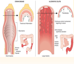
Chronic transmural!!! (full-wall thickness) disease that can affect any part of the GI tract (mouth to anus)
-most commonly involves the terminal ileum.
There is no cure, and recurrence is common even after surgery.

Crohn's disease - sxs
HALLMARK SYMPTOMS:
-abdominal pain RLQ
-fevers
-weight loss
**Crohn's is more likely to present with non-bloody diarrhea compared to UC
"skip lesions" with a normal section of bowel between diseased sections
•Fistula and stricture development
Noncaseating granulomas (macrophages, with/without multinucleate giant cells).
Crohn's disease
-labs
LABS
-Increased C-reactive protein and sedimentation rate
-Leukocytosis
-Anemia
-Thrombocytosis
-Fecal calprotectin elevated**
-Stool studies for ova and parasites, culture, and C. difficile should be obtained to rule out infection
•**Anti-Saccharomyces antibody is elevated in 80% of CD patients. ASCA
Crohn's disease
-diagnostic test
DIAGNOSTIC TEST
-Ileocolonoscopy to distinguish between UC and colonic CD
--> Allows for biopsy
--> Ability to visualize "cobblestoning", strictures, or ulcers
-Capsule endoscopy is useful for staging or identifying small-bowel CD.
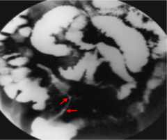
barium swallow (small bowel)
-may show ulcerations in between normal and abnormal mucosa, including fistulas, cobblestoning, strictures, or ulcers.
--> "string sign" on barium swallow, which signifies passage of contrast through a narrowed lumen.
Magnetic resonance imaging (MRI) with enterography is also with evaluating the small intestine.
--> Helpful with evaluating rectal or perirectal disease and fistulas.
CT scan of abdomen and pelvis or CT enterography can also be useful in some cases.
Crohn's disease
-acute flares treatment/side effects
Acute flares
-Supplement calcium and vitamin D.
-Corticosteroids are effective in reducing inflammation quickly, but they come with a range of potential side-effects
--> Commonly used as initial therapy for low-risk patients
--> Due to side effects, not used as a long-term therapy
•Side effects include:
•Osteoporosis, hyperglycemia, PUD, increase susceptibility to infections, cataracts, pancreatitis, weight gain, avascular necrosis, mood disorders, and growth retardation in children.
Crohn's disease
-chronic maintenance treatment
severe: Azathioprine- an immunosuppressive agent.
-Azathioprine inhibits the proliferation of immune cells, providing an immunosuppressive effect.
--> Induce and maintain remission
•Steroid-sparing effect, minimizing potential steroid-associated side effects
--> May promote healing of fistulas in Crohn's disease
mild: to moderate: 5-ASA derivatives (mesalamine and sulfasalazine)
-Reduces inflammation
-Used with mild-to-moderate disease activity
-More commonly used in UC compared to Crohn's
Crohn's disease
-Tumor Necrosis Factor (TNF) inhibitors
TNF-α promotes inflammation, tissue damage, and fistulas
- promote mucosal healing, closing these fistulas, and reducing the output from enterocutaneous fistulas.
-Induce remission and maintain remission.
--> Infliximab (Remicade), Adalimumab (Humira), Certolizumab pegol (Cimzia)
Side effects include:
•Increased risk of infections
•Increased risk of lymphoma
•Potential development of antibodies against the medication (which can reduce its effectiveness)
•Reactivation of latent tuberculosis.
Crohn's disease
-surgical management
Reserved for complications of Crohn's
-segmental resection of the involved portion of bowel
•Up to 50% of patients experience disease recurrence 10 years postoperatively.
Indications for surgery include: SBO, fistulae, perforation or abscess.
Nutritional supplementation and support—parenteral nutrition is sometimes necessary.
Crohn's disease
-complications 1
Fistula Formation
-A fistula is an abnormal connection between a hollow organ like the bowel and another hollow organ or the skin.
•Fistulas that occur between colon and other segments of intestine (enteroenteral), bladder (enterovesical), vagina (enterovaginal), and skin (enterocutaneous).
•Disruptions in bowel mucosa can lead to abscesses, which can cause infection and peritonitis

Crohn's disease
-complications 2
Perianal Disease
-Common manifestations include perianal pain, anal fissures, perirectal abscesses, and anal fistula.
-Anal fissures from Crohn disease are typically posterior to the pectinate line.
-Perianal abscesses and fistulas are treated with ciprofloxacin and metronidazole.

Crohn's disease
-complications general
Malabsorption
-The presence of extra bile acids in the lumen increases its osmotic gradient and causes diarrhea.
•Lack of fat absorption can also cause steatorrhea (fat in the stool).
Stricture Formation
•When strictures become complete or near complete, they can lead to small bowel obstruction), which is a common indication for surgery in patients with Crohn disease.
Malignancy—increased risk of colonic and small bowel tumors
--> Colon cancer screening should occur after 8 years of colonic involvement, with colonoscopy every 1 to 2 years.
•Endoscopy is the most accurate test when the disease can be reached by a scope.
Cholelithiasis may occur secondary to decreased bile acid absorption.
Ulcerative Colitis
-patho
-skip lesions?
-typical symptoms
UC is a chronic inflammatory condition of the mucosal layer of the colon.
-UC involves the rectum in all cases and can involve the colon either partially or entirely but is always continuous.
-There are no skip lesions as are seen in Crohn disease.
-UC typically presents with diarrhea that is more often bloody than the diarrhea seen with Crohn disease.
Ulcerative Colitis
-sxs
-bowel movements are small and frequent.
-Colicky abdominal pain, tenesmus (sensation of incomplete defecation/feeling of needing to evacuate the bowels frequently), and fecal incontinence are common.
-fevers, fatigue, and weight loss
•It may occur at any age (usually begins in adolescence or young adulthood).
Ulcerative Colitis
-bowel involved
•The small bowel is not usually involved in UC, but it may reach the distal ileum in a small percentage of patients ("backwash ileitis" in 10% of cases)
Ulcerative Colitis
-clinical features
-Hematochezia (bloody diarrhea)
-Abdominal pain
-Bowel movements are frequent but small
-May have fever, anorexia, and weight loss (severe cases)
-Tenesmus (rectal dry heaves)
-Extraintestinal symptoms (jaundice, uveitis, arthritis, skin lesions)—

Ulcerative Colitis
-diagnosis tests
+ Antineutrophil cytoplasmic antibodies (ANCA)!!!!!
Initial studies:
-Stool cultures for Clostridium difficile, ova, and parasites to rule out infectious diarrhea.
-Fecal leukocytes can appear in UC, ischemic colitis, or infectious diarrhea.
Colonoscopy to assess the extent of disease and the presence of any complications.
Ulcerative Colitis
-treatment
5-ASA medications (sulfasalazine, mesalamine, olsalazine)
-Mainstay for mild to moderate UC
-Can be used on an ongoing basis.
Corticosteroids
-Initial therapy for most moderate cases and all severe cases
-Can be used intravenously, orally, or as an enema (for isolated rectal involvement or proctitis)
-Oral budesonide undergoes extensive first-pass metabolism and has fewer side effects.
TNF inhibitors (infliximab, adalimumab)
-Alone or in combination with azathioprine can be used in patients who do not respond to corticosteroids or for severe disease.
Ulcerative Colitis
-complications 1 of 2
Iron deficiency anemia:
-UC more likely presents in the form of attacks that last weeks to months.
-Due to rectal blood loss, some patients with UC may occasionally experience dyspnea and palpitations due to iron-deficiency anemia.
Hemorrhage:
-Massive hemorrhage can occur in up to 3% of patients and is an indication for urgent colectomy.
Fulminant Colitis:
-Fulminant colitis is a complication of UC that presents with >10 stools a day and is associated with bleeding, abdominal pain, and distension.
-Patients appear toxic with fever and anorexia
Ulcerative Colitis
-complications 2 of 2
Toxic Megacolon
-The colon becomes grossly dilated (>6m) and inflamed, losing its ability to function normally
-Leading cause of death in UC and affects
Ulcerative Colitis
-indications for surgery
Surgery—often curative (unlike Crohn's) and involves total colectomy. Indications include:
-Toxic megacolon (risk of perforation).
-Stricture
-Massive bleeding
-Obstruction (due to stricture)
-Severe hemorrhage perforation
-Fulminant exacerbation that does not respond to steroids.
-Evidence of colon cancer or increased risk of colon cancer.
-Refractory severe inflammation
Diverticular Disease
-patho
-cause
-A sac-like protrusion of the colonic wall created by the herniation of the lining mucosa through a defect in the muscular layer of the gastrointestinal (GI) tract.
-occurs due to diverticular bleeding, diverticulitis, segmental colitis, or symptomatic uncomplicated diverticular disease.

Diverticulosis
-patho
-risk
-common location
Caused by increased intraluminal pressure through the focal area of weakness in the colon wall. (no pain or inflammation)
-> Out-pocketing of the colon
-MCC of acute lower GI bleeds in adults (not in slides)
Risk factors:
-Low-fiber diets
-Constipation causes intraluminal pressures to increase.
-Positive family history.
-Prevalence increases with age.
The most common location is the sigmoid colon but can occur anywhere in the colon.
Diverticulosis Disease
-life style factors associated with disease
-High intake of red meat
-Low dietary fiber
-Lack of exercise
-Overweight (body mass index >25)
-Smoking.
Diverticulosis Disease
-sxs
-asymptomatic and discovered incidentally on imaging or when a complication occurs (diverticular bleed or diverticulitis).
-vague left lower quadrant (LLQ) discomfort, bloating, constipation/diarrhea.
-painless rectal bleeding (up to 40% of patients)
-Bleeding is usually stop spontaneously.
-Bleeding can be severe in about 5% of patients, requiring hospitalization.
Diverticulosis Disease
-complications
-testing
**If bleeding is persistent and/or recurrent, surgery (segmental colectomy) may be needed.
Complications -abscess formation, obstruction, perforation, and fistula formation.
TESTING
-most accurate test is colonoscopy.
-Barium studies are acceptable, but not as accurate.
Diverticulitis
-patho
-sxs
Occurs when feces become impacted in the diverticulum, leading to erosion and micro-perforation.
-LLQ, tenderness, fever, and leukocytosis
-pericolic fat stranding and colonic wall thickening
-alteration in bowel habits (constipation or diarrhea), vomiting, and sometimes a painful mass on rectal examination
•Nausea, constipation, and bleeding may be present
Diverticulitis
-testing
The best initial test is a CT scan.
Colonoscopy and barium enema are dangerous in acute diverticulitis because of increased risk of perforation. Infection weakens the colonic wall.
Diverticulitis
-treatment
Uncomplicated diverticulitis is managed with:
Ciprofloxacin with metronidazole for 7-10 days, bowel rest (NPO), IV fluids.
Mild episodes may be treated outpatient if patient is reliable and have few comorbid conditions.
If symptoms persist after 3 to 4 days, surgical resection of the involved segment, may be necessary.
After successful treatment, about one-third has recurrence.
Surgery recommended for recurrent episodes or complications
Diverticulitis
-surgical indications
-No response to medical therapy
-Frequent recurrences of infection
-Perforation
-Fistula formation
-Abscess
-Strictures
-Obstruction