Dental Anatomy and Occlusion
 Call Kai
 Learn
Learn Practice Test
 Spaced Repetition
 Match
Match Flashcards
 Knowt Play
1/58

There's no tags or description
Looks like no tags are added yet.
Name | Mastery | Learn | Test | Matching | Spaced | Call with Kai | Chat |
|---|
No analytics yet
Send a link to your students to track their progress
59 Terms
Maxilla
upper jaw
mandible
lower jaw
labial
refers to the lips
nasolabial fold
the crease on the sides of nose
vermillion border
the top edge of lips
vermillion
the lips themselves, pink
philtrum
column that is right under nose
cupid’s bow
the fold on the top of lips
Oral commissure
the corder of lips
labiomental crease
under lips and above the chin
Rugae
ridges on the hard palate
palatine raphae
the midline that runs through the hard palate and the soft palate
Palatoglossal arch
the outer curve around the uvula, the more lateral of the two
palatopharyngeal arch
the curve that is closes to the uvula, nearest the throat
uvula
the suspended ball towards the back of throat
Retromolar pad
the area that is behind the third molar
Pterygomandibular raphae
the area where we inject patients, it is close to the third molar
lingual
refers to the tongue
ventral tonge
underside of tonge
lingual frenum
small tissue that secures the tonge
wharton’s duct
thing under the tongue that secretes saliva
Buccal
refers to the cheeks
labial frenum
connective tissue that is right under the lips, inside of the mouth
buccal frenum
connective tissue of the cheek
tubercle
small bump, projection
diastema
A gap between two teeth
Mucosa
the moist, protective tissue lining internal body cavities and organs exposed to the outside environment
Alveolar mucosa
top layer of gum, darker red color
Mucogingical junction
the line between the alveolar mucosa and the attahced gingiva
Attached gingiva
the pinkish gums right above the teeth
Buccal corridor
The visible shadow seen from the outside when you smile
Interdental Papilla
the small, triangular piece of gum tissue that fills the space between two adjacent teeth
Buccal Vesitbule
The physical pocket inside the mouth between the cheek and the teeth
Labil mucosa
protective tissue of the lips
wide smile, narrow buccal corridor
Preferred smile
Gingival margin
the curved line where the teeth and the gums meet
gingival sulcus
the free gingiva over the potential space
popcorn kernels get stuck here all the time
Alveolar crest fibers
periodontal ligament fibers that are located at the top
Horizontal fibers
periodontal fibers that are right under, their oritation is perpendicular to the tooth
Oblique fibers
slanted periodontal fibers
actively resisting the forces of chewing.
Apical fibers
periodontal fibers that are furthest down, nearest deep gums
Transseptal
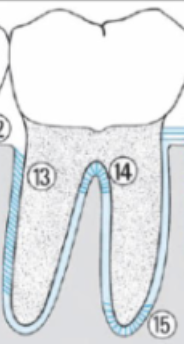
periodontal ligament that exists between teeth, connects one tooth to anotherI
Interradicular
located only between roots of multirooted teeth, not every tooth has it.
It is that number 14 in the photo
Enamel
Protective external surface layer of the anatomic crown
highly calcified/mineralized and is the hardest substance in the body
Thickest at the occlusal/ incisal portion (biting and
chewing)
• Thinnest at cementoenamel junction where it
terminates
• Made up of rods that are perpendicular to the
dentinoenamel junction except at the gingival/
cervical area
Dentinoenamel junction
the interace where the enamel (outer crown) meets the dentin (inner tooth structure)
junction between the enamel inner surface where it joins dentin
Cementoenamel Junction
the external boundary at the tooth’s neck where the enamel stops and the root’s cementum begins
junction between the enamel covering the anatomic crown and the cementum covering the anatomic root
Cementum
external layer of the anatomic
root
• very thin, especially next to the
part of the root where the
crown joins with the root
50-60% mineralized
periodontal ligaments are embedded hear and attach to alveolar bone
Dentin
• tissue underlying the enamel and cementum
• makes up the bulk of the inner portion of each
tooth crown and root
• about 70% mineralized
• less hard (and less brittle) than enamel
• Has tubules where processes of odontoblasts lie
• area that allows teeth to have sensitivity
Pulp
soft, nonminrealized connective tissue containing a rush supply of blood vessels contained in its chamber (in the crown) and root canals
it’s alive
dentil-producing cells (odontoblasts) produce dentin thorughout the life of a tooth
anatomic crown
portion of the tooth covered by enamel
anatomic root
portion of the tooth covered by cementum
Clinical Crown
the portion of the tooth that is visible in the oral cavity ( not covered with gingiva) this includes the anatomic crown
Clinical root
the portion of the tooth that is not visible since it is covered with gingiva
Palatal surface
inside surface that applies to maxillary teeth ONLY (anterior or posterior)
Incisal Edge
cutting surfaces of anterior teeth
Occlusal surface
chewing surface of posterior teeth
Mesial surface
the proximal tooth surface faces towards the arch midline (closer to the midline)
Distal surface
the proximal tooth surface faces further from the midline
lingual surface
the surface of a maxillary or mandibular tooth nearest to the tongue (anterior and posterior)