nervous system part 1
1/69
There's no tags or description
Looks like no tags are added yet.
Name | Mastery | Learn | Test | Matching | Spaced |
|---|
No study sessions yet.
70 Terms
The nervous system
The nervous system contains all neural tissue in the body and is compromised of two different cell types
Neurons
Neuroglia
There are also two main divisions of the nervous system
Central nervous system (CNS)
Peripheral nervous system (PNS)
Neurons
Cells that send and receive signals
Neuroglia
Cells that support and protect neurons
Central nervous system (CNS)
Contains the brain and spinal cord
Peripheral nervous system
Delivers sensory information to the CNS; transmits motor info from the CNS
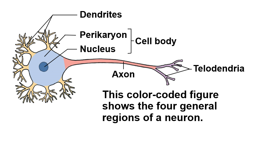
The neuron
Can be split up into four general regions or areas
Cell body
Dendrites
Axons
Telodendria
Cell body
Contains the perikaryon (cytoplasm) and nucleus
Dendrites
Receive info from other neurons
Axon
Carries electrical signal (action potential) towards targets
Telodendria
Extensions of the axons that end in synaptic terminals
Four distinct neuron types
Anaxonic neuron
Bipolar neuron
Unipolar neuron
Multipolar neuron
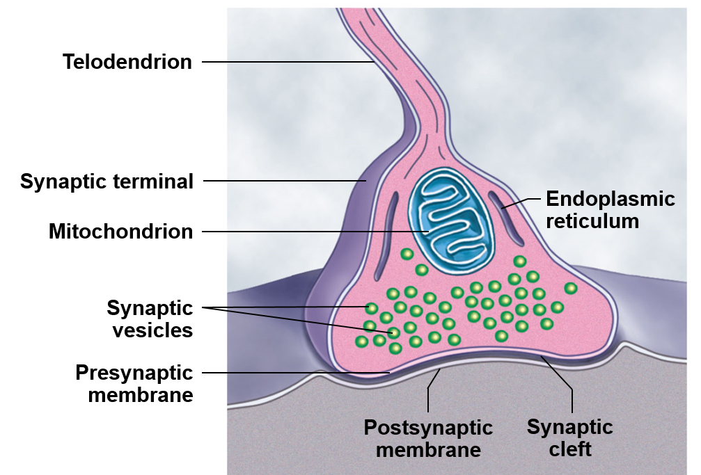
The synapse
Where the action potential is converted into a chemical signal
When this involves a muscle, we call the synapse the neuromuscular junction
When a gland is involved, we might say the neuroglandular junction
Myelination
Increases the velocity of an action potential
The most important Neuroglia are involved with myelination
Oligodendrocytes
Myelinate axons of the CNS
without myelin, the action potential wouldn’t travel quick enough from one side of a neuron to the other side of the neuron, leading to a loss of signal
Shwann cells
Myelinate axons of the PNS
Action potential
The electrical propagation through a neuron in order to initiate a response
Two factors that action potentials use to work
Permeability
Electrochemical gradient
Two channels that action potentials use to work
Sodium voltage gated ion channel
Potassium voltage gated ion channel
Depolarization
Making the membrane potential less negative
Polarization
Making the membrane potential more negative
Action potential process
At rest, the membrane of a neuron isn’t permeable to sodium, and slightly permeable to potassium
When an electrical stimulation causes a threshold to be reached, first sodium voltage gated channels open, allowing an infux of sodium
After the peak of an action potential, sodium voltage gated channels open, causing a release of potassium
After a short afterhyperpolarization, the resting potential re-establishes

Absolute refractory period in action potential
An amount of time in which under no circumstances another action potential can be triggered
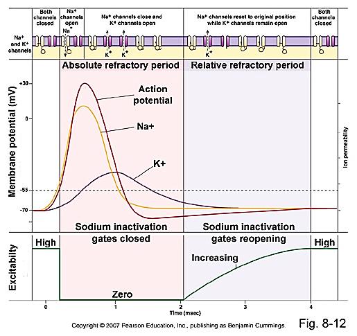
Relative refractory period in action potential
An amount of time in which an increased stimulus can trigger an action potential

When is an action potential termed an all or none phenomenon
If the threshold is reached, an action potential has to occur
If the threshold is not reached, an action potential never occurs
Saltatory propagation
The velocity of the action potential is quicker in myelinated segments, with restoration and slowing of the action potential in unmyelinated segments called the Nodes of Ranvier
Propagation along a myelinate nerve is often termed saltatory propagation
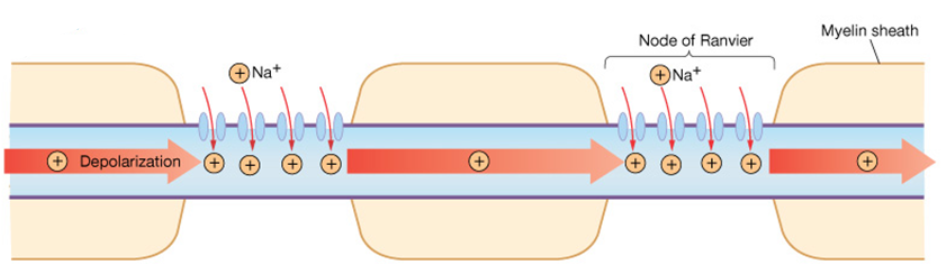
An action potential undergoing salutatory propagation
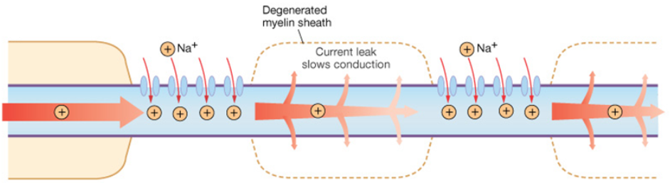
Action potential loss with demyelinating disease
Synapse
When the action potential reaches a synapse, the electrical signal is changed into a chemical signal with the help of vestibular transport. Chemicals are used in the synaptic cleft to introduce the desired effect on the target tissue.
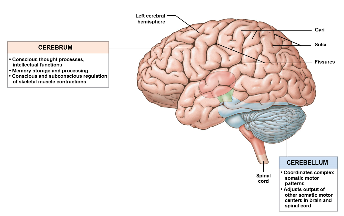
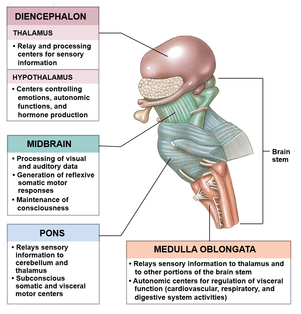
Brain
Contains 97% of the body’s neural tissue in the adult; exerts centralized control of the other organs in the human body
Spinal cord
Continuation of neural tissue off of the medulla oblongata; transmits information to and from the brain; independently responsible for numerous reflexes
Begins past the medulla oblongata and tapers at the conus medulla ribs (L1 or L2)
Frontal lobe
Involves the ability to recognize future consequences resulting from current actions and helps with the determination of similarities and differences between things or events
Temporal lobe
Involves the retention of visual memories, the processing of sensory input, the comprehension of language, emotion, and the storage of new memories
Parietal lobe
Helps integrate sensory information from various parts of the body; involved with the knowledge of numbers and their relations
Occipital lobe
Primarily involved with processing visual information
Precentral gurus
Directs voluntary voluntary movement
Postcentral gurus
Receives somatic sensory information
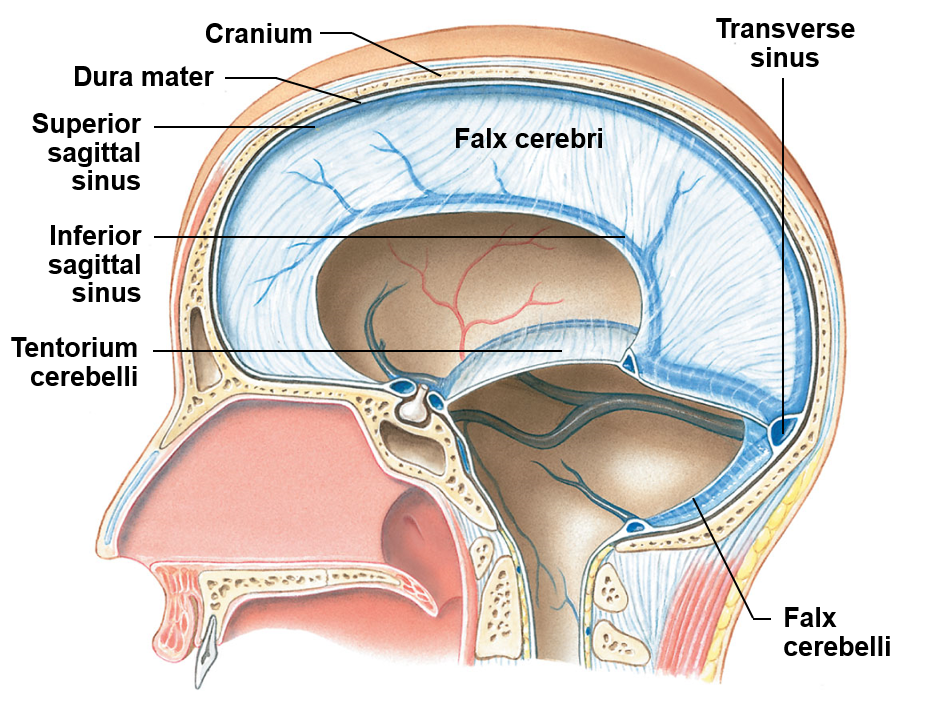
Meninges
Membranes that surround the central nervous system
Dura matter
Thick membrane and outermost of the meninges
Arachnoid matter
Middle, web-like meningeal layer
Pia mater
The innermost and delicate meningeal layer; clings on to the brain and spinal cord
Dura folds
Brain ventricles
Create and help transport cerebrospinal fluid (CSF)
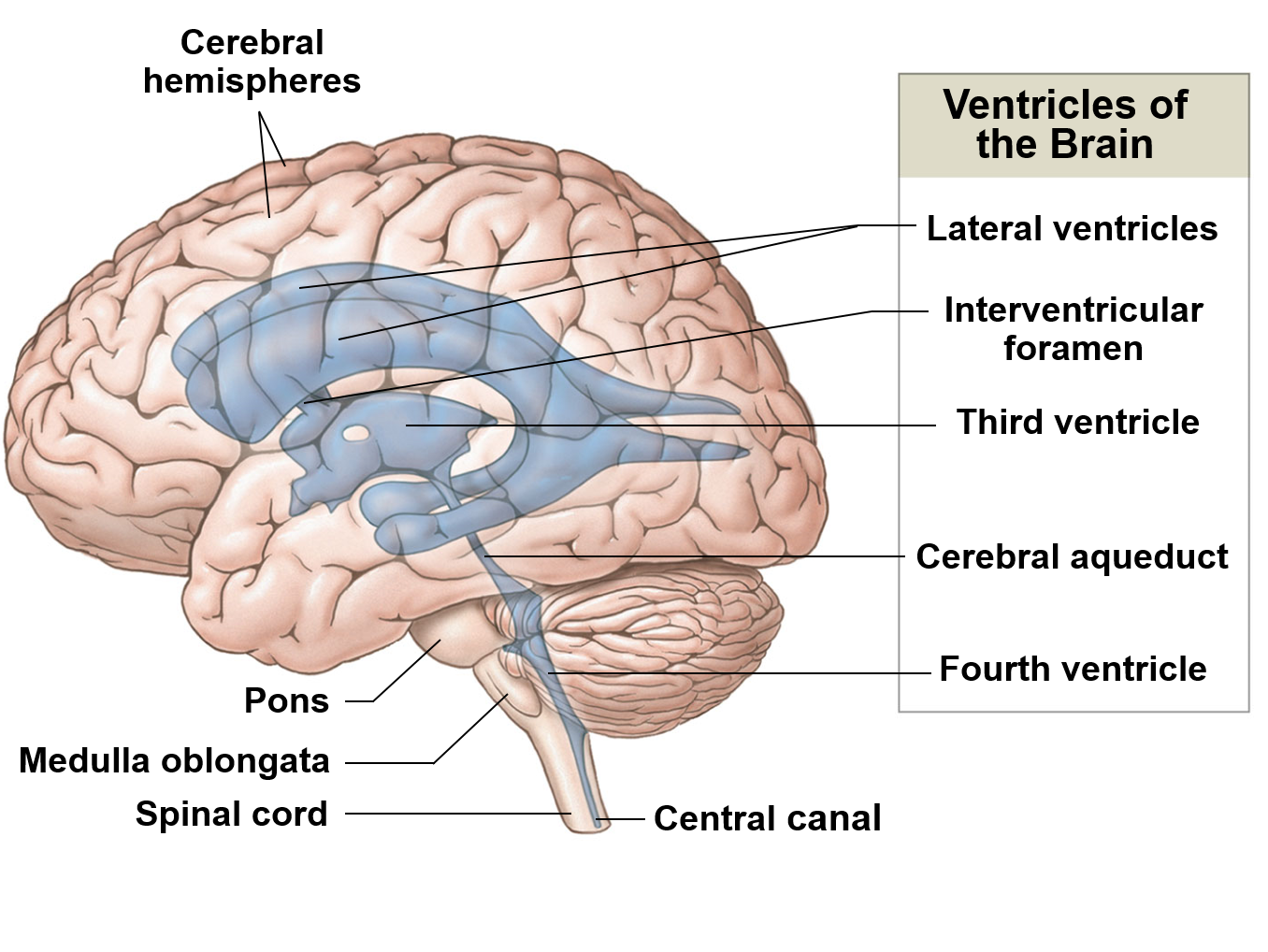
Cerebrospinal fluid
Clear, colorless, bodily fluid that helps protect the brain and the spinal cord mechanically and chemically; found in the subarachnoid space (between pia mater and arachnoid mater)
Arachnoid granulations
Projections of arachnoid mater that allow cerebrospinal fluid to enter the rural venous sinuses
Rural venous sinus
Channel found between layers of the dura mater that receive blood from the veins of the brain and cerebrospinal fluid from the subarachnoid space; empty into the internal jugular veins
Pituitary gland
Endocrine gland responsible for interfacing the nervous system and the endocrine system
Cauda equine
Bundle of spinal nerves that continue off of the conus medulla ribs
Filum terminate
Modification of pia mater that attaches the conus medullaris to the coccyx
Spinal cords seven branches
8 cervical spinal nerve pairs
12 thoracic spinal nerve pairs
5 lumbar nerve pairs
5 sacral nerve pairs
1 coccygeal nerve pairs
Efferent nerves
Those that carry motor information from the CNS
Automatic nervous system
sympathetic division
Parasympathetic division
Sympathetic division
Generally, causes stimulatory effects on the body
Parasympathetic division
Generally, causes relaxing effects on the body
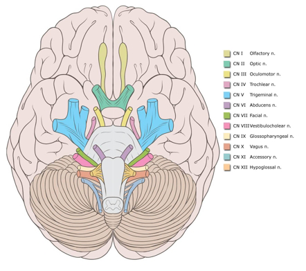
the cranial nerves are 12 pairs of nerves that arise from the inferior aspect of the brain
The cranial nerves are numbered from anterior to posterior as they arise from the brain
The functions of each cranial nerve varies considerably from nerve to nerve
(1) CN I - Olfactory nerve
Function to facilitate smell (sensory)
To move from the olfactory to epithelium, the nerves pass through the foramina of the cribriform plate and back towards the brain to convey sensory information
(2) CN II - Optic nerve
Facilities vision (sensory)
Nerve travels from the retina of each eye and through the optic canals of the skull in order to reach the optic chasm and optics tracts before finally reaching the brain
(3) CN III - Oculomotor nerve
Innervates muscles associated with eye movement, accommodation, and pupil constriction (motor)
inferior rectus muscle
Superior rectus muscle
Medial rectus muscle
Interior oblique muscle
Ciliary muscle- controls accommodation
Iris- responsible for controlling the diameter of the pupil
Lavatory palpebrae superioris muscle- elevates the superior eyelid
(4) CN IV- trochlear nerve
Innervates the superior oblique muscle (motor)
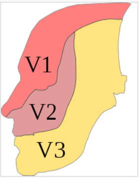
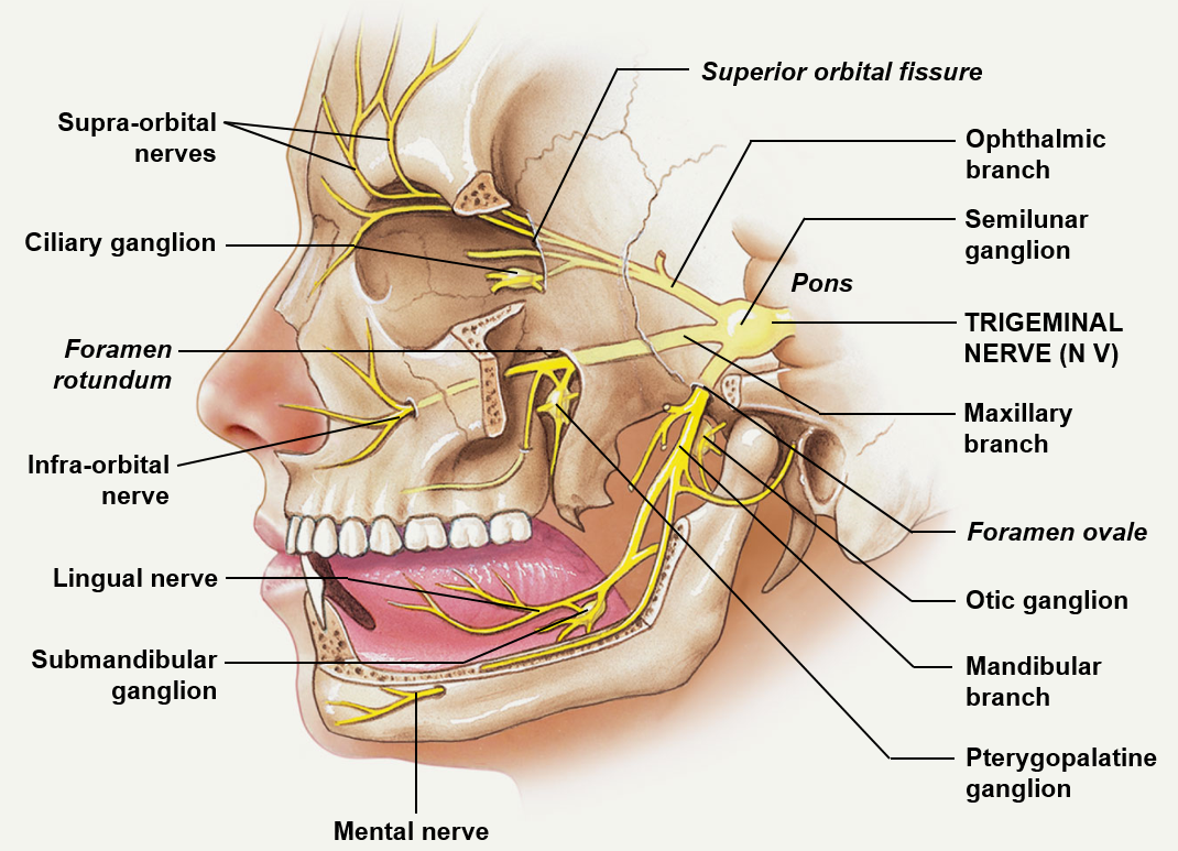
(5) CN V- trigeminal nerve
Has both sensory and motor function (mixed)
There are three sensory branches of this nerve1
Opthalmic (V1)- conveys information from the scalp, forehead, upper eyelid, nasal mucosa, tip of the nose, and frontal sinus
Maxillary nerve (V2)- conveys information from the lower eyelid, cheek, upper teeth and gums, the palate, and the sphenoid, ethmoid, and maxillary sinuses
Mandibular (V3)- conveys information from the lower lip, lower teeth and gums, chin, jaw, and areas of the ear
(5) CN V- Trigeminal nerve pt.2
The motor aspect of the trigeminal nerve controls eight muscles
Tensor veil palatine muscle
Tensor tympani muscle
Mylohoid muscle
Anterior belly of the digastric
Temporalis muscle
Masseter muscle
Lateral pterygoid
Medial pterygoid
(6) CN VI- abducens nerve
Innervates the lateral rectus muscle (motor)
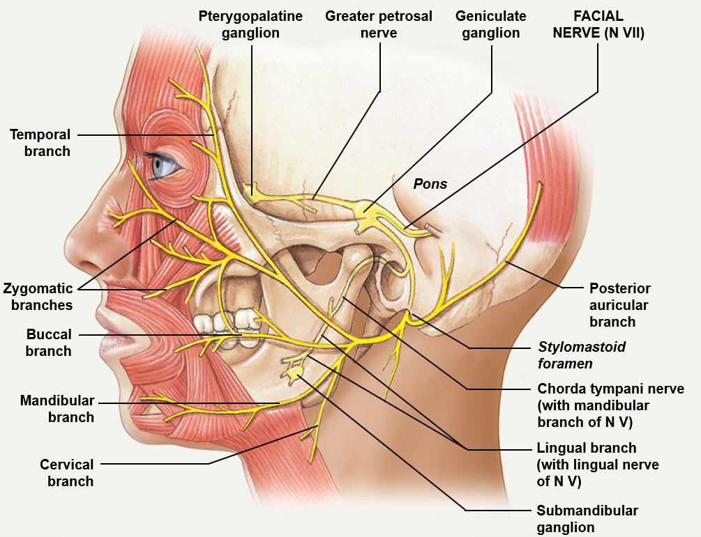
(7) CN VII- facial nerve
Carries both sensory and motor information (mixed) and leaves the cranium via the stylomastoid foramen
Carries sensory information concerning taste from the anterior 2/3 of the tongue, specifically from taste receptors which send information to the chorda tympani
The facial nerve sends motor (somatic) information to the muscles of the facial expression
The facial nerve sends motor (visceral) information to the sublingual and submandibular glands
(8) VIII- vestibulocochlear nerve
Made of a vestibular branch that conveys information about balance (sensory) and a cochlear branch that conveys information about hearing (sensory)
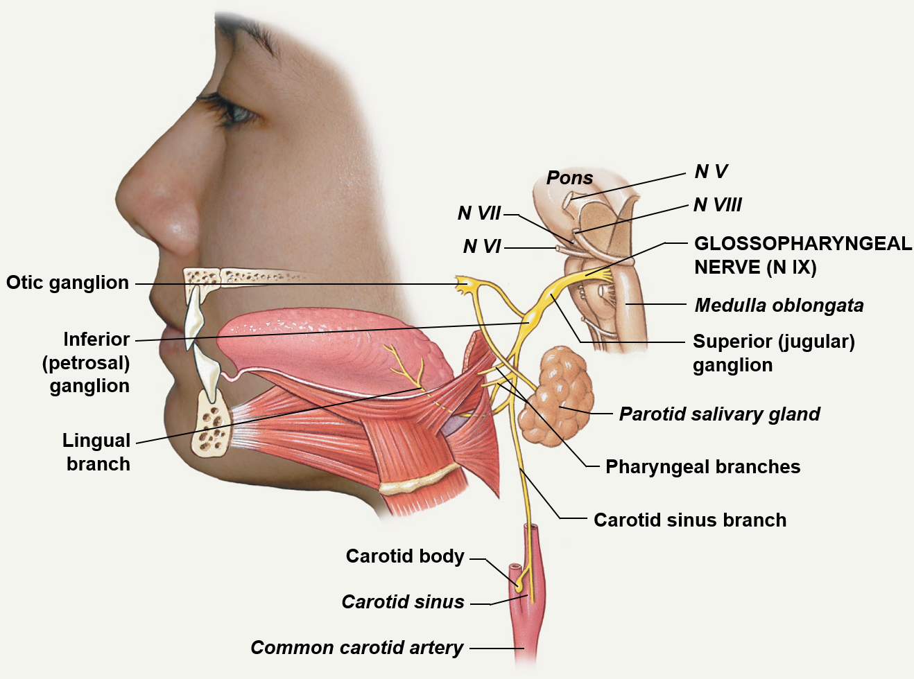
(9) CN IX- Glossopharyngeal nerve
Mixed nerve that involves the head and neck; travels through the jugular foramen
This nerve sends sensory information back to the brain, including information concerning taste from the posterior 1/3 of the tongue (lingual branch), information from the pharynx and palate, and information about blood pressure from the carotid arteries
Motor (somatic) information travels via pharyngeal branches to control muscles associated with the pharynx
Motor (visceral) information travels towards the parotid gland in order to stimulate salivation
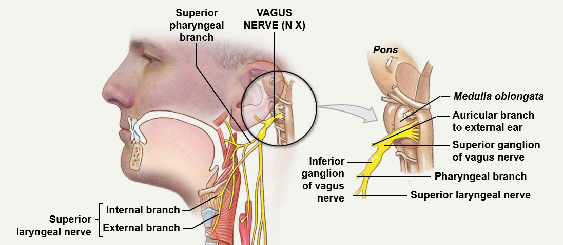
(10) CN X- Vagus nerve
The longest of the cranial nerves which happens to be a mixed nerve; travels through the jugular foramen
Sends info to the brain concerning the pharynx and epiglottis, along with information concerning the visceral organs (sensory)
Additionally, the nerve transmits motor information to the palate and pharynx (swallowing and coughing) and the viscera
(11) CN XI- spinal cord accessory
A motor nerve; sends and internal branches towards voluntary muscles of the palate, pharynx, and larynx; sends an external branch to the sternocleidomastoid muscle and trapezius muscle; travels through the jugular foramen
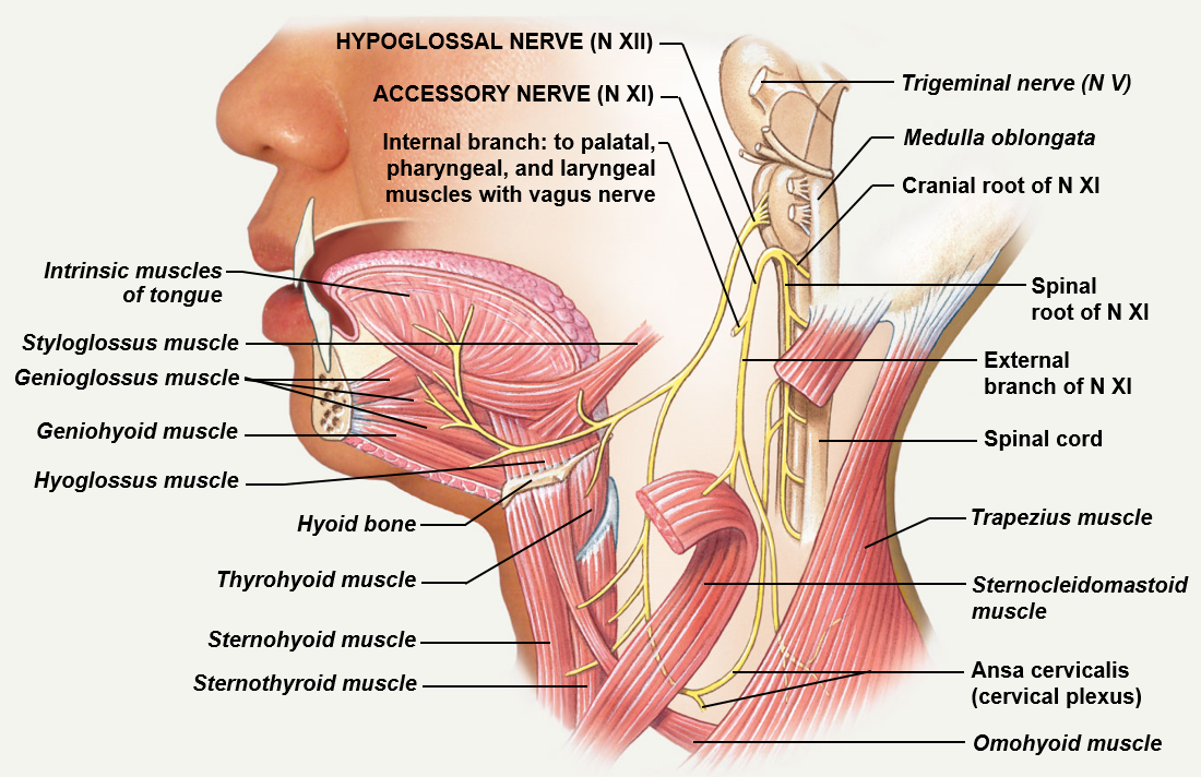
(12) CN XII- hypoglossal nerve
A motor nerve associated with movement of the tongue; travels through the hypoglossal canal
