Molecular techniques - tools for regulation
 Call Kai
 Learn
Learn Practice Test
 Spaced Repetition
 Match
Match Flashcards
 Knowt Play
1/47

Earn XP
Description and Tags
plamids, cloning, regulation
Name | Mastery | Learn | Test | Matching | Spaced | Call with Kai | Chat |
|---|
No analytics yet
Send a link to your students to track their progress
48 Terms
Plamids
extra chromosomal DNA elements used in DNA exchange
thorugh transformation
Plasmid maintenance
1. high copy number
2. partitioning genes
3. resistance markers
4. addiction molecules
Plasmid key features
origin of replication and an area to clone
want to have many plasmids per bacteria (40-50)
allows daughter cell to have plasmids
High copy number
Want bacterial cell to have many plasmid copies to transfer to daughter cells
substantially increases chances
forces bacteria to replicate plasmid
Partitioning genes
Help carry individual plasmids with chromosome during division
Resistance markers
often utilize antibiotic resitance
keep growing only bacteria with plasmid
can also use metals
Addiction modules
having a poion encoded on plasmid; plasmid also has antidote
1. poison is long lasting while antidote is short lasting
2. each daughter cell gets equal dose of poison
3. cell must replicate plasmid to survive (produce antidote)
Restrictive modification
Protective mechnism against harmful DNA integration (bacteriophage DNA)
use of exonucleases
Guest lecture: Yersinia Pestis
Plasmids determine virulence
chromosome encoded virulence
Lipoproteins/proteins assiciated with adhesion
T3SS delivers YOPs to trigger apoptosis, inhibit phagocytosis, and block cytokine production
F1 encoded by caf gene: immune evasion
exonucleases (function)
cleave (retrict) foregin DNA at specific sequences
DNA considered foregin if it has not been properly modifed (methylation)
Types of restriction endonucleases
Class I: Multisubunit enzymes with specific requirments (ATP/Mg/S-adenosylmethionine) and cut DNA at a site distant from sequence (~100 basepairs)
Class II: only require Mg and typically cut within palidromic sequence Ex) EcoRI
Class III: Requires ATP and cleaves DNA at sites 25-27bp away
Class I restriction endonucleases (mechnism)
Act like molecular motors that cut ~100bp away from the recognition site [ cut cannot be predicted]
translocates DNA at a rate of 400bp/sec (reason for ATP)
Methylase and nuclease are part of the same complex (reason for SAM)
Class II restriction endonucleases (mechnism)
cuts within or near the (usually palidromic) recognition sequence (only Mg needed for cut)
shift cut away from site always at same distance
Class III restriction endonucleases (mechnism)
Wors similar to type I but cuts closer to recogistion site (predictable)
requires ATP but not SAM
currently no commerically avaible type IIIs
Dr. Olsons favorite cloning vector
pBluescript II KS (-)
lac site, ampicillin resistance, 3.0 kb long
Cloning with restriction enzymes
Extract DNA
cut DNA with restriction enxyme
cut plasmid with same restriction enzyme
Incubate the DNA fragments with plasmid→ add enzyme DNA ligase (closes nicks in dna seq)
Transform the ligated products into e. coli
grow abcteria on plates containing antibiotic (whichever resistance your vector encodes for)
Screen the colonies for inserted DNA
Incubating DNA with Plasmid
Allow the DNA to sit for hours so that ligase can connect all the DNA fragments
Both plasmid and DNA have similar ends (lots of plasmids/DNA go back together)
Screening colonies (why?)
Important because cell should have plasmid but unknown if insert in the plasmid correctly
Blue/White screening
uses lacZ gene product (b-glactosidase) to determine if piece of DNA has been clonded into the multiple cloning site (MCS) of the plasmid
good colonies = white (lacz imparied)
addx-gal and if cut, blue forms
Ways to measure regulation
measure enzyme activity
activity of a reporter
mRNA concentration
northern blot
qRT-PCR
Measure protein level
SDS page
Western Blot/ ELISA
Reporter Genes
Beta- galactosidase
GFP
beta-glactosidase assay
fuse the promoter of the gene of interest to lacZ (on plamis or in the chromosome)
Grow cells under conditions you want to compare
add colir reagent to cells (x-gal=blue ONPG=yellow)
Measure color - sepctrophotometer: more color, more expression
Trp operon - regulation with lacZ
(-) tryptophan = activation of trpEDCBA genes to make trp
(+) tryptophan = reppresion of trpEDCBA genes
TrpE is an anthanilate synthase
how can we tell if trp genes are on
measure anthanilate synthase too hard
OR replease the trpE gene with lacZ - B-gal → easy to measure
grow cells (±) trptophan→ add ONPG or x-gal→measure amount of product with spectrophotometer
+ trp - absorbance reading - 10 units
- trp - absorbance reading - 10,000 units
Green flurorecent protein
GPF naturally fluorescent protein from jelly fish aequoreus victoria
good reporter since you dont have to fix cells or add color reagnet to see expression
GFP reporter
substrate is light so you can shine UV light on live vells and check for fluorescence
Direct mRNA measurement
Northern blot - uses DNA:RNA hybrid to measure mRNA level
RT PCR: two steps
1st - make DNA copy of mRNA with reverse transcriptase
2nd - amplify DNA copy by PCR
Northern Blot
DNA:RNA hybrid to measure mRNA level
labeled DNA sequence to probe for RNA
only tells you presence of mRNA, not quantity
RT-PCR - steps
Process of making DNA copy from mRNA
use reverse transcriptase and a DNA primer to produce complementary DNA strand
add taq polymerase, second primers, dNTPs, and heat
Initiate PCR process and camera measures fluorescence
Quantification with PCR process
reaction takes place in the presence of sybr green dye that fluoresces when bound to dsDNA
therocycler measures the flurorescence
computer graphs according to cycle #
the more mRNA, the more DNA, higher fluorescnece
Quantification 10th round
detecttion limit; acts as threshold for camera
Cycle # vs mRNA values
The more mRNA you start with, the earlier the increase in fluroreence
in early cycles → very little DNA
Measure protein: western blot
measures the amount of protein expressed
Disadvantage: uses an antibody against protein
advanatge: sometimes the amount of protein expressed is not entirely dependent on the amount of mRNA expressed
translational regulation
protein turnover rates are not always equal
SDS-PAGE
First step of western blot: proteins are separated by size via SDS gel electrophoresis
each protein is denatured and give negative charge by treatment with detergent SDS
Smaller proteins travel further down the gel
Antibody detection w/ western blot
want to find protein of interest out of all bands
must use a antibody that only recognizes your protein
Antibody gets detected by blotting
radioactive (film)
enzyme reaction (color)
Enzyme reaction (light, film)
Genome wide regulation
gene arrays: used to measure the amount of mRNA from all the genes in an organims
RNA seq: uses next generation DNA sequencing to determine transcriptome
2-D gels: used to measure the amount of all the proteins expressed
LC-MS-MS: mass spectrophy identifies proteins
Gene arrays
each gene of a sequences organims is ficed to a slide in a n ordered array
The array is then proped with cDNA library of mRNA from cell (two conditions)
upregualated (green)
down regulated (red)
Gene array steps
Experiment with two conditions
sample a and b
Isolate all RNA from both samples
generate cDNA from this bc - block cDNAse
label probes - two different colors
take probes and hybridize to an array
mixed color - 50/50 each
not quantitative
RNA seq work flow
better than gene array for transcriptomics
1. isolate total RNA
2. eukaryote vs prokaryote
easier in eukaryote
need to get rid of rRNA(will be overexpressed and get very little signal for mRNA)→
if eukaryote → polyadenylate their rRNA (3’end they add As) → put over oligoDT column (has DNA and mRNA will hybridze to TTT (poly ADT)-….→ postive selection
Prokaryote dont so remove rRNA with a kit →…negative selection
removed rRNA and have mostly mRNA left
will fragment it between 300nt and 700nt - illumina
hexmer primers and cDNA synthesis
Ends replacement, adapter → have primers for sequencing rxn
sequence all mRNA
how cDNA is islated that corrsepnd to mRNA
RNA seq protocol
sample rRNA—amplify it→cDNA—make fragments→ have reads—reads→ just count the reads
Counts to expression levels
RPKM for gene i = 10^9 x(ci/l’N)
tag count 1,000,000/ total number of tags kilobase of transcript
ci# tags (reads) length * total tags
Proteins and proteomics
gene arrays can tell about transcriptional regulation, but there are lots of conditions by which you want to know about the levels of proteins in the cell
They study of all proteins in a cell is called proteomics
Advantages: Detects non-transcriptional control, can be cheaper
Disadavnatges: cheap methods miss low abundance proteins
2D gel electrophoresis
separates by charges (depends on pH)
all proteins hav particular pH at which they have no charge, which is called their isoelectric point
separates by size (SDS Page)
acid proteins on one side, basic proteins on the other. Large at top and small at bottom
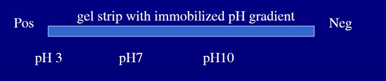
1st Dimension - isoelectric focusing
charge on a protein depends on the pH
All proteins have a particular pH at whihc they have no charge, which is called their isoelectric point
pH<pI→ positive charge
pH>pI → negative charge
pH=pI → no charge
would have a strip with gel and have immoblized pH graident
proteins with positve migrate toward negative electrode until they lose enough protons to lose charge
Each stops at its isoelectric point → no charge
Put this gel on top on regular SDS-page gel
run a current and separate by size
Tryptic digest
trypsin cuts proteins after R and K residues [Arginine and Lysine]
Excise the spot, treat the spot with trypsin to cut it up.
Weigh the fragments by Mass spectroscopy
Eliminate the gel with LC-MS workflow
proteins goes through tryptic digestion to create peptide mixture
LC/MS and MS/MS spectroscopy creates a gas flow which leads to an elution time
full MS spectrum leads to quantification
fragmetn selected peptide leads to spectrum levels that have peptide identifications
Peptides→ digest them→ liquid chromatography→Dual mass spec→ get protein spectra→ figure out what peptides each one of these
Guest Lecture: bordetella pertussis
Key virulence: exotoxins
Hfq is a post-transcriptional regularotr that b. pertussis has
regulates abundanec of protien
When iron starved, must adapt
Changes in BvgAS two component system →regulates turning off and on virulent, avirulent, and intermediate genes
Regulation of Hfq increaed abundance of Ptx (toxin) in iorn staved conditions
Hfg decreased abundance of two fimbrae proteins (fim2 and fimC) and one adhesion protein
Hfq regulates abundance of FhaB in low iron conditions
Basically hfq is involved in post-transcriptional regulation of certain B. pertussis virulence factors in iron starved conditions
Hfq helps with tolerance to oxidative stress that was originally induced by low iron levels
Computer put the sizes together
computer program determines the zies of all proteolytic fragments from the predicted genome sequence
computer compares experimental mass spec to predicted mass spec from database
then identifies porteins