Pathology Exam 1
 Call Kai
 Learn
Learn Practice Test
 Spaced Repetition
 Match
Match Flashcards
 Knowt Play
1/54

There's no tags or description
Looks like no tags are added yet.
Name | Mastery | Learn | Test | Matching | Spaced | Call with Kai | Chat |
|---|
No analytics yet
Send a link to your students to track their progress
55 Terms
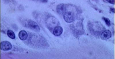
What kind of chromatin is pictured in these nuclei
euchromatin
What type of chromatin is tightly coiled and inactive
heterochromatin
What type of chromatin is loosely coiled and allows for active transcription to mRNA
euchromatin
What is the function of the nucleolus
rRNA synthesis
Where is the nucleolus
within the nucleus
Mitotically active or cancerous cells tend to have an abundance of which organelle
nucleolus
The nucleus is covered in a bilayered nuclear membrane with
pores
Why is it called the rough ER
it is covered in ribosomes
Function of the rough ER
protein synthesis
The basophilia of a cell’s cytoplasm is caused by the
rough ER
The smooth ER has NO
ribosomes
Function of the smooth ER
metabolizing steroids, lipids, carbohydrates, and glycogen
Function of the Golgi apparatus
synthesis and packaging of proteins for export out of the cell
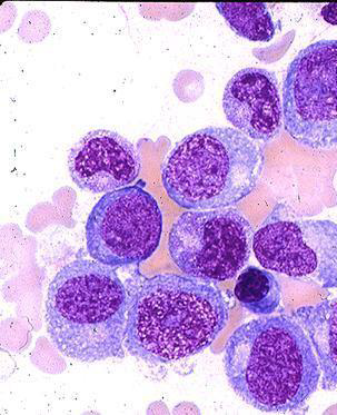
The white circular area outside of the nucleus is what organelle
Golgi
Perinuclear clear zone is used to describe which organelle
Golgi
Which organelle plays roles in producing ATP, apoptosis, cell signaling, cell differentiation, and cell growth
Mitochondria
What kind of enzymes are found in lysosomes
hydrolytic
Function of lysosomes
Intracellular digestion
The structural network of a cell that helps in movement, cell division, and bicohemical pathways
cytoplasm
Microfilaments
facilitate cell motility
Intermediate filaments
provide physical strength to cells via junctional complexes
Microtubules
move organelles and vesicles within the cytosol and chromosomes
4 major mechanisms of cell injury
depletion of ATP
permeabilization of cell membranes
DNA damage
disruption of biochemical pathways
The most common and important cause of cell injury
hypoxia
DAMNITV is an acronym used to describe
causes of cell injury
Hypoxia
Deficiency of oxygen that reduces aerobic respiration but still allows for anaerobic glycolysis to occur
Ischemia
Interruption of blood flow that results in inadequate oxygenation, nutrient and substrate delivery to cells, and inadequate removal of waste affecting both aerobic and anaerobic respiration
Short term solution to hypoxia
anaerobic glycolysis
ATP deficiency means that the Na/K pump is
impaired
Causes of hypoxia
cardiac/respiratory failure
reduced vascular perfusion
reduced oxygen transport by RBCs
Inhibition of respiratory enzymes
Reperfusion injury causes an increase in cell death after blood flow is restored due to
free radical formation
Intracellular calcium overload is associated with
cell membrane damage
Function of free radical scavengers
neutralize the free radical’s harmful effects on cells
Free radical scavengers
superoxide dismutase, glutathione peroxidase, ceruloplasmin, vitamin A, C, E
Reversible cell injury can either revert back to normal or cause
cellular adaptation
Irreversible cell injury leads to
cell death
1st response to cell injury
acute cell swelling
Causes of acute cell swelling
hypoxia
direct injury to cell membrane
damage to ion channels
binding of toxins
Appearance of acutely swollen cells
distinct cell borders and pale cytoplasm
Tissue with acute cell swelling is
heavier, pale
Cell swelling in parenchymal tissue
hydropic degeneration
Cell swelling in keratinocytes
ballooning degeneration
Cell swelling in astrocytes
cytotoxic edema
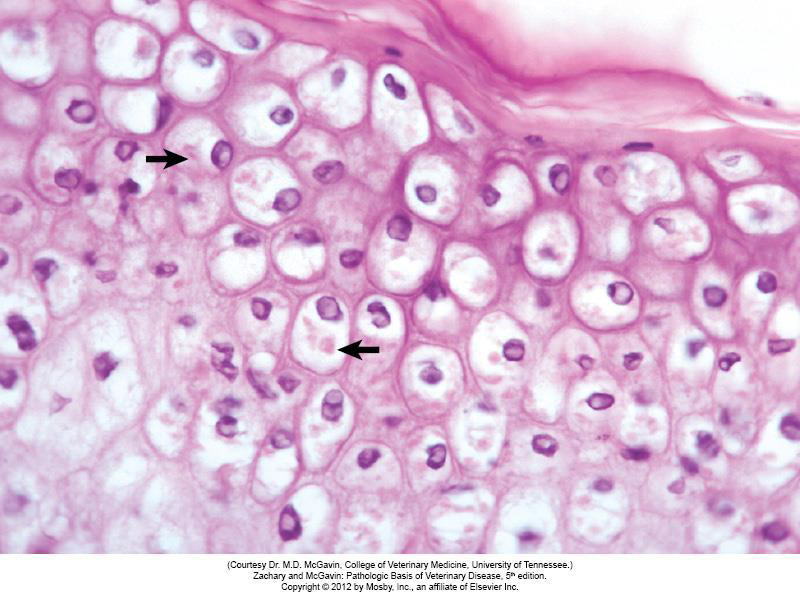
Do these cells look normal or abnormal
Abnormal due to their distinctive borders and pale cytoplasm
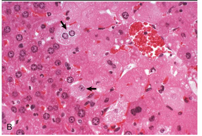
Which side of the image contains normal cells
left
Cell stress
altered physiologic state resulting from nonlethal injurious stimuli that disrupts homeostasis
Cell injury
alteration in cell structure or biochemical function resulting from some stress that EXCEEDS the ability of the cell to compensate through normal physiologic adaptive mechanisms
A cell is considered injured when
the adaptation is exceeded so that critical systems within the cell cannot maintain normal function
A cells response to stress and injury depends on
cell type, nature and intensity of injury, and metabolic state at the time of injury
When damage is reversible cells can become rapidly non-functional but remain
viable
What leads to an irreversible injury
longer duration or higher intensity injury
Histologic and morphologic changes can lag behind
functional change
What is the 1st manifestation of almost all forms of cellular injury
acute cellular swelling
Acute cellular swelling is associated with the failure of the
Na/K pump
Na/K pump failure with acute cellular