Disease process & Immunity Test 2
 Call Kai
 Learn
Learn Practice Test
 Spaced Repetition
 Match
Match Flashcards
 Knowt Play
1/84

There's no tags or description
Looks like no tags are added yet.
Name | Mastery | Learn | Test | Matching | Spaced | Call with Kai | Chat |
|---|
No analytics yet
Send a link to your students to track their progress
85 Terms
When does adaptive immunity activate?
When innate immunity has failed to prevent infection
Both systems work together to eliminate the pathogens
What are the characteristics of adaptive immunity?
Highly specific
Memory - can provide long-lasting protection
Able to distinguish self-antigen from pathogens
What are the immune cells involved in adaptive immunity?
Lymphocytes
T cells
B cells
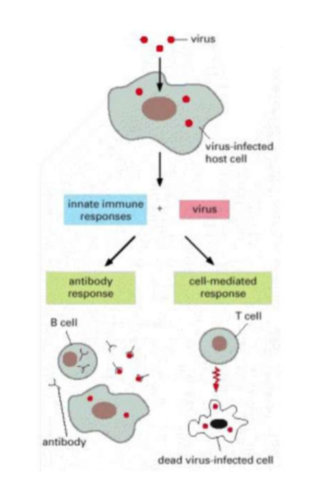
What are the two types of responses for adaptive immunity?
Cell mediated immune responses
Antibody responses
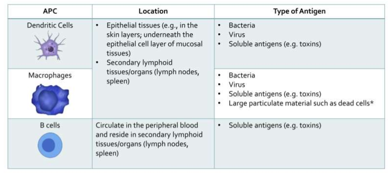
What are the APC (Antigen presenting cells)?
Dendritic cells
Macrophages
B cells
What are the types of T cells? What MHC class do the effector T cells have?
Effector T cells
CD4+ Helper T Cells - MHC II
CD8+ Cytotoxic T Cells - MHC I
Memory T Cells
Naive T Cells
What immune cell activates naive T cells
Dendritic cells
How do APC present antigen fragments?
Ingest microbe antigen
ingested antigens are broken down
A fragment of the broken down antigen is presented with MHC molecule on the cell surface
How does T Cells fight against pathogen?
An APC presents the antigen to the CD4+ Helper T Cell
CD4+ Helper T Cell releases cytokines
Cytokines activate CD8+ cytotoxic T cells and cytotoxic T cells duplicate
Cytotoxic T cells bind to infected cell body and causes cell lysis by release of perforins.
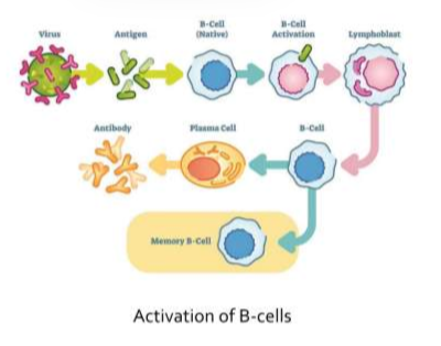
How does antibody mediated immunity work?
Activated B cells produce antibodies that bind to free antigens in body fluids
Some B cells become memory cells which become active in future encounters of this same antigen and confer immunity to that antigen
What are the 2 ways B cells are activated?
T Cell dependent
T Cell independent
How does T-cell independent B cell activation happen?
B cell receptors (BCRs) interact with the antigen
BCRs for naive B cells are membrane-bound
An antigen binds to the BCR on only one type of B cell
B cell then produces clones of itself
Clones of B cells becomes plasma cells → produce antibodies that are specific to the same type of antigen
The response is short lived and does not result in the production of memory B cells
How does T cell dependent B cell activation happen?
Antigen binds to BCR on naive B cell and stimulates an internalization of the antigen.
After internalization, antigen is processed and presented with MHC II to CD4+ Helper T.
Presented antigen is recognised by CD4+ Helper T cell
CD4+ Helper T produce and secrete cytokines that activate B cell
B cell proliferates → plasma cell → antibodies
B cell clones also proliferate into memory B cells
Quickly responds to subsequent exposures to same antigen.
What does MHC stand for, and what is its primary function?
Major Histocompatibility Complex
It presents antigens to T cells to trigger an immune response
Why is antigen presentation necessary for adaptive immunity?
T cells cannot recognize whole viruses; they need antigen fragments presented on MHC molecules.
What types of cells express MHC molecules?
All nucleated cells express MHC I APCs (Dendritic cells, macrophages, B cells) express MHC II.
What type of antigens are presented by MHC Class I?
Endogenous antigens — viral proteins made inside infected cells.
Which T cell recognizes MHC Class I?
CD8+ cytotoxic T cells.
What immune response is triggered by MHC I presentation?
CD8+ cytotoxic T cells kill infected cells to stop viral replication.
Which cells express MHC Class II molecules?
Antigen presenting cell (APC): Dendritic cells, macrophages, and B cells.
What type of antigens are presented by MHC Class II?
Exogenous antigens — viral particles taken up by APCs.
Compare MHC Class I and II molecules
Feature | MHC Class I | MHC Class II |
Expressed on | All Nucleated cells | Professional APCs only |
Presents | Endogenous (intracellular) antigens | Exogenous (extracellular) antigens |
Recognized by | CD8+ Cytotoxic T cells | CD4+ Helper T cells |
Immune Response | Kills infected cells | · Activate B cells, macrophages |
Describe the immune response triggered by MHC I antigen presentation?
MHC I-antigen complex is transported to the surface of the cell. CD8+ cytotoxic TCR (T-cell receptor) recognises a foreign peptide presented by MHC 1 and activates. The CD8+ cytotoxic T cells release perforins and granzymes. Which induce apoptosis in the infected cell. Afterwards some CD8+ cytotoxic T cells differentiate into memory T cells to provide long-term immunity by enabling a faster stringer response if the same antigen is encountered again
Name an example of PAMPS (Pathogen-Associated Molecular Patterns)
Flagellin
LPS (lipopolysaccharide)
Double stranded RNA
Unmethylated CpG motifs
How are T cells activated?
CD4+ T cells recognize the MHC II–antigen complex via their TCR and become activated.
What happens when CD4+ Helper T cells release cytokines?
Stimulate B cells
Enhance activity to phagocytose and kill bacteria.
macrophages
Recruit other to the site of infection.
immune cells
What cell activates Naive CD4+ T cells?
Dendritic cells
How do antibodies help eliminate the bacteria from the body? (Name the mechanisms)
Neutralization: Antibodies bind to bacterial toxins or surface proteins, blocking their ability to infect host cells.
Opsonisation: Antibodies coat the bacteria, making them easier for phagocytes like to recognize and engulf.
Complement Activation: Antibodies trigger the complement system, leading to bacterial lysis.
Table for hypersensitivity
Type | Name | Immune Mechanism | Onset | Examples | Key Cells Involved | Clinical Relevance / Feature |
I | Immediate (Allergic) | IgE-mediated activation of mast cells | Seconds to minutes | Asthma, anaphylaxis, hay fever | Mast cells, basophils, IgE | Can be life-threatening; rapid onset; allergy-related |
II | Antibody-mediated Cytotoxic | IgG/IgM antibodies bind to cell surface antigens | Minutes to hours | Hemolytic anemia, blood transfusion reactions | IgG, IgM, complement, NK cells | Targets specific cells; often involves blood cells |
III | Immune Complex-mediated | Antigen-antibody complexes deposit in tissues | Hours to days | Serum sickness, lupus, rheumatoid arthritis | Neutrophils, complement | Causes inflammation in tissues; systemic effects |
IV | Delayed-type (Cell-mediated) | T-cell mediated response | 48–72 hours | Contact dermatitis, TB skin test, graft rejection | T cells (CD4+, CD8+) | No antibodies involved; delayed response |
What does IgE bind to?
Mast cells
Basophils
What antigen is involved in Type I Hypersensitivity?
IgE
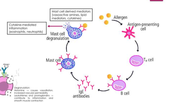
Define Type I hypersensitivity
Type I hypersensitivity is an immediate allergic reaction mediated by IgE.
What is the immunological mechanism involved in Type I Hypersensitivity.
IgE-mediated activation of Mast cells and Basophils
Releases Histamine
Why do type I hypersensitivity symptoms appear so rapidly upon re-exposure to allergen?
Body was already sensitised to allergen
IgE antibodies specific allergen protein were previously produced.
IgE antigens were already bound to mast cells
Upon re-exposure, the allergen cross-linking, causing immediate degranulation and release of histamine, leading to rapid onset of symptoms
What are the two emergency treatments And it's commonly used for anaphylaxis?
Epinephrine
Anti-histamines
How does epinephrine help with Anaphylaxis?
Reverses airway constriction
Reduces swelling
Raises blood pressure
How does antihistamines help with anaphylaxis?
Blocks histamine receptors
Reducing symptoms like hives and itching
What are the key cells involved in Type II Hypersensitivity?
IgG
IgM
Complement
NK Cells
Define Type II hypersensitivity
Type II hypersensitivity is Antibody-mediated Cytotoxic with IgG or IgM antibodies binding to cell surface
What is the immunological mechanism involved in Type II Hypersensitivity.
IgG/IgM antibodies bind to cell surface antigens
binding activates the complement system or attracts phagocytic cells, leading to the destruction of the red blood cells.
What is the onset of type I hypersensitivity?
Seconds to minutes
What is the onset of type II hypersensitivity?
Minutes to hours
What is the onset of type III hypersensitivity?
Hours to days
What is the onset of type IV hypersensitivity?
48–72 hours
What are the key cells involved in Type III Hypersensitivity?
Neutrophils
Complement
What are the key cells involved in Type IV Hypersensitivity?
T cells (CD4+, CD8+)
What is the immunological mechanism involved in Type III Hypersensitivity.
Antigen-antibody complexes deposit in tissues
Immune complexes can deposit in small blood vessels → Activate complement system → inflammation and tissue damage
Organs that have dense capillary networks and high filtration networks make them prone to immune complex deposition
What is the immunological mechanism involved in Type IV Hypersensitivity.
T-cell mediated response
CD4+ helper T cells and CD8+ cytotoxic T cells recognize antigens presented by APC → release cytokines that recruit macrophages and cause tissue inflammation
What are the symptoms of Type I hypersensitivity?
Asthma
anaphylaxis
hay fever
What are the symptoms of Type II hypersensitivity?
Hemolytic anemia
Blood transfusion reactions
What is the clinical relevance of Type I Hypersensitivity?
Can be life threatening
rapid onset
allergy related
What is the clinical relevance of type II hypersensitivity?
target specific cells
often involve blood cells
What is the clinical relevance of type III hypersensitivity?
cause this inflammation in tissues
systemic side effects
What is the clinical relevance all type IV hypersensitivity?
No antibodies involved
delayed response
What are the symptoms for type III hypersensitivity?
Serum sickness
lupus
rheumatoid arthritis
What are the symptoms for type IV hypersensitivity?
Contact Dermatitis
TB skin test
graft rejection
Why are type IV hypersensitivity reactions delayed?
Involve cell mediated immunity
takes time for T cells to recognize antigen become activated and recruit other immune cells to the site
unlike antibody mediated reactions which occur within minutes
Why are complement levels often low in patients experiencing type III hypersensitivity reactions like lupus?
Complement system is continuously activated by immune complexes deposited in tissues
Persistent activation leads to consumption of complement proteins
Low complement levels are a marker of ongoing immune complex mediated inflammation
Why is it important to perform blood typing and cross matching before transfusions?
Ensure donor blood is compatible with recipient's immune system
If incompatible blood is transfused recipient's antibodies may recognize the donor red cells as foreign and attack them
leading to type II hypersensitivity reaction like hemolysis
What is myocardial infraction?
Sudden and complete interruption of blood supply to a portion of the heart
An area of tissue death due to a lack of oxygen
What can cause myocardial Ischemia or infraction?
Partial obstruction of artery
Atherosclerotic plaques can cause partial or complete narrowing (stenosis)
Coronary vasospasm (vasoconstriction)
Acute Coronary vasospasm can be relieved by nitrates \
Restoring normal coronary blood flow
Thrombus (stationary clot)
plaques occasionally rupture
induces platelet activity and coagulation cascade to form clot or thrombus
Embolus (floating clot)
thrombus detached from vessel wall
carried in bloodstream as embolus
What are the complications arising from uncontrolled hypertension?
Renal complications
End stage Renal Disease
Cerebral complications
Hemorrhagic stroke
Ischemic stroke
Left ventricular hyperhtrophy
Coronary artery disease (CAD)
Myocardial infraction, angina
Left ventricularsystolic failure
heart failure
Peripheral vessel damage
Leads to peripheral vascular disease stiffening of blood vessel
accelerates atherosclerosis
State the formula for defining blood pressure and explain its individual component.
BP = CO (Cardiac Output) x PVR (Peripheral Vascular Resistance)
Cardiac output is a measurement of blood flow through the heart to the systemic and pulmonary circulation per min (ml/minute)
What is the difference between myocardial ischemia and myocardial infarction?
myocardial ischemia
When the lumen is significantly blocked (not complete) , oxygen supply to heart muscle is diminished
Myocardial infraction
When there is a sudden and complete interruption of blood supply to a portion of the heart
What is the difference between Atherosclerosis and Arteriosclerosis?
Atherosclerosis: formation of intimal lesions that protrude into and obstruct vascular lumina
Arteriosclerosis: Hardening of the arteries resulting in thickening and loss of elasticity
What are the invasive methods used to assess atherosclerosis?
Intravascular ultrasound
Coronary Angiography
What are the clinical manifestations of atherosclerosis?
Coronary heart disease
Angina pectoris, myocardial infraction, sudden cardiac death, congestive heart failure (CHF) and arrhythmias
Cerebrovascular disease
Transient ischemic attack, stroke
Peripheral vascular disease
Intermittent claudication, gangrene, cold feet, painful feet, impotence
What are the different types of stroke?
Transient Ischemic attack
Ischemic stroke
Hemorrhagic stroke
State 3 symptoms of stroke?
Slurred speech
Weakness or numbness in face, arm and leg on one side of the body
Double vision
Describe the role of kidney in blood pressure regulation.
Renin-angiotensin system (RAS)
restore normal blood pressure and fluid balance by
regulating sodium and water retention
vascular tone
systemic vascular resistance
State 3 consequences of myocardial infarction.
Sudden cardiac death
cardiac arrythmia
vascular remodeling
deep vein thrombosis
recurrent myocardial infraction
Explain why edema(redness) is a common symptom of Heart Failure.
Presence of edema suggests congestive heart failure
failure to properly eject blood from LV cause back flow of blood to lungs. This results in pulmornary edema
When the right side of the heart fails, it cause blood to be back up in the venous system and cause edema in the periphery i.e legs
What are the 2 categories of heart failure?
Systolic dysfunction
Reduced contractility (left ventricular) is the principal mechanism of heart failure in coronary heart failure in coronary heart disease and dilated cardiomyopathy
Diastolic dysfunction
Disorders in which the filling of the ventricle is impaired either by inflow obstruction or by reductions in ventricular compliance
What are the 3 types of angina and define each type.
Stable symptoms that occur during physical exertion and are relieved by rest or medication
unstable angina - angina at rest well as with minimal exertion
Vasospastic or Prinzmetal’s angina resulting from vasospasm of coronary artery, occurs most commonly at rest and is not precipitated by exertion of stress.
If premature beat occurs in the ventricular, it will affect ____?
Ventricular
If premature beat occurs in the atrial, it will affect _____?
Atria and Ventricular
Explain why ventricular fibrillation has higher mortality as compared to atrial fibrillation.
Ventricular Fibritillation | Atrial fibrilation |
• Life threatening/emergency, required immediate defibrillation | • Can be managed chronically with medication |
• Signal is pass from atrial to ventricular; ventricular is the end stage of contraction | • If fibrillation occurs at atrial, atrial will beat irregularly & inefficiently |
◦ Sudden death | • Risk of stroke & heart failure |
Explain how the following influences the firing rate of myocyte causing arrhythmia.
Circulating hormone (thyroid hormone/ adrenaline)
Thyroid hormone/ adrenaline: increase heart rate
Serum ion concentration
Electrolytes maintain action potential
Na/K: influence depolarisation & repolarization
Cellular hypoxia
Ocygen is essential for the normal electrical activity of cardiac muscle cells
Hypoxia reduces ATP production; failure of ATP- dependent ion pump (Na/K-ATPase pump)
causing partial depolarisation
Drugs
Affecting ion channels, altering electrolyte balance, increasing sympathetic or slowing electrical condition in the heart
eg. digoxin/bronchodilators/ anti psychotics
What is the pathology of Erectile Dysfunction?
Nerve impulses in the brain,spinal column and area around the penis
response in muscles, fibrous tissues, veins and arteries in and near the corpora cavernosa
ED occurs when any of the events are disrupted
What diseases contribute to Erectile dysfunction?
Diabetes (Thicker blood)
High blood pressure
nerve disease or nerve damage
multiple sclerosis
atherosclerosis
heart disease
affect nerves, arteries, smooth muscles and fibrous tissues
What lifestyle factors contribute to erectile dysfunction?
Smoking
Drinking alcohol
Obesity
Not exercising
Contributes to heart disease and vascular problems
What types of surgeries contribute to erectile dysfunction?
Prostate and bladder surgery for cancer
Can injure nerves and arteries near the penis → could result in ED
Wh
What are the side effects of medication that can contribute to erectile dysfunction?
Blood pressure drugs
Anti histamines
Anti depressants
Appetite suppressants
Cimetidine
Ulcer drug
W
What are the psychological factors that can contribute to erectile dysfunction?
Stress
anxiety
depression
low self esteem
fear of sexual failure
What hormonal abnormality contributes to erectile dysfunction?
Low levels of testosterone