2. GENERAL CHARACTERISTICS OF BACTERIA
1/100
There's no tags or description
Looks like no tags are added yet.
Name | Mastery | Learn | Test | Matching | Spaced | Call with Kai |
|---|
No analytics yet
Send a link to your students to track their progress
101 Terms
True
T or F
prokaryotes and eukaryotes use the same kinds of chemical reactions to metabolize food, build proteins, and store energy
peptidoglycan
what is the composition if cell wall for prokaryotes?
cellulose and chitin
what is the composition if cell wall for eukaryotes?
False; there is an exception “miss u”
mycoplasma
urea plasma
T or F
all bacteria does not contain carbohydrate and sterols
they lack cell wall
why does mycoplasma and urea plasma incorporate sterols in their cell composition?
histones ; nonhistone
DNA is consistently associated with chromosomal proteins called ___________ and with _______________
mitosis ; binary fision
METHOD OF CELL DIVISION?
eukaryote ; _____________
prokaryote ; _____________
EUKARYOTE
PROKARYOTE OR EUKARYOTE
Have a number of membrane-enclosed organelles
EUKARYOTE
PROKARYOTE OR EUKARYOTE
Cell walls, when present, are chemically simple
PROKARYOTES
PROKARYOTE OR EUKARYOTE
DNA is not enclosed within a membrane and is usually a singular circularly arranged chromosome
PROKARYOTE
PROKARYOTE OR EUKARYOTE
Cell walls almost always contain the complex polysaccharide peptidoglycan
plasma membrane
periplasmic space
outer membrane
ENUMERATE
cell wall inclusion of G (-) bacte.
plasma membrane
cell wall
ENUMERATE
cell wall inclusion of G (+) bacte.
G (+)
which gram stain bacte. has a thicker peptidoglycan layer?
False
T or F
all bacteria has a capsule
cell membrane
what is being used as energy in bacteria since they dont have mitochondria?
Bacterial cell wall
what bacte. chararcteristic provide the basis for the Gram stain?
0 .25 to 1 ; 1 to 3
Most clinically relevant bacterial species range in size from__________ μm in width and __________ μm in length
Haemophilus ducreyi
smallest bacteria causing the disease chanchroid and is <1 micrometer
Bacillus anthracis
considered as the largest bacte. causing anthrax, and is 3-10 micrometer
Cocci ; Staphylococci
BACTERIAL SHAPE
circular ; give example
Coccobacilli ; (ex: normal flora found in female vagina (Gardnerella)
BACTERIAL SHAPE
ovoid ; give example
Bacillus ; (ex: Bacillus anthracis)
BACTERIAL SHAPE
rod shaped ; give example
Fusiform
BACTERIAL SHAPE
tapered, pointed ends
Curved
BACTERIAL SHAPE
(just like vibrio) i.e cholera
Spiral ; spirochetes (Treponema pallidum, Borrela spp., Leptospira interrogans)
BACTERIAL SHAPE
helical, like corkscrew ; give example
Pleomorphic
BACTERIAL SHAPE
no defined shape (because of their cell envelope)
Staphylococci ; Streptococci ; Diplococci ; Micrococcus
Cluster: ___________
Chains: _________
Pairs: __________
Tetrads: ___________
outer membrane
periplasmic space (periplasm)
what are the cell envelope components that is only found in G (-) bacte. only?
plasmid
this cell structure is important for the bacteria to be able to cleave because it codes for cellular function. thus it codes for special characteristics such as drug resistance which gives advantage to the bacteria
pilus (attachment)
sex pilus (transfer of gen material via conjugation) (ie. E. coli)
ENUMERATE
two types of pilus
True
T or F
plasmid is optional for bacterias
True
T or F
other bacterias do not have capsule
lipopolysaccharide (LPS)
the outer membrane is a bilayered structure composed of ____________________ Which gives the surface of gram-negative bacteria a net negative charge
PORINS
Protein structures scattered throughout the lipopolysaccharide macromolecules
PORINS
Water-filled structures that control the passage of nutrients and other solutes, including antibiotics, through the outer membrane
MUREIN LIPOPROTEINS
They act as anchors that connect the outer membrane to the peptidoglycan cell walls
CELL WALL ( MUREIN LAYER)
Gives the bacterial cell shape and strength to withstand changes in environmental osmotic pressures that would otherwise result in cell lysis
a. G (+)
it has a higher resistance to physical disruption since it has something to do with thickness of its peptidoglycan
a. G (+)
b. G. (-)
Penicillins
Cephalosporins
what are the drugs that target cell wall, this also inhibits cell wall sensitivity thus G(+) bacterias are more targetted
disaccharide-pentapeptide subunits.
Peptidoglycan is made up of repeating _____________________
N-acetyl-D-glucosamine (NAG)
N-acetyl-D-muramic acid (NAM)
Alternating sugar components (moieties),with the amino acid chain linked to N-acetylmuramic acid molecules
Mycobacteria
which bacteria contain mycolic acid?
mycolic acid
what is the waxy layer that coats the bacterial cell wall thus gram staining is pale and it stains lightly or “gram ghost”
Acid-Fast stain
since mycobacteria contains modified cell wall, what is the staining method used?
1. Ziehl–Neelsen Stain (hot acid-fast stain)
2. Kinyoun Stain (cold acid-fast stain)
ENUMERATE
2 AFS technique
mycobacteria, nocardia, Legionella micdadei
Give examples of acid-fast bacteria
Rhodococcus
Tsukamurella
Gordonia
Corynebacteria
ENUMERATE
bacteria that dies not utilize AFS but has small amnt of mycolic acid
carbol fuchsin
Primary stain use in acid-fast stain
2
how many rings around flagellum if it is a G(+) bacteria?
4
how many rings around flagellum if it is a G(-) bacteria?
motility ; attachment
flagella; __________
pili; ___________
TEICHOIC ACID
this is a special component of G(+) bacteria that is anchored to the peptidoglycan (N-acetylmuramic acid)
LIPOTEICHOIC ACID
this is a special component of G(+) bacteria that is anchored to the PM
TEICHURONIC ACID
in a G(+) bacteria is low on phosphate it produces ____________________ in place of theichoic acids. thus is has similar polymers, but the repeat units include sugar acids (eg, N-acetylmannosuronic or d-glucosuronic acid) instead of phosphoric acids
A. O-specific polysaccharide
B. Core polysaccharide
C. Lipid A (also called endotoxin)
ENUMERATE
the three regions of LPS
O-specific polysaccharide
(the three regions of LPS)
Outermost part
Highly variable among bacteria
Antigenic → stimulates immune response
Used for bacterial identification/serotyping
Core polysaccharide
(the three regions of LPS)
Connects O antigen to Lipid A
Contains:
KDO (Ketodeoxyoctanoic acid)
Heptose sugars
Lipid A (also called endotoxin)
(the three regions of LPS)
Innermost portion
Embedded in the outer membrane
aka “ENDOTOXIN”
O antigen ; O-specific polysaccharide of LPS
H antigen ; Flagella
K antigen ; Capsule
ENUMERATE
bacterial antigens used for serotyping
positive ; negative
exotoxin ; G (__)
exotoxin & endotoxin ; G (__)
periplasmic space
Space that separates cell wall and plasma membrane:
LPS
• Vital in evading the host defenses
• Contribute to the negative charge of the bacterial
surface, which stabilizes the membrane structure
• Considered as an endotoxin
Lipid A moiety
The phosphate groups in LPS contribute to the bacterium's negative surface charge.
Lipid A moiety
responsible for producing fever and shock conditions in patients infected with gram-negative bacteria
Porins
are protein channels found in the outer membrane of Gram-negative bacteria.
G(-)
what gram stain bacteria can u find periplasmic space?
faint blue (gram-positive) color
what is the color of the stain of Mycobacterium and Nocardia in AFS?
if the bacteria lost its rigid cell wall
how are L-form bacterias formed?
serum
high sugar concentration
how do L-formed bacteria survive?
cytoplasmic membrane / PM
what characteristic of bacterial cell wall contributes to the generation of chemical energy (ie. ATP)
RIBOSOMES
Site of protein biosynthesis and give the cytoplasm a granular structure
Streptomycin and Gentamicin
what are the antibiotic that attaches to the 30S subunit and interfere with protein synthesis
Erythromycin and Chloramphenicol
what are the antibiotic that interfere with protein synthesis by attaching to the 50S subunit
Large Plasmid
responsible for the production of B lactamase that provide resistance to B lactam antibiotics (penicillin and oxacillin)
Small Plasmid
resistant to tetracyclines an Chloramphenicol
INCLUSION BODIES
Serve as the energy source or food reserve of the bacteria or as a reservoir of structural building blocks
Babes–Ernst Bodies
a metachromatic inclusion body that is associated with Corynebacterium diphtheriae
Halberstaedter Bodies
an inclusion body associated with Chlamydia trachomatis
Levinthal–Cole–Lillie bodies
an inclusion body associated with Haemophilus influenzae
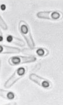
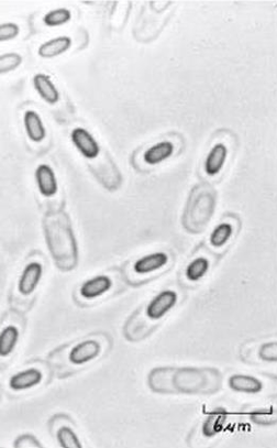
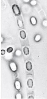
ENDOSPORES/ ASEXUAL SPORES
Small, dormant structures located inside the bacterial cell
a. Terminal spore
b. Subterminal spore
c. Central spore
ENUMERATE
Types of Spores according to location
Clostridium tetani
what is the bacteria id spore location is terminal spore
Clostridium botulinum
what is the bacteria id spore location is Subterminal spore
Bacillus anthracis
what is the bacteria id spore location is Central spore
Terminal spore
Clostridium tetani
identify;
spore location
bacteria
Clostridium botulinum
identify;
spore location
bacteria
Bacillus anthracis
identify;
spore location
bacteria
GLYCOCALYX
outward complex of polysaccharide on the bacterial surface and other cells
GLYCOCALYX
appears as a capsule or a slime layer
FLAGELLA
exterior protein filaments that rotate and cause bacteria to be motile
Proteus
can "swarm," or show rapid wavelike movement across a solid culture medium since it has a lot of flagella
a. Atrichous
b. Mnotrichous
c. Amphitrichous
d. Lophotrichous
e. Peritrichous
ENUMERATE
arrangement of the flagella
Atrichous
what do you call the arrangement if the bacteria does not have any flagellum?
Monotrichous
what do you call the arrangement if the bacteria has single flagellum at one end
Lophotrichous
what do you call the arrangement if the bacteria has tuff or group of flagella on one end or both ends
Peritrichous
what do you call the arrangement if the bacteria is covered with flagella
Axial Filaments
bundles of fibrils that arise at the ends of the cell beneath an outer sheath and spiral around the cell
hanging drop method
what method in demonstrating motility is used to best observe brownian movement?
PILI (FIMBRIA)
hairlike, proteinaceous structures that serve as adhesins that help bacteria attach to animal host cell surfaces, often as the first step in establishing infection