AOV - MST1
 Call Kai
 Learn
Learn Practice Test
 Spaced Repetition
 Match
Match Flashcards
 Knowt Play
1/197

There's no tags or description
Looks like no tags are added yet.
Name | Mastery | Learn | Test | Matching | Spaced | Call with Kai | Chat |
|---|
No analytics yet
Send a link to your students to track their progress
198 Terms
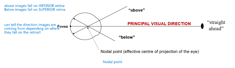
What is the principle sense of direction
direction signalled by the fovea (straight ahead)
above objects fall on INFERIOR retina
below objects fall on SUPERIOR retina
define binocular vision
vision that results from the combined input from the two eyes
advantages of binocular vision (4)
1. stereopsis: greatly enhances depth perception
2. larger visual field (150 v 180 - coverage of blind spot )
3. spare eye
4. binocular summation (2 eyes better than one)
Disadvantages of binocular vision
Neurophysiology required to move both eyes and combine both input is more complex and therefore prone to problems.
Common cause of eyestrain, headaches etc. Refractive imbalance, incorrect alignment can cause dissimilar images in both eyes. Need for monovision contact lenses
misconceptions about bino vision
1. stereopsis is the only advantage
2. without binocular vision you have no depth perception (monocular cues like overlap, colour)
3. stereopsis is equally useful at distance and near (only useful at short distances!! up to 6m)
what does the foveal line represent?
the principle visual direction
What is the monocular sense of direction? what is it organised around?
It is oculocentric
It is organised around the fovea (principle visual direction)
Define oculocentric direction
Visual direction relative to the eye
determined by where the image falls on the retina
What gives rise to our oculocentric direction
Retinal local sign
What would displacement of the retina effect?
would impact occulocentric direction and give rise to perceived distortions
what will superimposed images on the retina give
same visual direction (but perhaps different distances)
what is the principle visual direction demonstrated by
Phosphenes
after images (despite eye movement sense of direction remains)
Can occulocentric direction account for our monocular sense of direction?
No
define egocentric direction
Visual direction relative to the observer (the head/body). Determined relative to a point called the egocentre
why are both oculocentric and egocentric sense of direction necessary
Oculocentric direction alone is insufficient because retinal position changes whenever the eye moves. However, objects do not appear to move when our eyes move.
Likewise, when we follow a moving object with our `fovea, we can still tell the object is moving even tho the retinal position is constant.
egocentric direction compensates for this by incorporating information about eye and head movements, allowing the brain to determine the object's true position in space
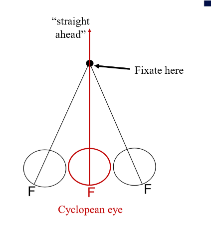
Define the cyclopean eye
The cyclopean eye is a hypothetical single eye located midway between the two eyes that represents how the brain combines visual direction signals from both eyes into one unified binocular sense of visual direction.
define corresponding points
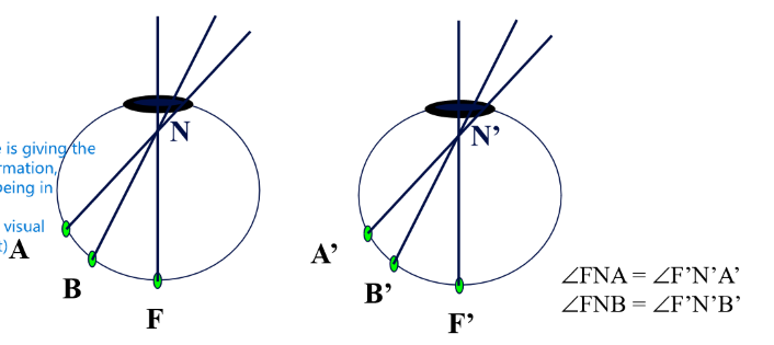
pairs of points on the left and right retina that have the same angular distance from the fovea (one temporal one nasal). In this case, we see the image as being in the same place.
they signal the same oculocentric direction in each eye
define the horopter (will need to draw this in an exam)
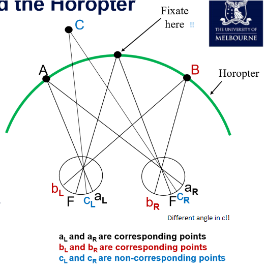
The locus of points in visual space that stimulate corresponding retinal points. If the object falls on the horopter, it will give rise to corresponding points (aka seen as a single object)
WILL NEED TO DRAW THIS IN AN EXAM. IT IS CRITICAL TO UNDESTAND THAT A RAY GOES THROUGH THE NODAL POINT (about 1/3 down the eye)
describe disparate points and corresponding points. where must corresponding points be?
Every retinal element (in the region of binocular overlap) has a corresponding point in the other eye. One is in the temporal retina and the other in the nasal retina.
All other points are non-corresponding or disparate points. Stimulating disparate points give rise to different visual direction signals (so produces diplopia).
define stereopsis
the perception of depth resulting from binocular disparity - the slight differences between the images seen by each eye
how does stereopsis work
Each eye views an object from different angle, this creates small differences in retinal image position (binocular disparities)
Brain uses these disparities as depth cues.
Disparity is processed in the visual cortex to calculate distance
doesn’t work past 6m due to decreasing disparity
what is the perception of these 3 retinal relationships
Retinal relationship |
Fovea + fovea |
Corresponding points |
Disparate points |
Perception |
single object straight ahead |
single object off-centre |
double vision (diplopia) |
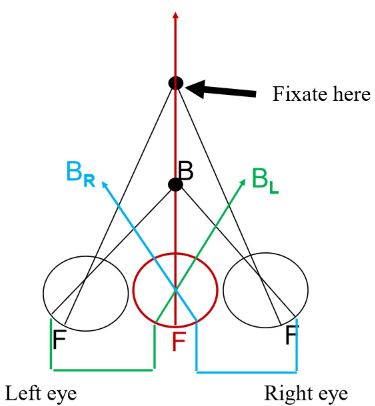
what do points imaged on the temporal retina of both eyes give rise to?
Crossed diplopea.
object is closer than fixation —> image on temporal retina of both eyes.
left eye image appears on right, right eye image appears on left.
Remember - corresponding points will give rise to one nasal and one temporal!
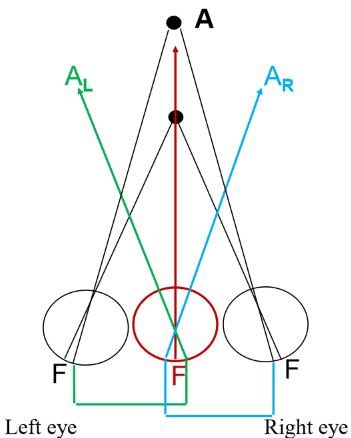
what do points imaged on the nasal retina of both eyes give rise to?
Uncrossed diplopea.
object is farther than fixation —> image on nasal retina of both eyes.
left eye image appears on left, right eye image appears on right.
Remember - corresponding points will give rise to one nasal and one temporal!
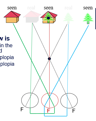
what did doctor Herring observe
Fly, tree and house appeared in the same visual direction.
Because the tree and house were beyond the fixation point, they produced uncrossed diplopia, so it appeared that each of the two images shared the same visual direction
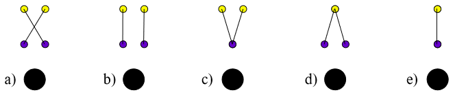
This image shows a Brock string. Assuming bifoveal fixation of the near bead on the Brock string, select the drawing that best depicts what the cyclopean eye would see. (one purple and yellow bead to begin with)
C
Brain receives two slightly different images of the world from each eye, why do we usually only see one version of the world?
physiological suppression!
The brain suppresses conflicting signals from the two eyes (mutual inhibition between cells)
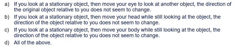
The following observations indicate that oculocentric visual direction is not sufficient to explain our monocular sense of visual direction?
A
B and C don’t involve a change in oculocentric direction 🙂
Which of the following is NOT an advantage of binocular vision:
a) larger field of view
b) superior depth perception under all viewing conditions
c) greater sensitivity because of binocular summation
d) a remaining eye if one is injured
B
don’t get superior depth perception under all viewing conditions!
Define and distinguish between versions and vergences as binocular eye movements
Vergence = the two eyes move in opposite directions.
Convergence → eyes rotate toward each other (for near)
Divergence → eyes rotate away from each other (for far)
Versions = the two eyes move in the same direction.
not as common to measure
only qualitative observation
Measuring versions (1)
perimeter using a moving target
fine target end point: subject unable to resolve because not on fovea
gross target end point: observe cessation of eye movement
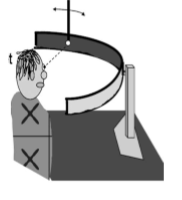
Ways to measure convergence (4)
Push up method - objective
Push up method - subjective
Prism method
Synoptophore method
push up = eye on pen and bringing in
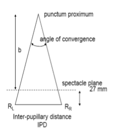
How to measure linear angle of convergence
Linear angle of convergence
d = b + 27 mm
Note: 27 mm comes from back vertex distance (~12 mm) + cornea to centre of rotation (~15 mm)
Then calculate convergence (see image)
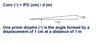
what can the angle of convergence be measured in
Degrees
Centrads (1/100 radian)
Prism diopters
Metre angles
what is a prism diopter and what is the symbol? How to calculate with convergence?
one prism diopter is the angle formed by a displacement of 1 cm at a distance of 1 m
symbol: triangle

what is a metre angle (ma)
the convergence for an object 1m away
conv (ma) = 1/d (m)

Calculate prism dioptres, and metre angles from this information
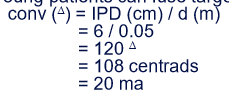
what distance defines convergence insufficiency
when d is larger than 10 cm
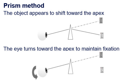
describe what a prism does
prism bends light towards the base
object appears to shift toward apex
eye turns toward the apex to maintain fixation
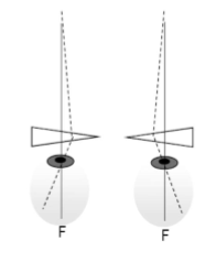
what will base out prism induce? Where is the retinal image in this scenario?
convergence. displaces the image to the temporal retina
can keep increasing base out prism power until the limit of convergence is reached (diplopia)
is our visual system better at diverging or converging?
converging.
limit of divergence about 8 prism diopters. limit of convergence is 20-30 prism diopters.
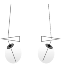
what will a base in prism induce? Where is the retinal image in this scenario?
divergence (outward, exo). displaces the image to the nasal retina.
what are fusional reserves
Fusional reserves are the maximum amount of vergence (convergence or divergence) a person can generate beyond their resting alignment to maintain single, fused vision.
this what we have been testing
what is the synoptophore method? What can it measure?
Synoptophore: a binocular instrument that presents separate images to each eye to measure ocular alignment, fusion, and vergence ranges by adjusting tube positions.
Convergence
Divergence
Supravergence (vertical changes in vergence)
Cyclovergence (rotational vergence)
what kind of binocular eye movement is primarily involved when a hunter follows a flock of ducks flying left to right at a distance of 100 m
version

vergence (prob a bit of accommodation as well)
What is the functional binocular position?
The fixation object is imaged at both foveae.
Requires vergence eye movements
How is the functional binocular position maintained?
anatomical forces (controls where eyes are pointing)
innervation of extraocular muscles
accommodation (closely linked to convergence)
attention
awareness of nearness
fusion reflux
what is the anatomic position of rest?
position when all innervation to extraocular muscles is removed (aka u dead)
17 prism diopters beyond parallel (diverged)
what is tonic vergence/ what it called?
position with normal innervation, but no stimulus (aka dark).
moves eyes to the physiological position of rest
define fusional vergence (disparity vergence). What is it called and what is it triggered by.
the spontaneous movement (reflex) of the eyes from the passive position to the active position to avoid diplopia.
the active position is maintained by tonic vergence together with fusion and fixation reflexes.
describe orthophoria. what this mean in terms of active and passive positions of the eyes
Ideal condition where the ocular motor apparatus is in perfect equilibrium so that both eyes retain their required positional relationship even when fusion is removed by dissociation of the eyes (one is covered)
the active and passive positions coincide the eyes are said to be in a state of orthophoria.
define initial vergence
movement by the eyes when they move from the physiological position of rest to the functional position of rest (passive position)
triggered by the fixation reflex
what is the functional position of rest. what is it triggered by
Passive position = where the eyes would point if fusion were removed (e.g., with cover test)
triggered by fixation reflex
define heterophoria. What is active and passive positions.
Present when a deviation of the visual axes is held latent by the power of the fusion reflex.
when the active and passive positions are different
** heterophoria = fusion reflex is doing work to keep eyes aligned
what is the fusion reflex
a neurological reflex that moves the eyes to eliminate disparity.
in heterophoria, the fusion reflex has to work to keep eye in the active position (aligned)
heterophoria represents strain on fusion reflex
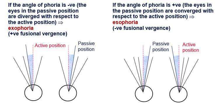
what is the angle of phoria? What does it mean when the angle of phoria is negative? what angle would the fusional vergence mean?
the angle the eyes move.
If angle of phoria is -ve (the eyes in the passive position are diverged in respect to the active position) —> exophoria (+ve fusional vergence)
If angle of phoria is +ve (the eyes in the passive position are converged in respect to the active position) —> esophoria (-ve fusional vergence)
Aetiology of heterophoria (origins)
globe size and shape
muscle/ligament abnormalities
innervation factors (accommodative-convergence synkinesis, binocular reflexes)
general fatigue, excessive use of alcohol/tobacco
lesions of ocular motor neural apparatus
what is cyclophoria
when eyes rotate about the visual axis when dissociated
inclophoria = rotate nasally
exclophoria = rotate temporally
How to label hyper/hypo phoria/tropia (up/down)
WE ALWAYS specify the eye UP!!
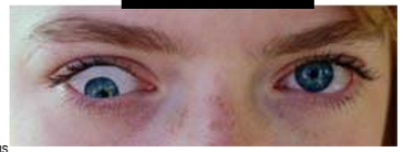
picture = left hypertropia
why is it common to be expohoric at near distances? what is this called?
Requires increased convergence (+ve fusional vergence). Common for the eye to undershoot this. Fusional reflex is working to turn the eye in more. when fusion is broken the eye turns OUT
called physiological exophoria
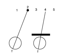
A person with a near exophoria is fixating a near point with the left eye while the right eye is covered (see figure). According to principles of visual direction, where would the fixated object (black dot) appear to be located?
a) Left of the true object (1)
b) In its true location (2)
c) Between the two visual axes (3)
d) On the right eye visual axis (4)
e) To the right of the right eye’s visual axis (5)
B
person won’t see the object move when the eye is covered but the eye will move (but visual information is blocked)
name for terms when:
heterophoria doesn’t lead to symptoms
heterophoria leads to symptoms
compensated phoria (no issue)
decompensated phoria (symptoms)
symptoms of decompensated heterophoria
headaches
aching eyes
diplopia
blurred vision
focusing difficulties
monocular comfort
difficulties with depth perception
sore eyes
general irritation
7 methods to measure heterophoria
cover test
maddox rod
maddox wing
von Graefe
prism bar
prentice card
synoptophore (this may be a duplicate)
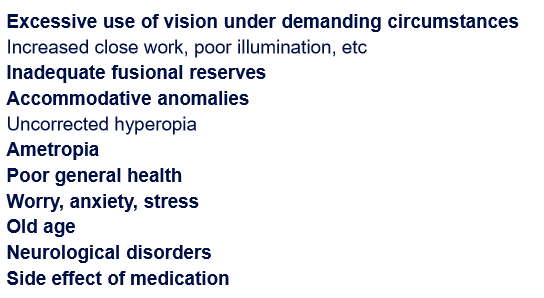
causes of decompensated heterotropia
What 5 steps make up a routine eye exam
History taking (how are you today, how can I help you)
Routine screening (VA, PD, cover test, etc)
Refraction (find prescription)
Anterior and posterior eye exam
Management
What 3 categories make up the routine screening portion of the eye exam
Refraction (2)
VA
PD
Binocular vision (4)
cover test
Near point accommodation (NPA)
Near point convergence (NPC)
stereopsis
Neurological (4)
ocular motility
pupils - bright, dim, RAPD, DCN
colour vision (CV) - Ishihara
visual field (VF) - confrontation, red cap and amsler grid
is the cover test objective or subjective?
objective
don’t need the patient to respond
what is myopia
nearsightedness
see near objects clearly but objects that are farther away appear blurry
this occurs because the eye focuses light in front of the retina instead of directly on it
need negative power lens
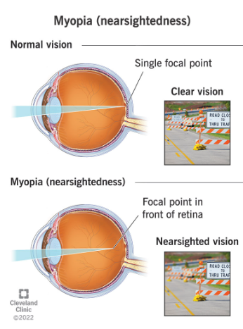
lenses in myopia, hyperopia and astigmatism
myopia and hyperopia = spherical lenses (± power) - myo need neg, hyper need pos
astigmatism = cylindrical lenses (negative power)
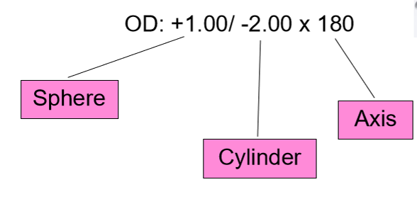
Difference between heterotropia (strabismus) and heterophoria
Hypertropia is a manifest deviation. Eyes are visibly misaligned even when fixating on an object
Heterophoria is when eyes are not properly aligned with each other when they are at rest.
orthophoria = eyes are aligned in the resting position
what does accommodation do to the power of the lens
increases the plus power of the lens (near)
more convex
eyes converge
What is NPC a measure of?
Explain:
set-up
which eye
target
distance
procedure
recording (units?)
normal reading
Near point convergence
binocular vision test - a measure of “how well you can bring your eyes inwards” - e.g. when reading\
always test from cornea
set-up: normal room, habitual Rx
which eye: binocularly only
target: 1 letter-size above best corrected vision. Clear and single?
distance: start at habitual reading distance (~40cm)
procedure (must do at least twice - take worst reading)
move inwards until sustained double vision is perceived (this is the break). Get patient to blink and try to correct.
then move out till clear and single image is achieved (this is the recovery)
Record: B/R 5/7cm with habitual Rx.
if to the nose record as TTN
Normal value: 6/8cm. Anything over 10 cm is an abnormal break.
what is it called when accommodation decreases with age
presbyopia
How will uncorrected myopia and hyperopia impact NPA
uncorrected myope: give a falsely high NPA reading
uncorrected hypermetrope: give a falsely low NPA reading
what declines with age (routine test)
Accommodation - directly age related. lens thickens. NPA more effected
Convergence - indirectly affected (small/variable change). NPC not effected much
what might abnormal values of CT/NPC/NPA/Stereopsis indicate?
abnormalities in binocular disfunction
need to determine underlying cause - congenital vs acquired?
investigate further - BV workup to assess accommodation, vergence and the interaction between both systems (more to come)
What is NPA a measure of?
Explain:
set-up
which eye
target
distance
procedure
recording (units?)
normal reading
Near point accommodation
binocular vision test - a measure of focusing ability/power
always test from cornea
measure in Diopters = 1/distance (in meters)
set-up: normal room, habitual Rx
which eye: monocularly only (always R first)
target: 1 letter-size above best corrected vision. Clear and single?
distance: start at habitual reading distance (~40cm)
procedure (must do at least twice for each eye)
move inwards until sustained blur. Get patient to blink to try to correct. measure in m.
repeat for the other eye
Record: R 10D, L 12D with habitual Rx
if to the nose record as TTN
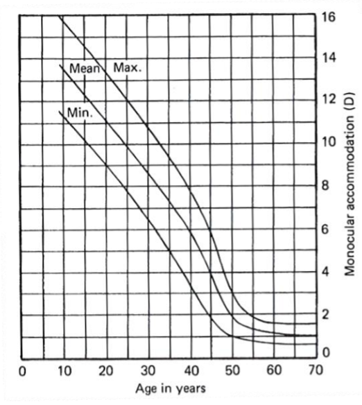
Normal value: 15-(1/4 age)
Monocular accommodation (D) vs age (graph)
Accommodation = 15 - (1/4 age)
If you’re 20, you should have about 11 Diopters of accommodation
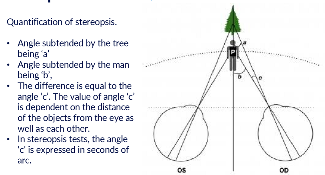
what is stereopsis tests recorded in?
seconds of arc
what is stereopsis doing exactly
determining the difference in the angles between two objects at different distances (i think). This angle is expressed in seconds of arc.
3 anatomical and physiological requirements for stereopsis
a large binocular over-lap of VFs
partial decussation of afferent visual fibres
coordinated conjugate eye movements
normal/abnormal stereopsis readings
measured in seconds of arc (“ arc) - smaller = better!
≤ 40” arc: eyes are aligned well. any ocular misalignment cannot be larger than Panum’s fusional area
60” arc: still within normal limits
> 60” arc: question whether accurate bifoveal fixation is present
What 3 things do you check for in stereopsis test?
How to record?
Suppression (R/L) - are both eyes working the same?
Global (random dot) - detect the shape in the dots
Local (contour) - identify what shape, animal or circle appears closer
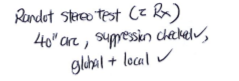
Randot stereotest (habitual Rx): No suppression, 40” arc, global pass,
What nerves innervate what extraoccular eye muscles
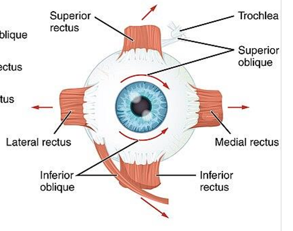
CN IV (4) = superior oblique
CN VI (6) = lateral rectus
CN III (3) = all others (superior rectus, medial rectus, inferior rectus, inferior oblique)
What test measures the eye muscle function
ocular motility
this is a neurological screening test.
if one of the muscles isn’t working, can get double vision cause the eye won’t be able to move to fixate (patient sometimes compensates by moving head)
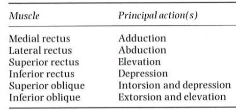
Principle action(s) of the 6 main extraocular muscles
adduction = toward nose (inwards)
abduction = away from nose (temporal) - outwards
intorsion = rotate toward nose
extorsion = rotate away from nose
Think of these in terms of what happens during cover test and what muscles may cause eso/exo etc.
What is ocular motility a measure of?
Explain:
set-up
which eye
target
distance
procedure
recording (units?)
normal reading
Measure: eye muscle function (or nerves i think)
set-up: normal room lights, Rx either on or off
which eye: binocularly only
target: H on PD ruler
distance: perform at 40 cm
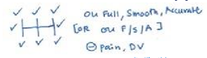
procedure: evaluate 9 positions of gaze (HH pattern)
Px to keep head still, and follow the target with eyes.
Px to report pain or diplopia at any time if present
check for overaction/underaction/restriction, any jerky eye movement.
Record: Full/Smooth/Accurate, -ve pain, DV
what is the term for when pupil sizes are different
Anisocoria
what is the sequence of evaluating pupil sizes and responses?
Sizes in light then dark
RAPD (relative afferent pupillary defect)
DCN (direct, consensual and near responses)
what may an afferent defect indicate?
Afferent = eye talk to brain
lesion in the retina, optic nerve head (ONH) defect
what may an efferent nerve defect indicate?
efferent = brain talk to eye
anisocoria (unequal pupil sizes)
lesion in the motor neuron system which carries signals from the CNS to the iris
what is a normal pupil size?
2-4 mm in bright
4-8 mm in dim
pupils get smaller (miosis) with age
what can pupil screening test evaluate
function and integrity of the iris, optic nerve, posterior visual pathways, 3rd CN, afferent and efferent pathways
Pupil test
Explain:
set-up
Rx
target
distance
procedure
recording (units?)
normal reading
Measure: “pupil size and reactivity”
set-up: Normal room lights to measure size first, then dim lighting to measure again. Use a light source for RAPD and direct/consensual/near testing. The sequence matters: bright → dim → RAPD (distant, light off) → DCN
Rx off.
target: distance: fixation light (or other non-accommodative target); near: near accommodative target (not specified)
distance: Distance: bright, dim and RAPD. Near: DCN
procedure:
Ask patient to fixate a distant non-accommodative target
Measure pupil size in bright
Measure again in dim
While in dim, perform swinging flashlight test for RAPD
Turn room lights back on
Assess direct, consensual, and near responses
Record: Bright: R: 4mm, L: 4mm; Dim: R: 6 mm, L: 7mm; (-)RAPD, DCN pass or PERRLA
PERRLA:
Pupils
Equal
Round
Reactive to
Light and
Accommodation

what is the consensual response when light is shone into an eye?
What would the response be in someone with an APD (afferent pupillary defect)?
normal: both pupils contract due to intact and consensual light reflexes
APD: pupil in normal eye dilates upon illumination of the pathological eye because of a lack of stimulus for the consensual light reflex
is colour vision congenital or acquired?
can be both. Can be congenital but diseases and drugs can alter colour vision as well.
If acquired there will be a difference in severity between both eyes!
what is the most common colour vision deficiency?
R-G most common
ishihara only tests red green!
Blue yellow less common
Protan = red-cone-type defect
Deutan = green-cone-type defect
Tritan = blue-cone-type defect
Colour vision test:
Explain:
set-up
Rx
which eyes
target
distance
procedure
recording (units?)
normal reading
set up: normal room lights
Rx: habitual
eyes: binocular for screening (/15), monocular for diagnostic (/17) - if more than 2 errors = FAIL, go on to monocular tests.
distance: hold at 75 cm
procedure:
3s per viewing plate
Recording:
15/15 OU
R 17/17 L 10/17