Class 1 - Extraoral radiology
1/15
There's no tags or description
Looks like no tags are added yet.
Name | Mastery | Learn | Test | Matching | Spaced | Call with Kai | Chat |
|---|
No analytics yet
Send a link to your students to track their progress
16 Terms
Lateral cephalogram
Used in ortho treatment
Assesses AP relationship b/w Mx & Md + skull
Assesses skeletal + soft tissue relationship
Monitors treatment progress & outcome
Continues orthognathic surgical treatment planning
CBCT use - Wisdom, Ortho & Implants
Wisdom teeth eval
Position inr el. to anatomical structures (mand canal)
Orthodontics
Diagnostic assessmen + analysis of facial jaw/anomalies
Root resorption
Position & location of impacted/supernumerary teeth in relation to anatomy
TMJ
Implantology
Eval height + width of AB
Precise distance to anatomical structures in planning
Possibility to use surgical guide - KSDT required in planning
Gold standard in planning
CBCT use - Perio, Oral, Facial, Maxo, TMJ & Endo
Perio
Bone height
Furcation
Period surgery
Maxo
Bening/Malignant formation
Calcification
SInuses
Injuries
Diagnosis of osteomyelitis
TMJ
Assess morphology of condyle + surrounding tissues
Asses joint space + position of condyle in joint cavity.
Condylar surfaces
Arthritis, ankylosing + condylar abnormalities
Endo
Additional canal ID
Root inclinations + anomalies
PA patho diagnosis in non-standard symptoms
Non-odontogenic patho diagnosis
Endo therapy complication diagnosis
Dentoalveolar injuries
Internal resoprtions
Apical surgery
PET-CT & Scintigraphy
Scintigraphy - 2D nuclear medicine tech depicting active bone remodeling.
PET- Tomographic nuclear medicine examination using positron-emitting radionuclides.
Clinical relevance
Detection of primary metastatic tumors
Diagnosis of osteomyelitis
Evaluation od osteonecrosis in jaw & skeletal growth disorders
Where the patient’s tissue itself becomes radio source, image interpretation is based on signal intensity & localization.
USG - Ultrasound
Principle - High frequency → different tissues have different echoes → echo perception
Indications
Salivary gland eval
TMJ disc eval
Lymph node eval
Blood flow eval (doppler)
Inflammation/infection (abscesses & phlegmons)
Soft tissue formations
MRI - Magnetic Resonance Imaging
Evaluation
TMJ + disc
Soft tissue
Leasions & inflammations
NOT PERFORMED ON:
PT´s w/ metallic devices & soft tissue implants
PERFORMED ON:
Dental implants, amalgam restorations.
Artifacts will be seen
Panoramic indications
Treatment planning
Ortho → Check presence/absence of teeth
Implant planning → Assess vertical bone height
3rd molar extraction → Position/condition before surgert.
Assessment/diagnosis
Poor oral condition (after clinical exam)
Unerupted teeth/bone formation not seen in intraorals.
Perio evaluation, pockets <5mm.
Mandibular fractures
Maxillary sinus pathologies (basr, posterior, lateral walls)
Review/Montior
TMJ joint changes → overview of condyles & joint area
Cons of OPG & CBCT
OPG
Lower resolution than intraorals, geometric distortion, positioning sensistive, not ideal for caries diagnosis.
CBCT
Higher radiodose than 2D imaging, more ecpensive, metal artifacts, Not for soft tissue imaging, overuse w/o indication (ALARA), training to interpret, Motion artifacts.
Analyzing OPG
PT age & gender
Age characteristics
Panoramic image → position & image formation.
Anatomical local. of lesion
Size, shape, borders & contours.
Lesion structure → opaque, lucent, mixed.
Effect on adjacent structures.
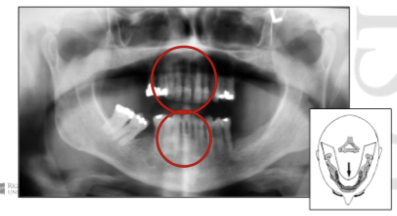
Patient position - Antero-posterior error
Patient too forward, too close to film. Teeth narrowed.
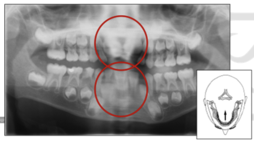
Patient position - Antero-posterior error
Patient too backwards, too far away from film. Teeth magnified & widened.
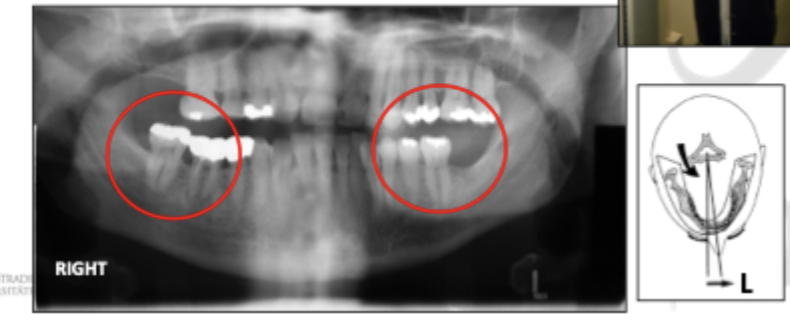
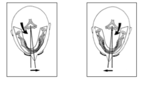
Patient position - Horizontal error
Pt rotated to right → left molar closer to film (smaller) & right molars further from film appear larger.
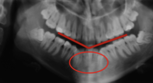
Patient position - Vertical error 1
Head & chin tipped down, frankfort plane not horizontal, occlusal plane distorted → Smiley
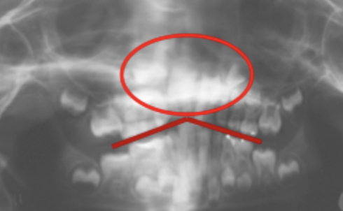
Patient position - Vertical error 2
Head & chini uppwards, frankfort not horizonral , occlusal plane distorted → Grumpy
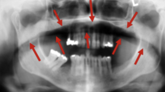
Patient position - Air shadow
Failure to instruct patient to press tongue to roof of mouth.
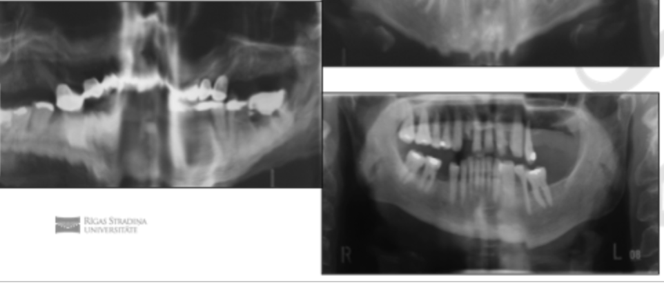
Patient position - Movement
Failure to instruct patient to keep still