LECTURE 2-MEIOSIS, GAMETOGENESIS AND SOURCES OF GENETIC VARIATION
1/47
There's no tags or description
Looks like no tags are added yet.
Name | Mastery | Learn | Test | Matching | Spaced |
|---|
No study sessions yet.
48 Terms
Quick Review of Main Concepts From Lecture 1-Chapter 16
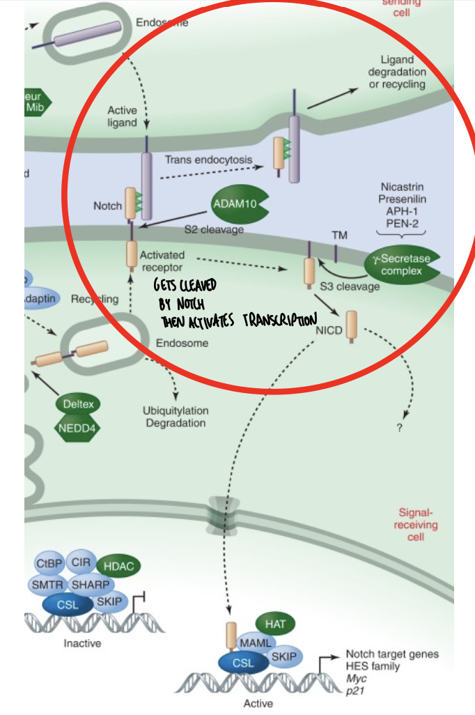
What happens to the cleaved notch receptor?
Lecture Outline: Meiosis, Gametogenesis, and sources of genetic variation
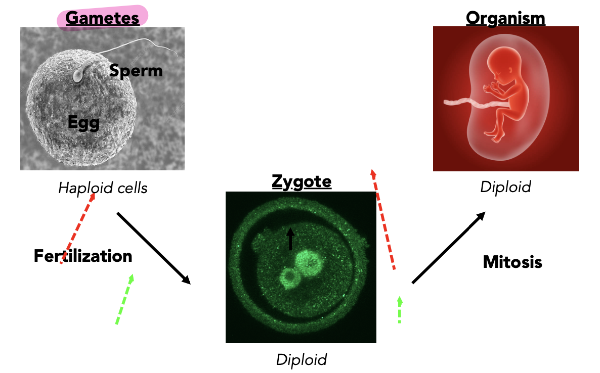
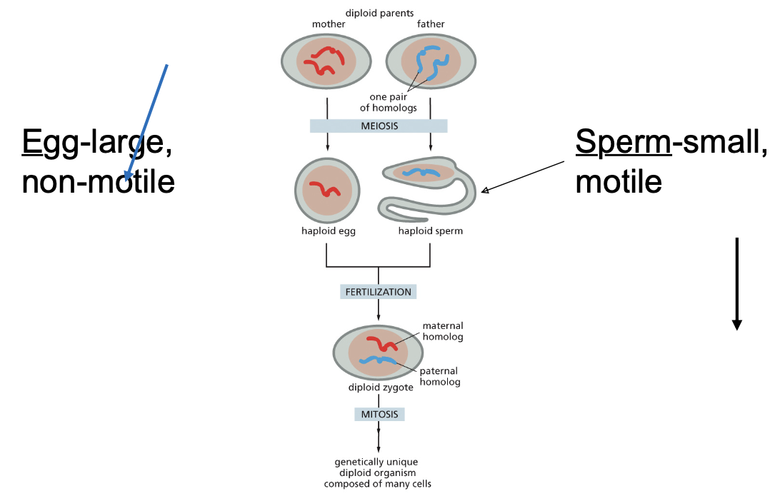
sexual reproduction: Fusing haploid gametes to establish diploid zygote
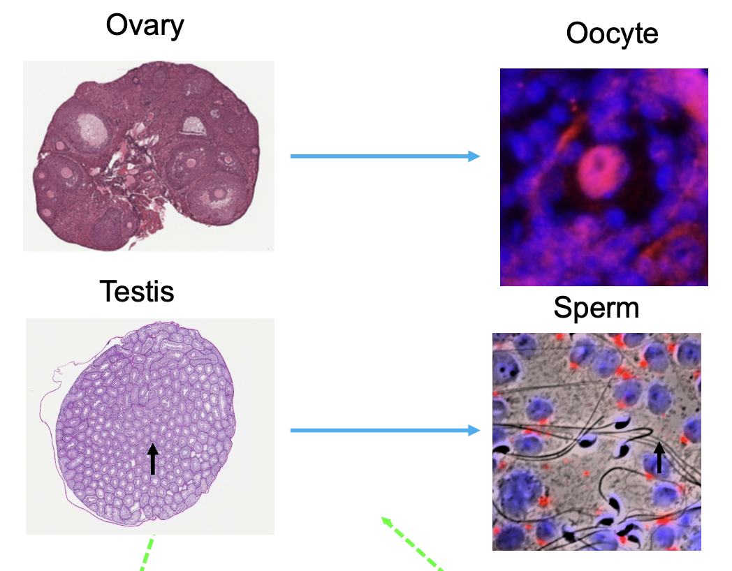
oocytes and sperm are produced in the ovary and the testis, respectively
Meiosis is a series of reductive cell divisions that yield haploid cells
human genetics of disease
What do the hydra vulgaris and the osbournes have in common?
Parent and offspring
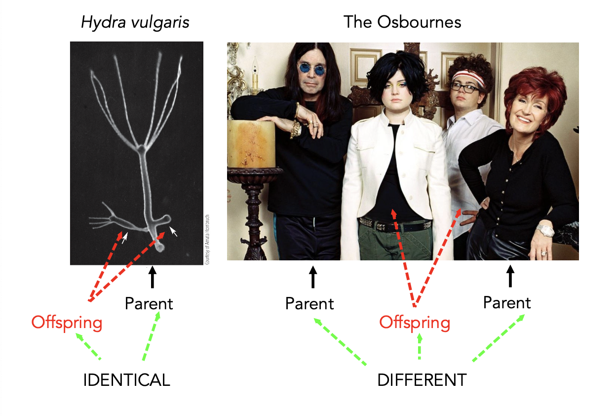
Sexual reproduction introduces genetic diversity by mixing parental DNA
Diploid organisms contain 2 copies of each chromosome and gene
Development is a cyclical process
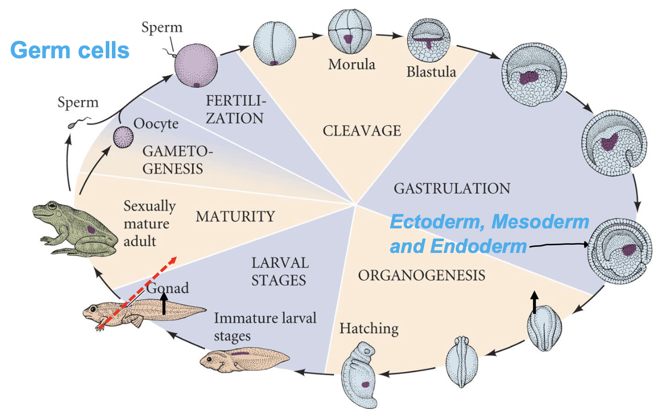
How does a single cell give rise to multiple cell types?
Gametes are produced within the ovary and testis
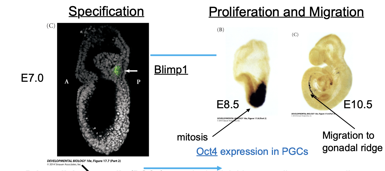
Primordial germ cells (PGCs) are mitotic diploid germ cell precursor cells that commit to meiosis (diploid to haploid-reductive system) to become gametes
BMP (important for differentiation) signaling in the posterior embryo induces PGC’s
Primordial germ cells (PGCs) are mitotic diploid germ cell precursor cells that commit to meiosis to become gametes
BMP4/BMP2 induce formation of PGCs in the embryo
Blimp1 is a transcriptional repressor important for PGC lineage specification
E=embryonic day
A=anterior
P=posterior
BMP=bone morphogenic protein
PGC=primordial germ cell
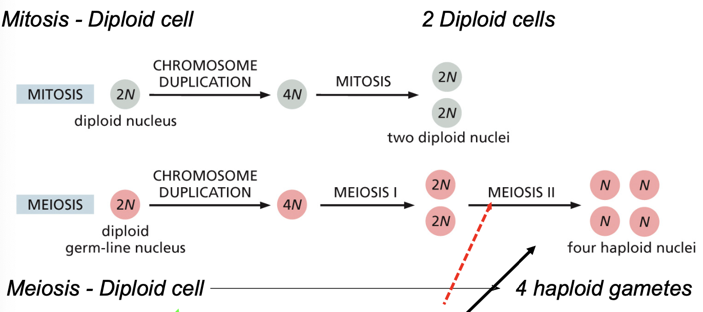
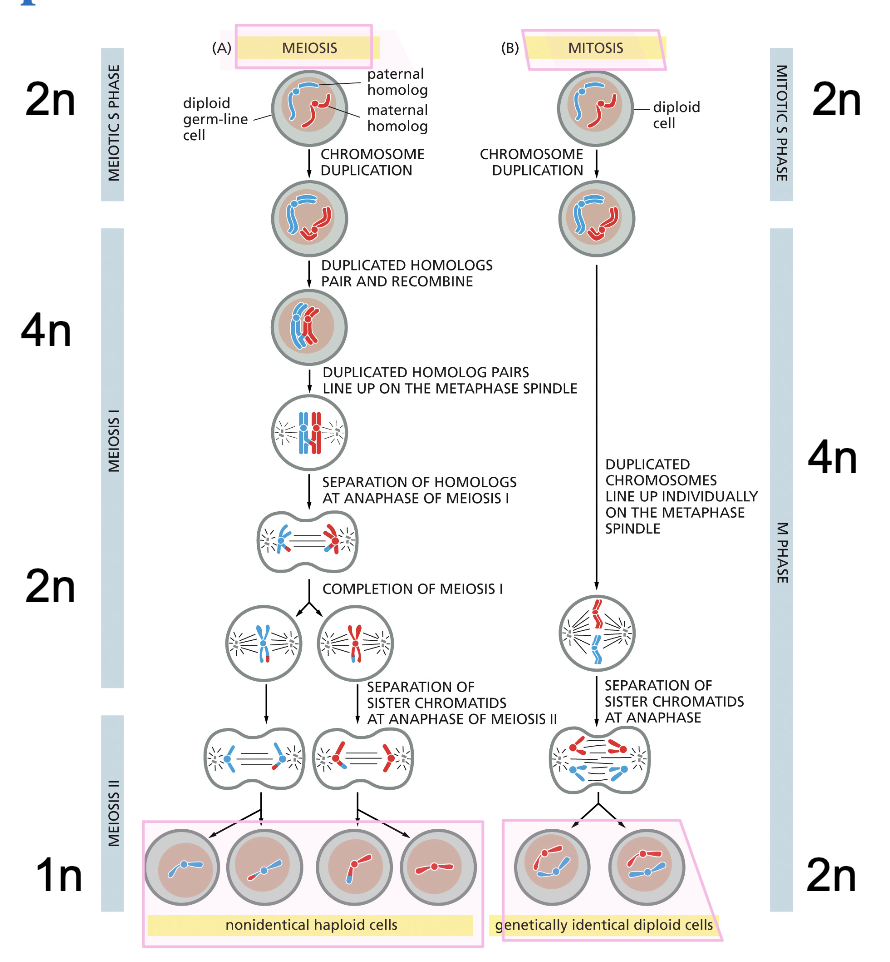
Mitosis and Meiosis both begin with chromosome duplication
Mitosis-diploid cell to 2 diploid cells
Meiosis-diploid cell to 4 haploid gametes
all somatic cells are diploid
only gametes are haploid
gametes are generated from diploid cells by reductive cell division-meiosis
Oogenesis in the Mammalian Ovary
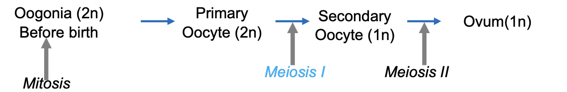
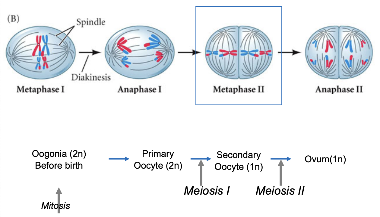
female gametes are produced in the ovaries during Oogenesis
1ry oocytes remains in Prophase I of meiosis (generated in the embryo) until puberty
2ry oocyte remains in Metaphase II of meiosis
@ fertilization 2ry oocyte (n) finishes Meiosis II producing the mature egg cell
OH: oogenesis is the process of making oocytes (diploid before meiosis I and II and haploid after so genetically distinct)
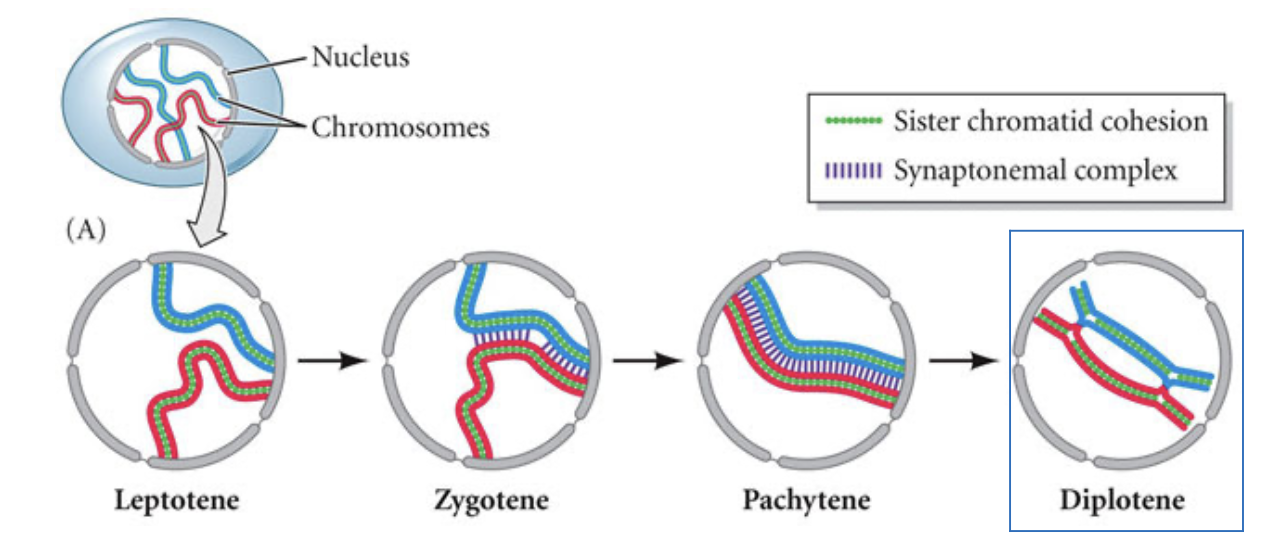
Prophase I has 4 key stages
Leptotene-duplicated chromosomes, each consisting of two sister chromatids condense
Zygotene-homologous chromosomes begin to pair up by formation of the synaptonemal complex-the bivalent is formed at this stage
Pachytene-homologous chromosomes are fully synapsed and crossing over (homologous recombination) occurs between non-sister chromatids
Diplotene-synaptonemal complex disassembles, homologous chromosomes begin to separate but remain connected at the chiasmata (points of crossing over)
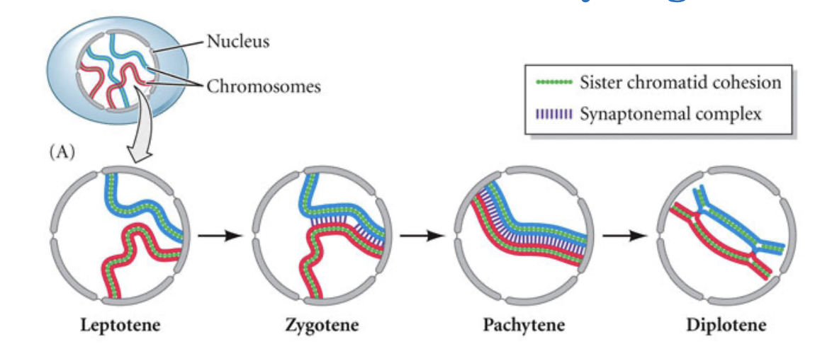
Primary oocytes are arrested at Prophase I of meiosis in the diplotene stage
Oocytes stay arrested in prophase I of meiosis at least until puberty
Secondary Oocytes are arrested at Metaphase II of meiosis
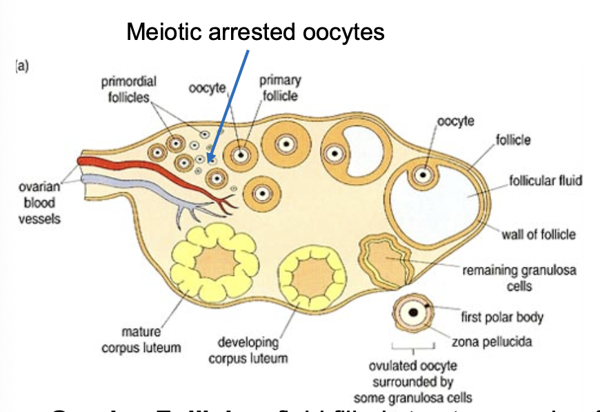
Major components of the ovarian follicle
oocyte=germ cell
somatic cells (granulosa cell and theca cell)—>estrogen
Ovarian follicle-fluid filled structure made of oocyte, theca, and granulosa cells
Theca and granulosa cells contribute to maturation of oocyte producing sex steroid hormones-estrogen
Ovulation-completing meiosis I
fertilization-completing meiosis II
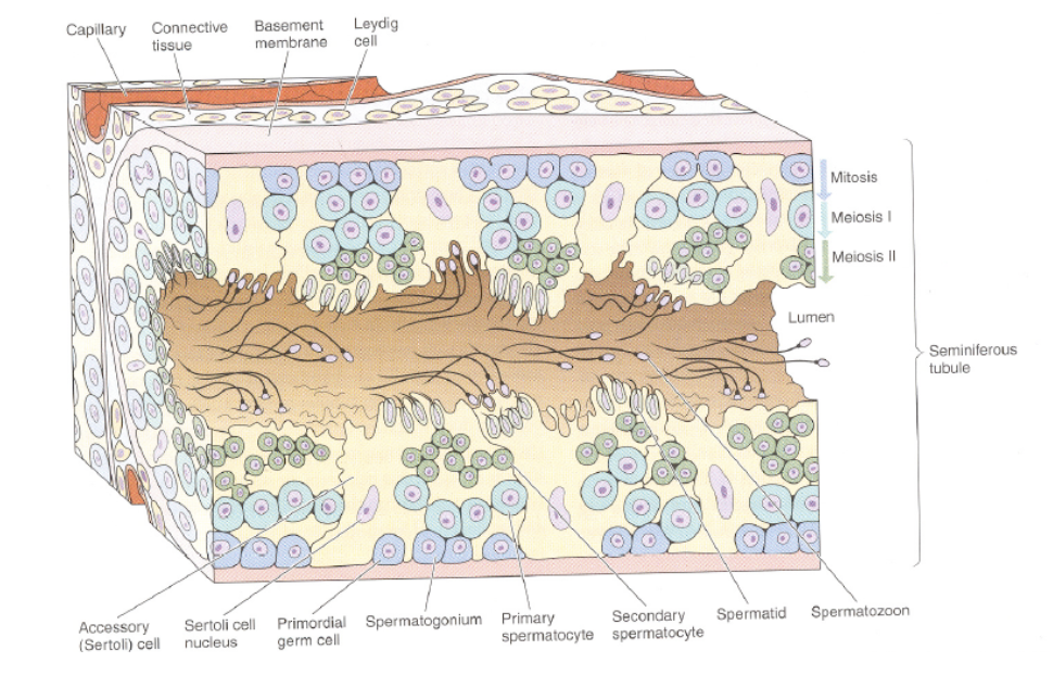
The seminiferous tubules of the adult mammalian testis contains true spermatogonial stem cells (SSCs)
spermatoza=germ cell
somatic cells (sertoli cells)—> leydig cells
Spermatogenesis-production of sperm cells within the seminiferous tubules of the male gonads (testes)
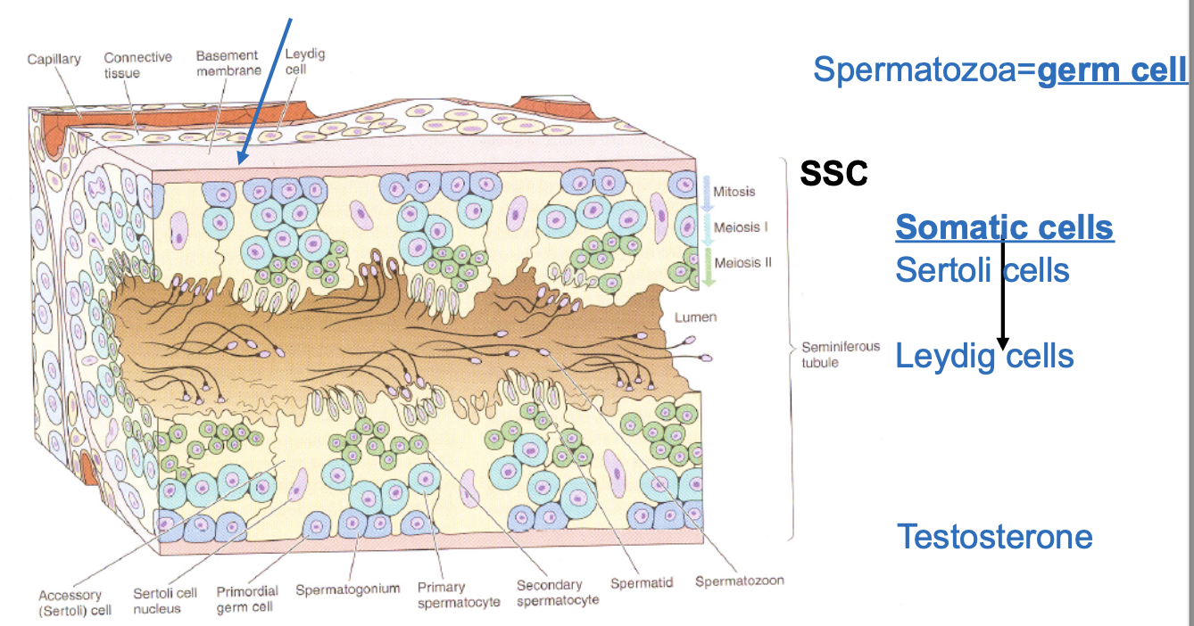
Spermatogonial stem cells (SSCs) undergo meiosis to generate stem cells
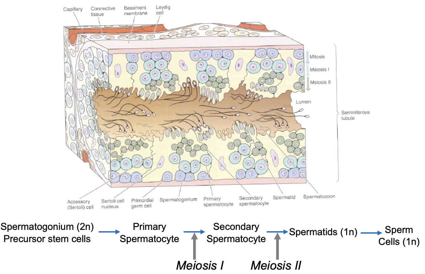
The seminiferous tubules contain support cells that also contribute to the differentiation of sperm cells
Leydig cells-release testosterone stimulating stem cells to differentiate into primary spermatocytes
Sertoli cells-provide nutrients to sperm cell + help reduce cytoplasm, sperm cells released from the seminiferous tubules mature and are stored in the “epididymus”
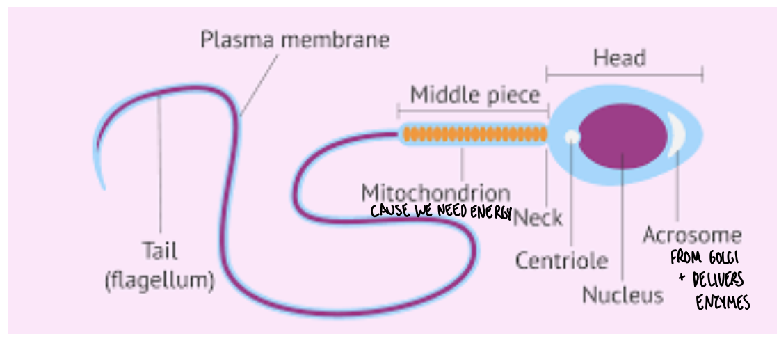
Sperm cells have a very specialized structure
Head-contains nucleus and acrosome (cap like organelle derived from golgi contains enzymes important to get through the eggs outer layer)
Middle piece-contains a lot of mitochondria
Tail-composed of a flagellum (MT structure) helps in locomotion
Sexual reproduction: Meiosis followed by Mitosis
Gametes are haploid (1n)
Males and females produce different gametes
Fertilized egg has homologous chromosomes from mother and father
Zygote produces individual with unique diploid (2n) set of chromosomes
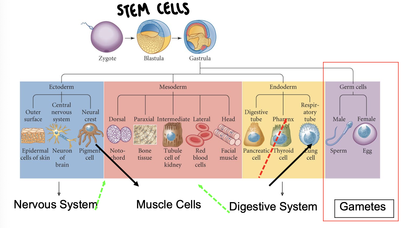
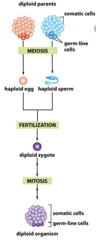
Differences between somatic and germ-line cells
Germ line cells are specified early in development and give rise to the haploid gametes by meiosis
Gametes propagate the genetic information to the next generation
Somatic cells form the body of the organism and are part of all our different tissues
Diploid germ line cells give rise to the haploid gametes
Mitosis and Meiosis both begin with chromosome duplication
all somatic cells are diploid
4N means 4 copies of each chromosome after replication
only gametes are haploid
gametes are generated from diploid cells by reductive cell division-meiosis
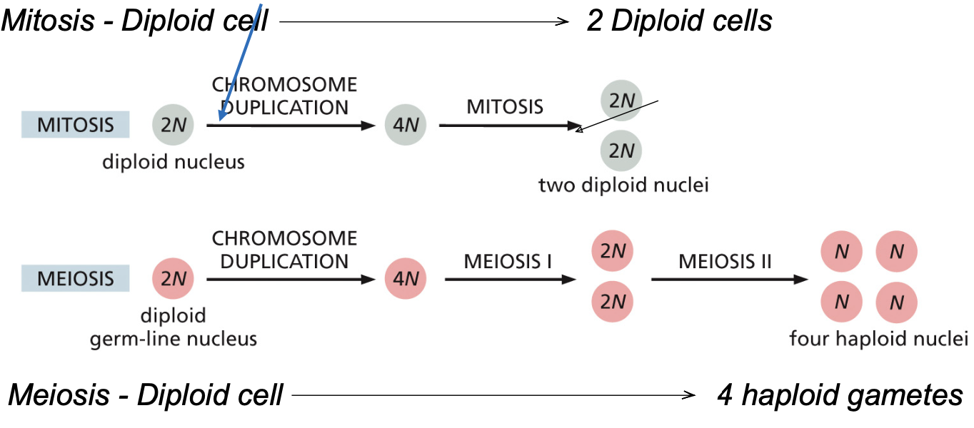
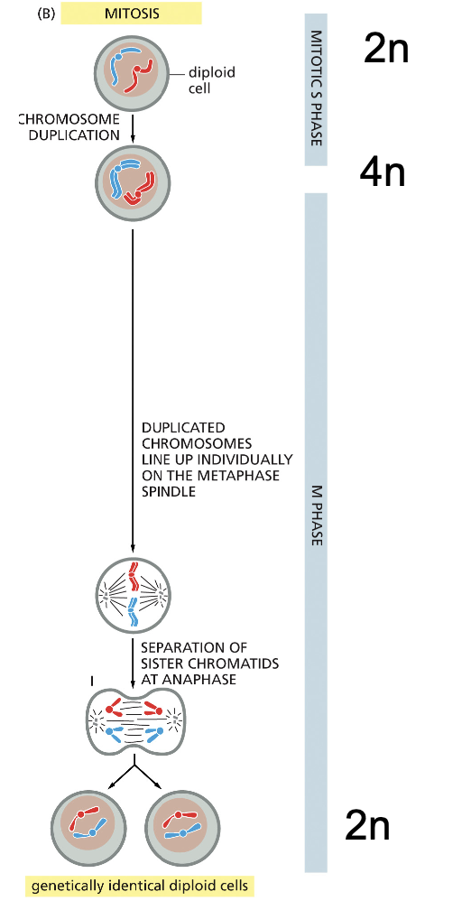
Mitosis: DNA replication and cell division
start with 2n cell
duplication of the genetic material (4n stage)
During M phase genetic material is equally separated
Two identical daughter 2n cells
Meiosis: DNA replication and two meiotic divisions
Start with 2n cell
duplication and recombination of genetic material (4n stage)
Meiosis I-produce 2n cells (reductionist division) that are genetically diverse
Meiosis II-reductionist division produces 1n cells that are genetically distinct from one another
Mitosis vs Meiosis
Meiosis generates for 4 haploid cells that are not identical to each other (like why me and Mary look different!)
Mitosis generates 2 diploid cells that are genetically identically identical to each other
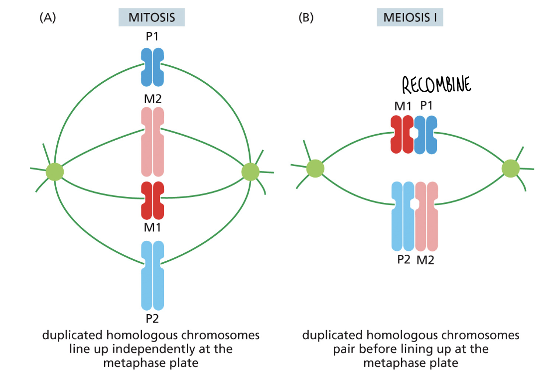
Mitosis vs Meiosis: pairing of homologous chromosomes
Mitosis: duplicated chromosomes line up at the metaphase plate and the sister chromatids separate in anaphase generating 2 identical daughter cells
Meiosis-paternal and maternal homologous chromosomes align with one another and inheritance by the daughter cells is random-daughter cells are not identical
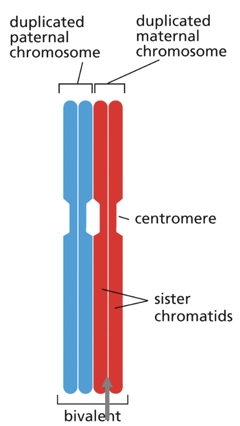
Duplicated maternal and paternal chromosomes pair during Meiosis I to from bivalents
each bivalent contains 4 sister chromatids
duplicated maternal and paternal homologous chromosomes are joint at the centromeres
during meiosis the chromosome arms are also interacting with each other providing the basis for exchange of genetic material
“swaping” genetic material leads to genetic variability
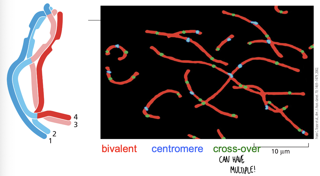
Chiasmata form between maternal and paternal chromosomes-crossing over between non-sister chromatids
Meiosis I-non-sister chromatids in each bivalent swap segments of DNA
exchange of material on inner chromosome arms is shown
region of crossing over is the chiasma
there are multiple sites of chiasma or crossing over
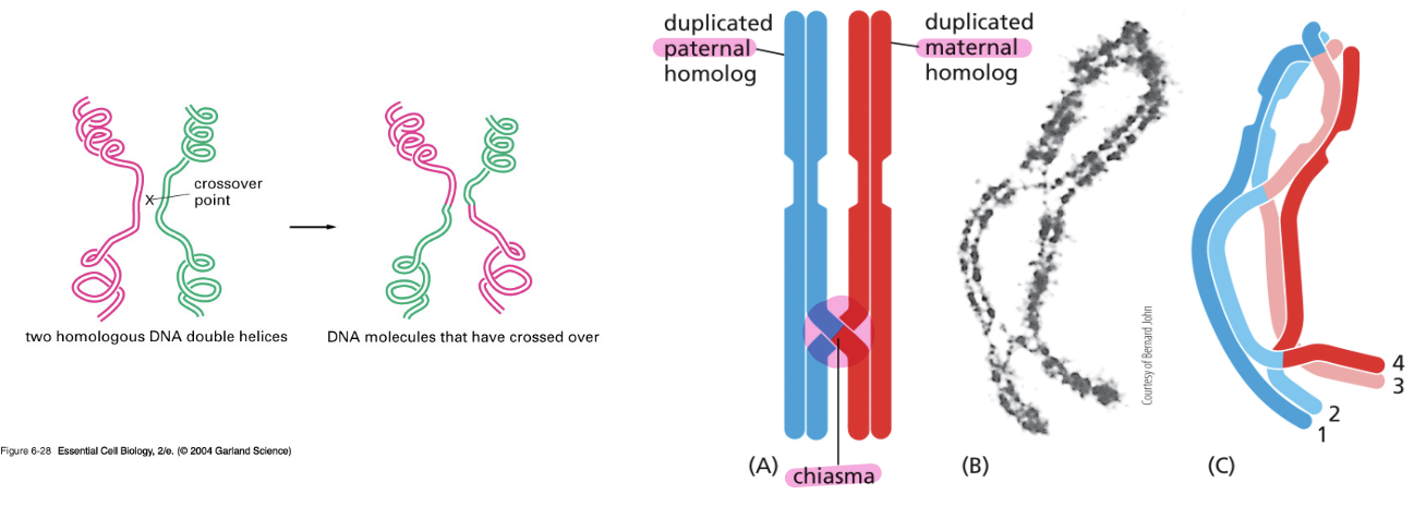
Chiasmata formed between maternal and paternal chromosomes allow for recombination between non-sister chromosomes
micrograph of a human oocyte that was stained with fluorescent markers
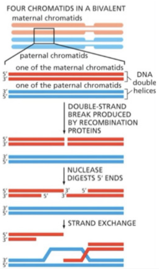
How does this “crossing over” happen?
recombination enzymes induce in either the maternal or paternal chromatid double strand breaks (DSB)
nucleases will digest the ends of strands leading to the formation of this overhangs
The template for homologous recombination repair will use the homologous chromatid from the other parent as a template for repair
Result-strand exchange where the maternal single strand will interact with the paternal homologous side
OH: in the end we repair the strand using enzymes and the template strand, so in this example the blue paternal chromatids count as the template strands
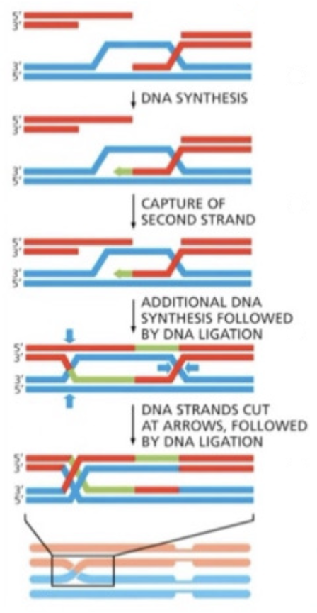
Homologous recombination repair requires DNA synthesis and DNA ligation
uses paternal as template to fill in the gaps by DNA synthesis (green)
results in a structure with 2 cross overs and nicks (breaks) are joined by DNA ligase
Once meiosis I finishes they are going to separate out from each other
Each chromatid is going to contain a segment of DNA from the original parent and from the other parent
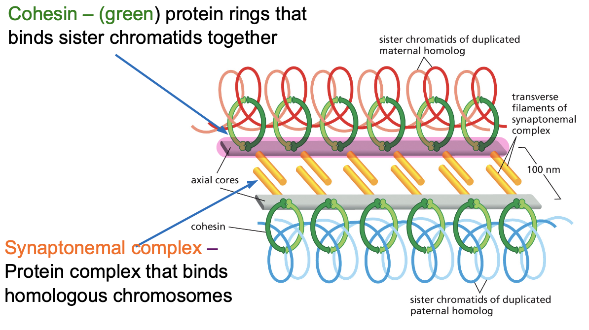
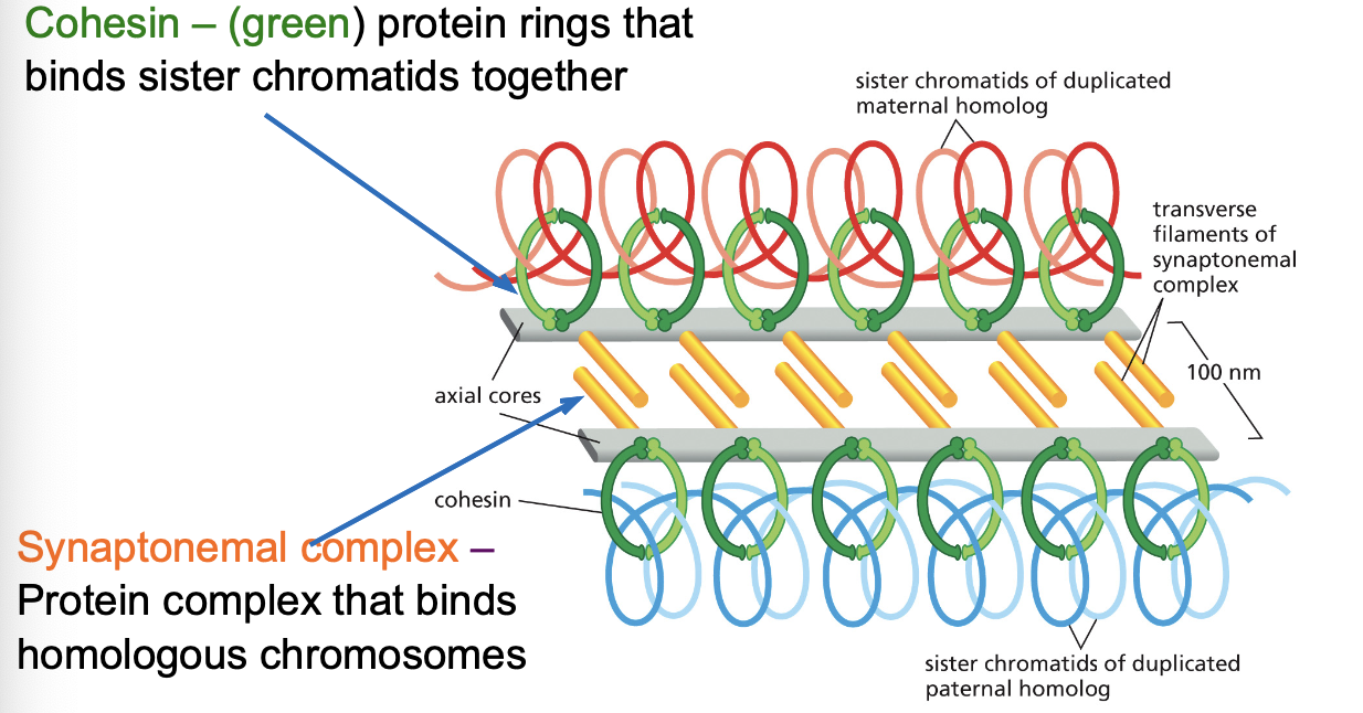
Cohesin and the Synaptonemal Complex: Two protein complexes important for proper Meiosis
Cohesin-(green) protein rings that binds sister chromatids together
Synaptonemal complex-protein complex that binds homologous chromosomes
Cohesin are attached to axial core proteins that also bind to transverse filaments
Cohesin and Synaptonemal Complex: Two protein complexes important for proper Meiosis
Transverse filaments form a zipper like structure that allows for inner arms of the chromosomes to interact with each other
Axial core and transverse filaments form the synaptonemal complex
Meiosis I: Separation of homologous chromosomes
Metaphase of meiosis I: Chiasmata hold maternal and paternal homologs together, cohesin keeps sister chromatids joined together along the arms, kinetochores of sister chromatid function as a single unit
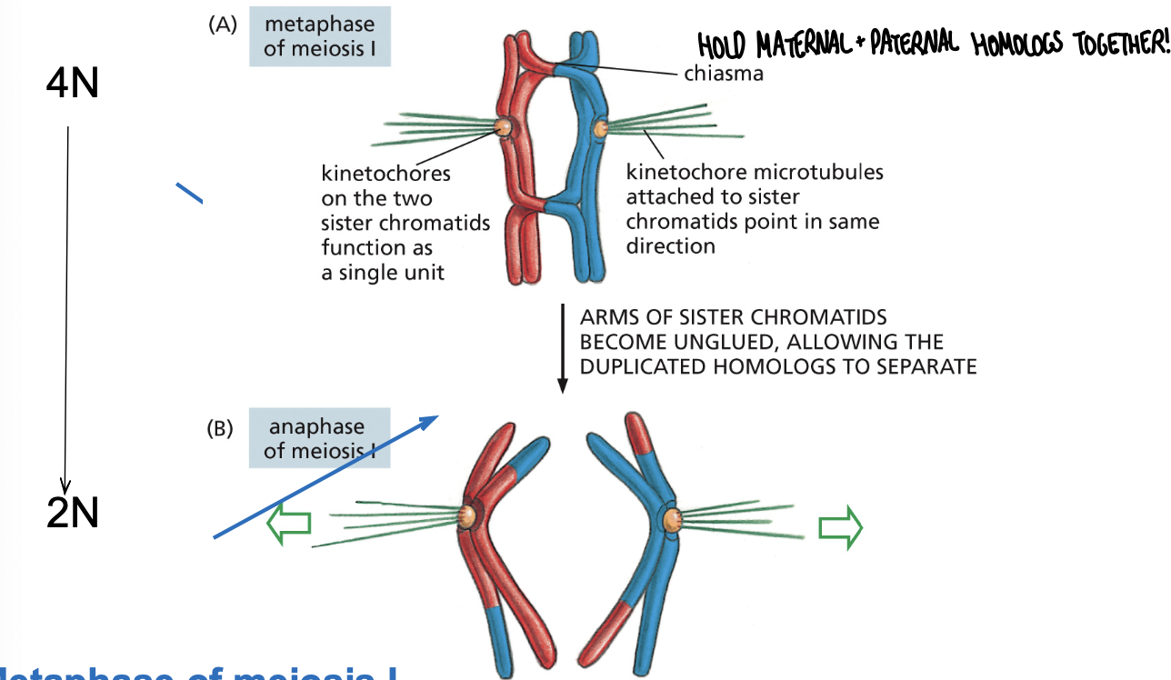
Meiosis I: Separation of homologous chromosomes
Anaphase of meiosis I: Cohesins holding the arms together are degraded allowing homologs to separate, cohesins at the centromeres are not degraded
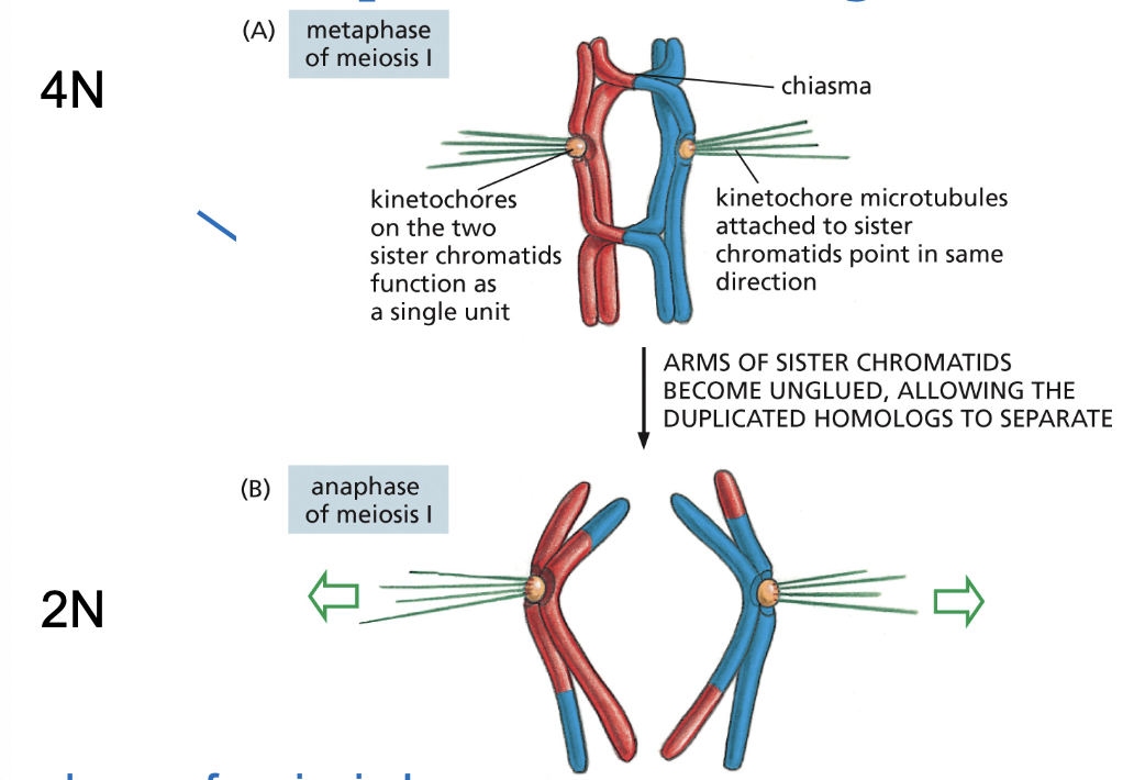
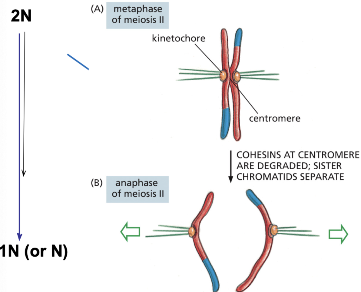
Meiosis II: Separation of sister chromatids
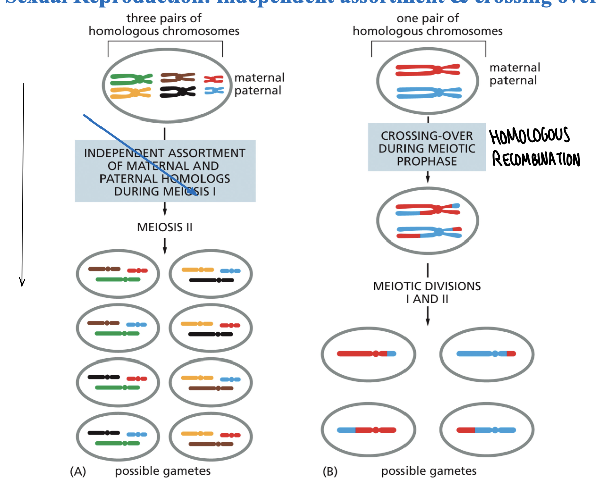
Sexual Reproduction: independent assortment and crossing over
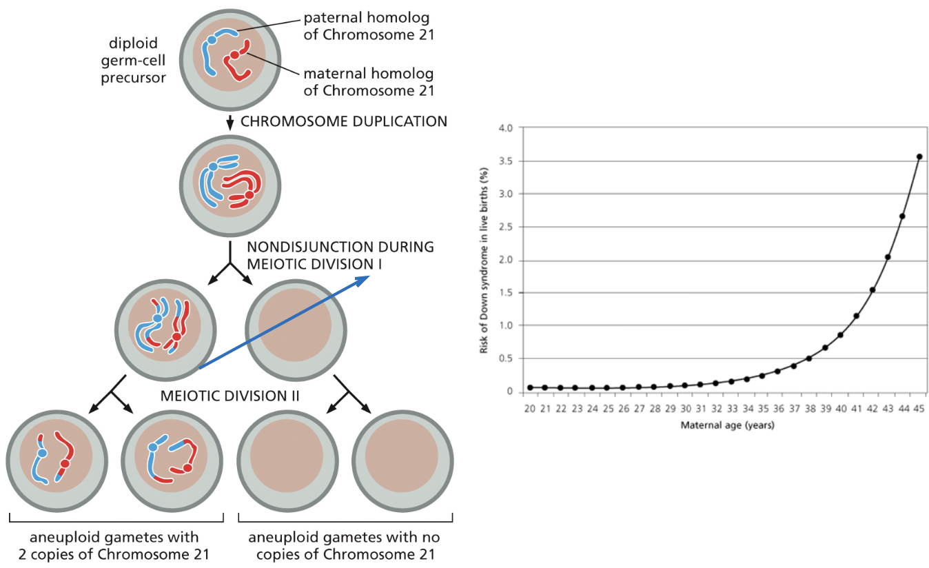
Abnormal chromosome segregation leads to aneuploid gametes
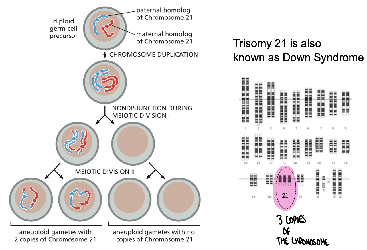
Abnormal chromosome segregation leads to aneuploid gametes
Genetics definitions (genotype, phenotype, wild-type, variants, mutants)
Genotype-specific set of alleles forming the genome of an individual
phenotype-visible trait, what we can see
wild-type-most common allelic form in a population
variants-alleles different in DNA sequence but showing wild type phenotype
Mutants-alleles with DNA sequence change that changes phenotype
Pathogenic sources of genetic variation
chromosomal abnormalities (inversions, deletions, duplications, rearrangements)
single gene mutations (RAS)
copy number variations
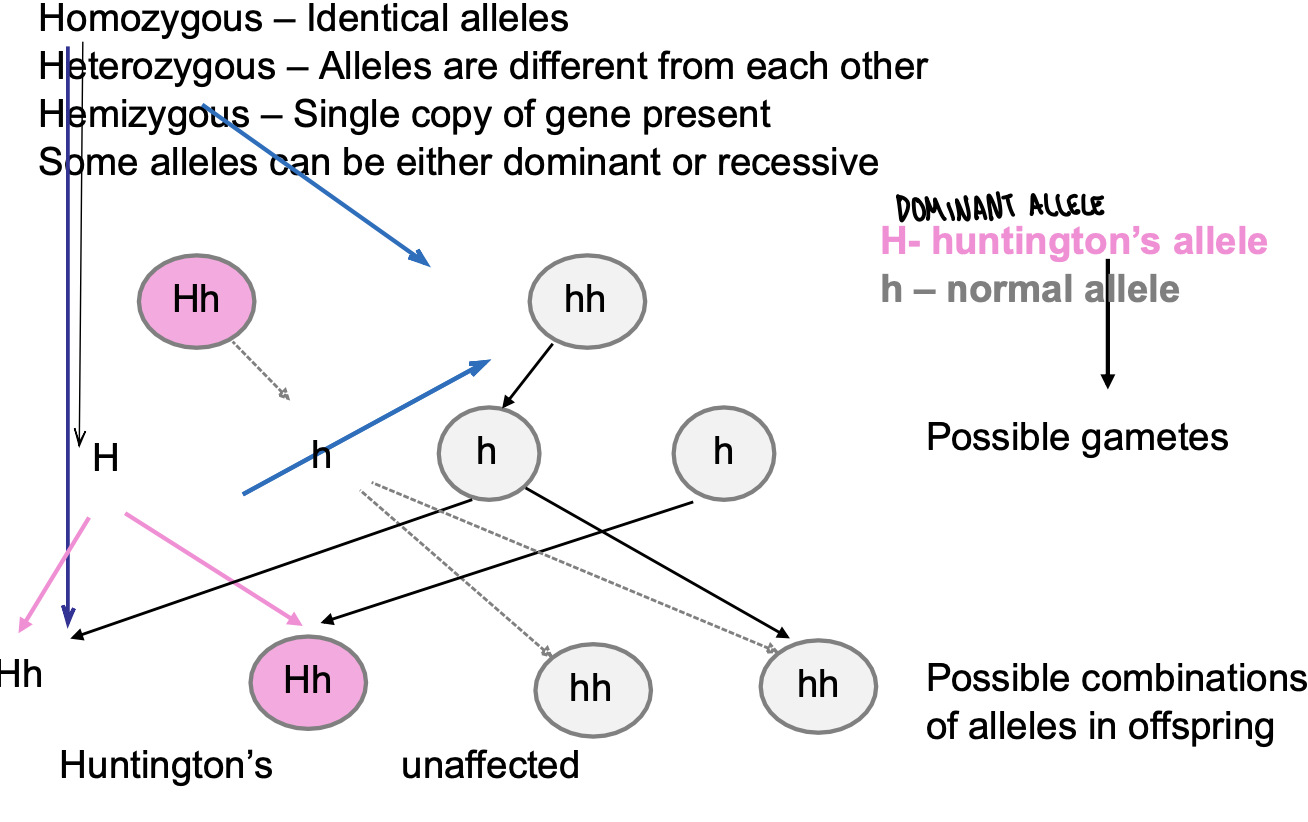
Types of alleles and what this means in terms of disease inheritance
homozygous-identical alleles
heterozygous-alleles are different from each other
hemizygous-single copy of gene present
some alleles can be either dominant or recessive
H-huntington’s allele
h-normal allele
possible gametes
possible combinations of alleles in offspring
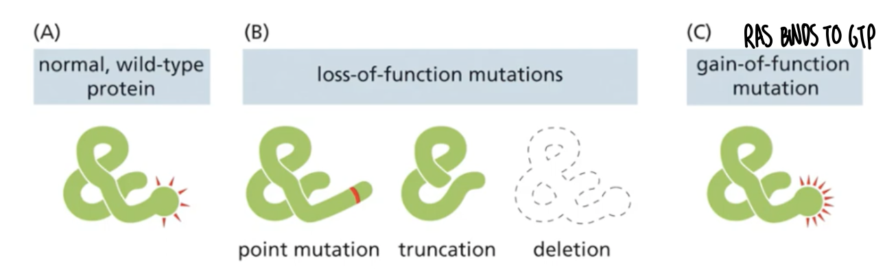
Mutations in protein coding genes can affect protein production in different manners
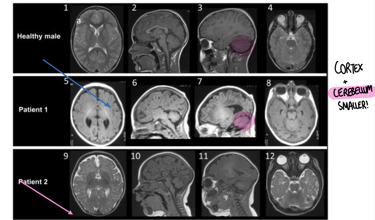
Human genetics in the study of brain disorders CDK5RAP2 mutations lead to reduced brain size
a single mutation of a recessive allele does not cause the disease
consanguineous marriage results in offspring inherits both recessive alleles
CDK5RAP2 is a centrosomal protein
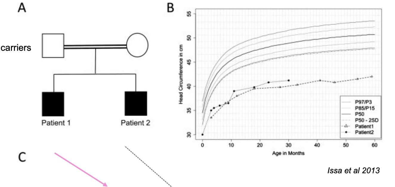
MRI OF CDK5RAP2 patients and healthy control
What type of genotype do these patients have?
Why are there brains so small?
How do we identify mutations in humans?
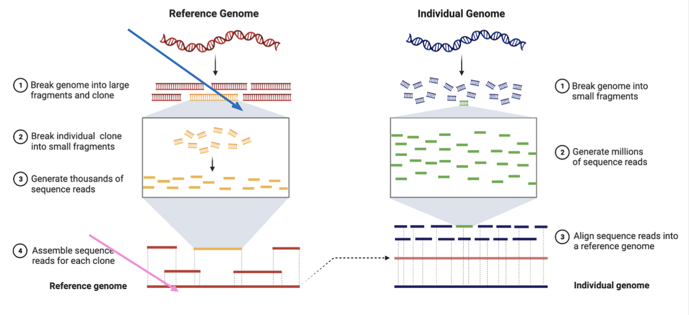
whole genome sequencing
How do we identify mutations in humans?
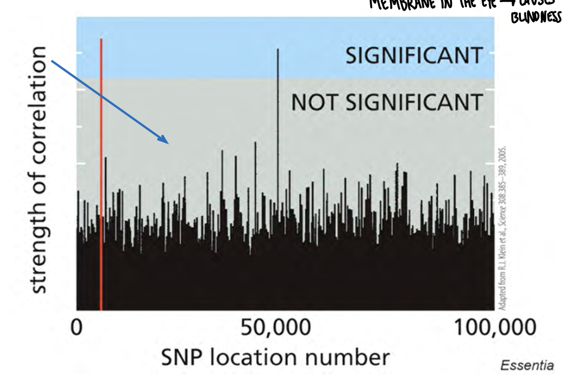
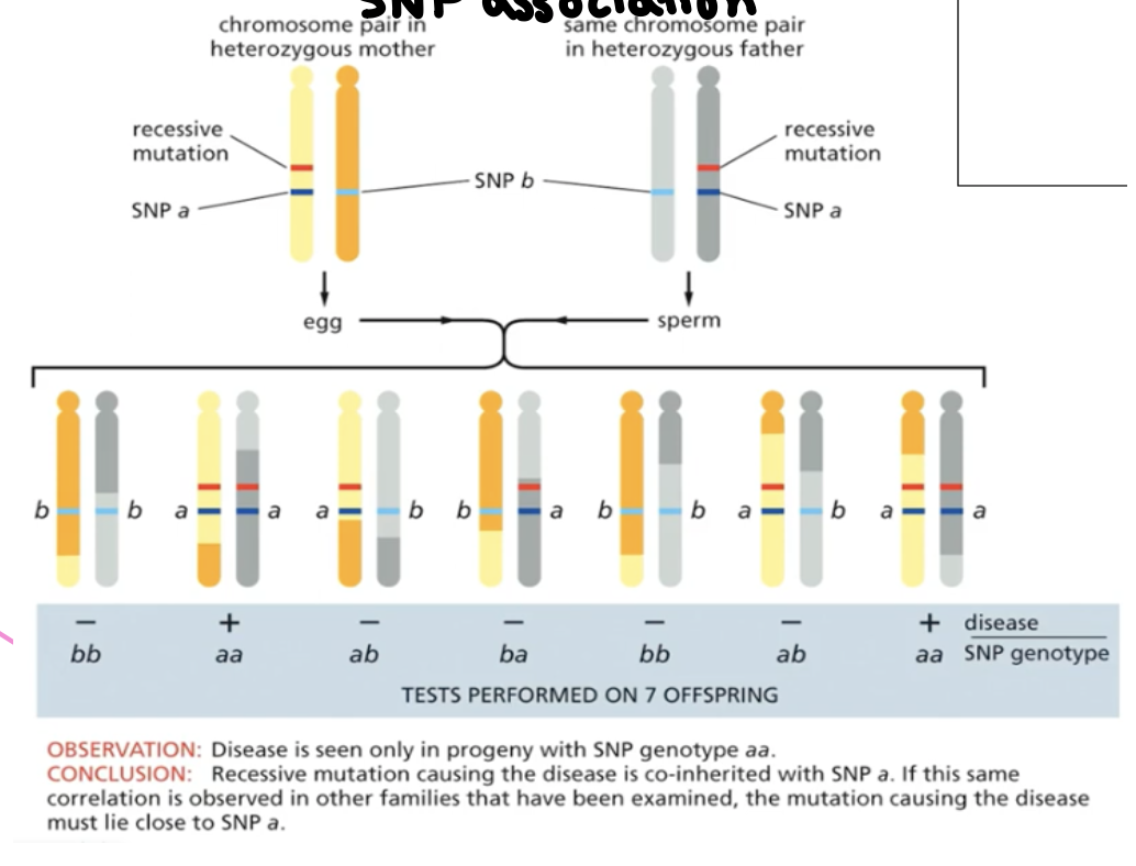
SNP association
SNP-single nucleotide polymorphism (can lead to area with the gene associated with the disease!)
Genome wide association studies in macular (membrane in the eye—>causes blindness) degeneration
identification of DNA variations associated with macular degeneration