Pathology Session #2 SLO and PowerPoint✅
 Call Kai
 Learn
Learn Practice Test
 Spaced Repetition
 Match
Match Flashcards
 Knowt Play
1/152

There's no tags or description
Looks like no tags are added yet.
Name | Mastery | Learn | Test | Matching | Spaced | Call with Kai | Chat |
|---|
No analytics yet
Send a link to your students to track their progress
153 Terms
injury
result of an alteration in the environment that causes tissue damage or necrosis. Less severe _______ may result in reversible responses such as hyperplasia, hypertrophy, and atrophy
Inflammation
_____________ allows the body to eliminate injurious agents, contain injuries and heal defects.
nonspecific
inflammation is __________ meaning it occurs in the same manner regardless of the injury
local
limited to the area of injury
systemic
involves the whole body
acute, chronic
inflammation can be ______ or _______
physical barrier, mechanical defense, antibacterial activity, removal of foreign substances, inflammation process
what are the innate defenses that protect our body?
redness and heat, swelling, pain, loss of normal tissue function
LOCALIZED Cardinal signs of inflammation?
fever, leukocytosis, lymphadenopathy, Elevated C-reactive protein CRP
SYSTEMIC cardinal signs of inflammation?
injury to tissue
what is the first part of the inflammation sequence of microscopic events?

constriction of small vessels in microcirculation (to stop the bleed)
what is the second part of the inflammation response following injury to tissue?
dilation of microcirculation, hyperemia, redness, heat
what is the third part of the inflammation response following constriction of small blood vessels of microcirculation?
hyperemia
increased blood flow
increase in permeability, vessels become leaky
what is the fourth part of the inflammation response following dilation of microcirculation, increased blood flow, redness and heat?
exudate leaves microcirculation and becomes transudate
what is the fifth part of the inflammation response following increase in permeability of vessels as they become leaky?
increased blood viscosity
what is the sixth part of the inflammation response following exudate leaves microcirculation and becomes transudate?
decreased blood flow
what is the seventh part of the inflammation response following increased blood viscosity?
margination of WBC's to the periphery (pavementing of WBC's and chemotaxis)
what is the eighth part of the inflammation response following decreased blood flow?
emigration, exudate, edema
following pavementIng and chemotaxis of the WBC's, the WBC's enter the injured tissue through _______ and cause ________ and _________
phagocytosis
white blood cells then eat the foreign mateiral through ___________?
hyperemia, redness and heat
constriction and dilation causes ________. Which causes?
exudate
fluid that flows into injured tissue after emigration. It contains cells and protein for injured tissue and helps dilute injurious agents, but results in edema
serous exudate
clear watery exudate that is mainly plasma fluids, proteins, and a few WBC's
purulent exudate
exudate containing plasma fluids and proteins, tissue debris, and many WBC's. Ex. abscess
emigration
the process by which WBC's escape from blood vessels through gaps in the endothelial cells.
pagocytosis
process by which WBC's ingest and then digest foreign substances
chemotaxis
directed movement of WBC's toward the site of injury
leukocytes
WBC's
Neutrophils (polymorphonuclear leukocytes)
phagocytotic cells with a multilobed nucleus and granular cytoplasm that contains lysosomal enzymes. Constitute 60-70% of the WBC population. Derive from stem cells in bone marrow
monocytes (blood), macrophages (tissue)
phagocytic cells with a single round nuclear that DO NOT have a granular cytoplasm. They constitute 3-8% of the WBC population and are derived from stem cells in bone marrow
Kinin System
biochemical mediator that is active in early phases of inflammation and is activated by substances in plasma and injured tissue. Causes increased dilation of blood vessels at the site of injury permeability of local blood vessels and induces pain
clotting mechanism
biochemical mediator that clots blood and mediates inflammation.
compliment system
biochemical mediator that produces a cascade of plasma protein. They are present in blood in an inactive form and are triggered by an antigen antibody complex.
mast cells
what cells release histamine
permeability, vasodilation
histamine causes an increase in vascular __________ and ______________
endotoxin
released by pathogenic microorganisms (produced by cell walls of gram negative bacteria) serves as a chemotactic factor; cam activate complement function as an antigen and damage bone and tissue
prostoglandins
biochemical mediator release by the body that increased vascular dilation and permeability tissue pain redness and changes in connective tissue
fever
systemic sign of inflammation that is caused by pyrogens that act on the hypothalamus.
pyrogens
fever producing substances produced by WBC's and pathogens that act on the hypothalamus
hypothalamus
the ______ increases body temperature
leukocytosis
increase in the number of white blood cells
Elevated C-reactive protein
-produced in the liver
-interacts with clotting and complement system
-levels can be used to help assess RA and SLE.
-Used to monitor healing
-used as early infection detection
-chronically increased level is associated with cardiovascular disease and may be a possible marker for periodontal disease
lymphadenopathy
enlarged lymph nodes due to changes in lymphocytes

viral infection
in a _______________ there is an increase in lymphocytes
bacterial infection
in a _______________ there is an increase in neutrophils
allergic reaction
in an ___________there is an increase in eosinophils
hyperplasia
an increase in the number of cells
hypertrophy
enlargement of individual cells
chronic inflammation
__________________ is caused by persistent injuries. Repair cannot be completed until the source of injury is removed.
granuloma
distinct form of chronic inflammation characterized by the formation of a _________________. which are microscopic groupings of macrophages surrounded by lymphocytes and plasma cells.
-group together to for multinucleated GIANT cells.
-associated with TB and foreign body reaction
atrophy
a decrease in size or function of a cell, tissue, organ, or entire body
repair
the restoration of damaged or diseased tissue
Day of injury
blood flows into injured tissue to produce a clot. what day is this?
regeneration
the process by which injured tissue is replaced with tissue identical to that present before the injury
fibrin, clumped RBC's, platelets
clots consist of?
day after injury
the ________________, neutrophils emigrate from the microcirculation into injured tissue in an acute inflammatory response and phagocytosis occurs
two days after injury
____________________
-monocytes change to macrophages in tissue
neutrophils are reduced in number
-granulation tissue is formed
blood clot acts as a scaffold for new CT
lymphocytes and plasma cells migrate to the area as chronic inflammation and the immune response begins
two weeks after injury
________________________
-initial granulation tissue and fibers have been remodeled
-matured fibrous CT is called scar tissue. it is white and laer due to increased collagen and decreased vascularity
healing by primary intention
type of repair:
healing of an injury in which there is little loss of tissue. The margins are close together and very little granulation tissue forms?
healing by secondary intention
type of repair:
the edges of the injury cannot be joined during healing. A large clot forms resulting in excess granulation tissue. May result in excess scar tissue (keloid)
osteoblasts
cells that create new bone tissue
factors delaying bone formation
blood supply at site, growth factors, edema, injury, infection
bruxism
grinding and clenching of teeth for nonfunction purposes
ex. stress, occlusal interferences, stress, tension, seizure disorders

factors influencing repair of bone
nutrition, age, tobacco use
attrition
tooth to tooth wear present in permanent and primary dentition. Bruxism can make teeth wear faster

signs and symptoms of bruxism
wear facets, abnormal rate of attrition, hypertrophy of masticary muscles, increased muscle tone, muscle tenderness, fatigue, cheek biting, pain in temporomandibular joint, tooth mobility, pulpal sensitivity to cold
Abrasion
pathological wearing away of tooth structure that results from repetitive mechanical habit. (notching on root surface accompanied with gingival recession)

abfraction
wedge shaped lesion at the cervical areas caused by microfractures of tooth structures in areas of concentrations of stress. may be related to fatigue, flexure, fracture, and deformation of tooth structure

bullimia
eating disorder characterized by food binges followed by induced vomiting. irritation of oral mucosa and lips.
traumatic lesions on backs of fingers

erosion
loss of tooth structure as a result of chemicals without bacterial involvement. tooth structure may be lost around a restoration making it stand out. Ask about patients history

erosion potential causes
Industrial factors
Intraorally applied cocaine hydrochloride drug abuse Overuse of soft drinks
Baby bottle caries
Sucking on lemon
Chronic vomiting
meth mouth
rapid destruction of teeth as a result of methamphetamine acid content, decreased salivary flow, cravings for high sugar, and lack of oral hygiene.

Aspirin burn
Caused by topical application of aspirin (misuse of product) tissue becomes necrotic and white the surface may slough off leaving a painful ulcer. Ulcer heals in 7-21 days.

Phenol burn
Used in dentistry as a cavity sterilizing agent and a cauterizing agent. Will cause whitening and sloughing of the area as a result of tissue destruction

Electric burn
May be seen in infants or young children who have chewed an electrical cord
May be quite extensive, damaging oral tissue and even tooth buds
May cause permanent disfigurement and scarring
Treatment
Plastic surgery
Oral surgery
Orthodontic therapy
Thermal burn
Hot food burns, Tx: therapeutic, agents that contain hydrogen peroxide or eugenol

Crack cocaine lesions
Lesions located on the midline of the hard palate may vary from ulcers to keratotic lesions to exophytic reactive lesions as a result of smoking crack cocaine

traumatic granuloma
The result of persistent trauma. Appears hard (indurated) and raised. Heals rapidly post biopsy

Self induced lesions
Chronic lip, cheek or tongue biting. Trauma to gingiva from fingernail Lesions may range from ulcer to epithelial hyperplasia and hyperkeratosis

Hematoma
Accumulation of blood trapped within tissue as a result of trauma. Appears as a red to purple to blush gray mass. Frequently seen on labial or buccal mucosa.

Frictional keratosis
Form of hyperkeratosis caused by chronic rubbing or friction against an oral mucosal. Resembles a callus on skin.
TX: identify and eliminate cause of lesion
CAN LOOK LIKE IDIOPATHIC LEUKOPLAKIA!!!!
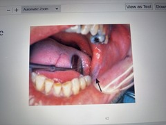
Nicotine stomatitis
Benign lesion associated with pipe/cigar smoking, or cigarette smoking. Initially appears: erythema
Keratinization occurs causing opacity.
Red raised areas occur at the openings of ducts of inflamed minor salivary glands.
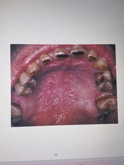
Smokeless tobacco keratosis
White lesion located where chewing tobacco is placed, most often the mucobuccal fold
Early lesions: granular wrinkled appearance
Long standing lesions: more opaquely white with corrugated surface

Traumatic ulcer
Caused by: Cheek lip or tongue biting, denture irritation, mucosal injury, or overzealous brushing
TX: heals within 7-14 days unless trauma persists.
May require biopsy
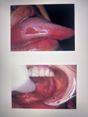
Tobacco pouch keratosis
Wrinkled, Corrugated, White lesion at site of tobacco placement. Long term exposure to _______ has been associated with increased risk of squamous cell carcinoma.
Tx: stop using ______, may need biopsy

Electronic cigarettes (e-cigarettes)
Can be damaging to oral tissues as regular smoking. Produces an aerosol or vapor to bring nicotine to the lungs. Vapor contains toxic chemicals and heavy metals
Traumatic neuroma
A lesion caused by an injury to a peripheral nerve.
The nerve sheath of Shwann cells is disrupted, occasionally the proximal end of the damaged nerve proliferates into a mass of nerve and Schwann cells mixed with dense fibrous scar tissue.
Diagnosis: need a biopsy and microscopic examination
TX: surgical excision

Palisaded Encapsulated Neuroma (PEN)
Benign lesion that appears clinically as a mucosal nodule. Under microscope, it is well circumscribed lesion that is composed of nerve tissue partially surrounded by fibrous CT.
Considered a reactive hyperplastic lesion
Amalgam tattoo
A flat bluish-gray lesion of the oral mucosa that results from the introduction of amalgam particles into the tissues.
Can occur during placement/removal of amalgam. Or during EXT. most commonly on gingiva or alveolar ridge
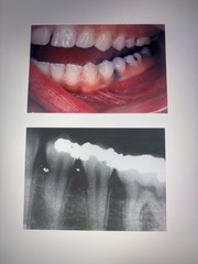
Malignant melanoma
Amalgam tattoo appears as a __________. So it is best to ensure pt history and a radiograph to diagnose an amalgam tattoo
melanosis
Normal physiologic pigmentation of oral mucosa. Can be genetic. May occur as a result of inflammation. If presenting as a macule, a biopsy is needed!

Solar Cheilitis (Actinic Cheilitis)
A degeneration of the tissue of the lips caused by exposure to the sun. Lips appear dry and cracked. Vermillion appears pale pink and indistinct. Microscopically, the epithelium is thinner than normal due to degenerative CT changes.
Smoking and alcohol use increases the risk for squamous cell carcinoma.
TX: avoid sun exposure, wear sunscreen

Mucous Retention Lesion: Mucocele
• A lesion formed when a salivary gland duct is *severed*; the mucous salivary gland secretion spills into the adjacent CT
• Not a true cyst because it is not lined with epithelium

Mucous retention lesion: mucous retention cyst
Dilated salivary gland ducts that developed as a result of duct obstruction.
TX: removal of affected minor salivary gland

Mucous retention lesion: ranula
A unilateral mucocele-like lesion that forms on the floor of the mouth
Associated with the ducts of submandibular and sublingual glands

sialolith
stone in the salivary gland. Can be found in both major and minor salivary glands. Formed by calcium salts around central core. Can be seen on radiographs (dense)
TX: sour candy to "milk" or surgical removal (risk damage)

Necrotizing Sialometaplasia
A benign condition of the salivary glands characterized by moderately painful swelling and ulceration in the affected area. Thought the be a result from low/blocked blood supply to the area. Salivary epithelium is replaced by squamous epithelium. Ulcer typically heals by secondary intentions.
Biopsy is needed to establish diagnosis

Acute and Chronic Sialadenitis
may occur as a result of obstruction of a slaivary gland duct or result of infection; presents as painful swelling and confirmed by radiograph of gland with radiopaque dye injected into it
reactive connective tissue hyperplasia
proliferating, exuberant granulation tissue and dense fibrous connective tissue that results from overzealous repair
May be a response to a single event or chronic low grade injury
Pyogenic granuloma
A proliferation of connective tissue containing numerous blood vessels and inflammatory cells occurring as a response to injury.
Name is a misnomer: this lesion is neither pyogenic NOR a true granuloma!
