Biopsychology
1/106
There's no tags or description
Looks like no tags are added yet.
Name | Mastery | Learn | Test | Matching | Spaced | Call with Kai |
|---|
No analytics yet
Send a link to your students to track their progress
107 Terms
assumes behaviour and experiences are caused by activity in the nervous system
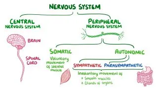
two main functions:
1) control of physical movement
2) regulation of homeostatic function
the brain receives information via sense organs and sends messages to the body using nerve cells in the spine
cerebellum
brain stem
diencephalon
divided into two cerebral hemispheres
involved in controlling motor skills, balance and coordinating muscles to allow for precise movements
motor and sensory neurons travel through the brain stem - allows impulses to travel between the brain and spinal cord
contains the thalamus and hypothalamus
the thalamus acts as a relay for incoming nerve impulses - directs them to the appropriate parts of the brain where they can be processes
the hypothalamus has functions like regulation of body temperature, hunger and thirst, and acts as a link between the endocrine system and nervous system
spinal nerves connect to specific muscles and glands
contains circuits of nerve cells that enable simple reflexes without direct involvement from the brain
relays impulses from the CNS to the rest of the body and back
made up of the somatic and autonomic nervous systems
somatic nerves have both sensory and motor neurons - sensory neurons relay messages to the CNS, motor neurons relay information from the CNS to other areas of the body
involved in reflex actions without the involvement of the CNS - allows reflexes to occur quickly
has two parts:
1) sympathetic nervous system
2) parasympathetic nervous system
both systems tend to regulate the same organs but have the opposite effects - due to the neurotransmitters associated with each system
sympathetic nervous systems generally use the neurotransmitter noradrenaline - it uses acetylcholine, which has inhibiting effects
neurons from the sympathetic nervous system travel to almost every organ and gland, preparing the body for rapid action
involved with energy conservation and digestion - called the rest and digest system

what people think and feel are caused by electrochemical events occurring within and between neurons
three main types: sensory, relay, motor
axons
myelin sheath
synaptic terminal
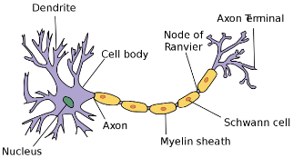
typically connected to the cell body
transmit away from the cell body towards the axon terminals
insulates the axon so electrical impulses travel faster along the axon
carry nerve impulses from the PNS to CNS
when the nerve impulses reach the brain they are translated to ‘sensations’
not all sensory neurons reach the brain - some stop at the spinal cord
have long dendrites and short axons
in the CNS
allow sensory and motor neurons to communicate
have short dendrites, long axons and no myelin sheath
control muscle movements and carry messages from the CNS to muscles and glands
release neurotransmitters that bind to receptors muscles to trigger a response and cause movement
the message reaches the CNS and connects to a relay neuron, and is then transferred to a motor neuron
it is then carries to an effector, which causes a muscle to contract
when a neuron is activated by a stimulus, the inside of the cell becomes positively charged - action potential
it creates the electrical impulses which travel through neurons
affects the transfer of an impulse to another nerve or muscle
‘taken back up’ into the terminal buttons of neurons - reuptake
can only travel in one direction at the synapse
2) synaptic vesicles at the axon terminals contain neurotransmitters
3) when action potential reaches the vesicles, they release their neurotransmitters into the synapse
4) neurotransmitters carry the signal across the synaptic gap
5) bind to receptor sites on the postsynaptic neurons - become activated and produces excitatory or inhibitory effects on the postsynaptic neuron
causes a positive electrical charge in the cell membrane - results in an excitatory postsynaptic potential (EPSP)
makes the postsynaptic cell more likely to fire
results in an inhibitory postsynaptic potential (IPSP)
makes the postsynaptic cell less likely to fire
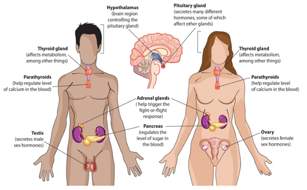
uses blood vessels to deliver hormones to target sites in the body
supplements the work of the nervous system - works with it to regulate the physiological processes of the body
regulated by feedback:
1) a signal is send from the hypothalamus to the pituitary gland - ‘releasing hormone’
2) this causes the pituitary gland to secrete a ‘stimulating hormone’ into the bloodstream
3) this hormone then signals the target gland to secrete its hormone
4) as the levels of this hormone rise in the bloodstream, the hypothalamus shuts down the secretion of the ‘stimulating hormone’
5) this slows the secretion of the target gland’s hormone - results in stable concentrations of hormones circulating in the bloodstream
affects target cells - cells with receptors for that hormone
when enough receptor cells are stimulated, there is a physiological reaction in the cell
timing of hormone release is critical for normal functioning, as well as the levels released
most common cause of excess cortisol is a pituitary gland tumour
controlled by the hypothalamus - hypothalamus receives information about basic functions and uses it to regulate the function
‘master gland’ - produces hormones to specific targets, directly causes changes in physiological processes in the body or stimulate other glands to produce hormones
high levels of hormones produced in other glands can stop the hypothalamus and pituitary gland from secreting hormones - negative feedback
two main parts - anterior pituitary and posterior pituitary
secretes luteinising hormone (LT) and follicle-stimulating hormone (FSH) - controls reproductive functioning and sexual characteristics
elabd et al (2014) - research in mice has found oxytocin is indispensable for healthy maintenance and repair, and declines with age
made of two parts:
1) adrenal cortex - outer part of the gland, hormones secreted are essential to life
2) adrenal medulla - inner part of the gland, hormones secreted are not essential to life
cortisol production is increased in response to stress
if cortisol levels are low, the individual will have low blood pressure, poor immune function and an inability to deal with stress
also produces aldosterone - responsible for maintaining blood volume and pressure
adrenaline helps the body respond to stressful situations by increasing heart rate and blood flow to the muscles and brain, and helping with the conversion of glycogen to glucose to provide energy
noradrenaline constricts blood vessels, causing blood pressure to increase
responsible for the production of eggs and the hormones oestrogen and progesterone
maner and miller (2014), progesterone is associated with the heightened sensitivity to social cues that indicate the presence of social opportunity or threat that would be significant during pregnancy
testosterone causes the development of facial hair, deepening of the voice and growth spurts that occur during puberty
testosterone production is controlled by the hypothalamus and the pituitary gland
controlled by the sympathetic nervous system, as it controls the necessary bodily functions needed when we are faced with a situation where we may need to fight or flee
every time a potentially threatening experience occurs, the body reacts - the constant shifting between the two systems keeps the body ready for any situation
the hypothalamus functions like a command centre in the brain - communicates with the rest of the body through the sympathetic nervous system
the type of response depends on the type of stressor
short term
results in the sympatho-adrenal medullary response (SAM)
long term
results in the pituitary-adrenal response (HPA)
2) the hypothalamus is activated
3) the sympathetic branch of the ANS is activated
4) the adrenal medulla is stimulated to enlarge and release adrenaline and noradrenaline into the bloodstream
2) the hypothalamus is activated
3) the pituitary gland is activated - releases ACTH into the bloodstream
4) ACTH causes the adrenal cortex to enlarge and release cortisol - has both positive (e.g. burst of energy, lower sensitivity to pain) and negative (e.g. impaired cognitive performance, lowered immune response) effects on the body
negative consequences - modern stressors rarely require physical activity, repeatedly activating the stress response can be negative, e.g. increased blood pressure can cause heart disease, cortisol suppresses immune response
alternative response - gray (1998), first reaction is to avoid confrontation, ‘freeze response’, adaptive advantage as it focuses attention to choose the best response
positive behaviours in response to stress - von dawans et al (2012), acute stress can lead to cooperative friendly behaviour, e.g. post 9/11, humans are fundamentally social
genetic basis for gender differences - lee and harley (2012), the male SRY gene promotes aggression and causes fight or flight, lack of SRY gene, oestrogen and oxytocin may prevent this response in females
what is localisation of functioning?
the idea that certain functions are localised to certain areas of the brain
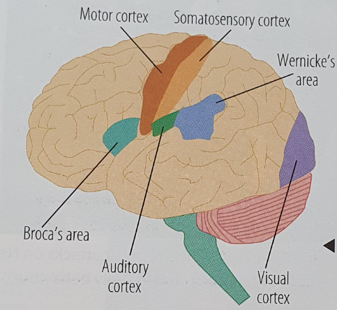
what is the motor cortex?
responsible for the generation of voluntary motor movements
located in the frontal lobe along the precentral gyrus
both hemispheres have a motor cortex - controls movements on the opposite side of the body
different parts of the motor cortex control different parts of the body - the regions are arranged logically next to each other
what is the somatosensory cortex?
detects sensory events arising from different regions of the body
located in the parietal lobe, along the postcentral gyrus - dedicated to the processing of sensory information relating to touch
produces sensations of touch, pressure, pain and temperature using sensory information from the skin
both hemispheres have a somatosensory cortex - receives information from the opposite side of the body
what is the visual centre?
visual processing begins in the retina, where light enters and hits the photoreceptors
nerve impulses from the retina are transmitted to the brain via the optic nerve - some travel to areas of the brain involved in coordination of circadian rhythms, most terminate in the thalamus, which acts as a relay station, passing information to the visual cortex
spans both hemispheres and contains several different areas that all process different types of visual information
what is the auditory centre?
mostly in the temporal lobes on both sides of the brain
auditory pathways begin in the cochlear - sound waves are converted to nerve impulses
the nerve impulses travel first to the brain stem, where decoding takes place and then to the thalamus which acts as a relay station and carries out further processing
by the time it arrives at the auditory cortex, sound is mostly decoded
what is broca’s area?
broca treated a patient named ‘tan’, who could understand language but not express it in writing or speaking
studied eight other patients with similar problems and found they all had similar lesions in their left frontal hemisphere
broca (1865) identified a ‘language centre’ in the posterior portion of the frontal lobe of the left hemisphere
fedorenko et al (2012), one part of broca’s area is selectively involved in language, the other in responding to many demanding cognitive tasks
what is wernicke’s area?
located another area of the brain involved in understanding language in the posterior portion of the left temporal lobe
those who had a lesion there could speak but not understand language
what did wernicke propose about the language centres?
language involves separate motor and sensory regions located in different cortical regions
the motor region (broca’s area) is close to the area that controls the mouth, tongue and vocal cords
the sensory region (wernicke’s area) is close to areas of the brain responsible for auditory and visual input
information is thought to be transferred to wernicke’s area where it is recognised as language and associated with meaning
there is a neural loop known as arcuate fasciculus between broca’s area and wernicke’s area
what is the evaluation for localisation of functioning?
individual differences - variation in areas of the brain activated in different individuals during the same task, women have proportionally larger broca’s and wernicke’s areas
equipotentiality theory - extent not location of damage is what is important in loss of functioning
aphasia patients - provide evidence for broca’s and wernicke’s areas
connections are more important than localisation
language production is not localised to just broca’s area
what is hemispheric lateralisation?
each hemisphere had functional specialisations
neural mechanisms for some functions are only found in one hemisphere
inspired by broca and wernicke’s research
what is the corpus callosum?
a bundle of nerve fibres that connects the two hemispheres
what is split brain research?
split brain patients are individuals who have had their corpus callosum cut - originally a treatment for severe epilepsy
what did sperry and gazzaniga (1967) investigate?
the extent to which the hemispheres are specialised for certain functions
tested the capabilities of separated hemispheres by sending information to one hemisphere at a time
what did sperry and gazzaniga (1967) do?
‘describe what you see’ test - when the picture was processed by the left hemisphere, the patient could describe it, but when it was processed by the right hemisphere, they could not describe it
tactile test - when the object was processed by the left hemisphere, the patient could verbally describe it, but when it was processed by the right hemisphere, they could not describe it
drawing task - when the object was processed by the left hemisphere, the picture drawn was not very clear, but when it was processed by the right hemisphere, the picture drawn was clearer
what did sperry and gazzaniga (1967) conclude?
the left hemisphere is dominant in terms of speech and language
the right hemisphere is dominant in terms of visual-motor tasks
connectivity and communication between the two hemispheres is important for effective functioning
what is the evaluation for lateralisation?
advantages of hemispheric lateralisation - multitasking, e.g. chickens looking for food and predators
split brain research is limited - due to small sample sizes and confounding variables reducing validity and reliability
lateralisation is linked to immune system function
individual differences in lateralisation - changes with age
language may not be confined to the left hemisphere - e.g. JW case study
what is plasticity?
the brain’s ability to change and adapt as a result of experience - plays an important role in brain development and behaviour
recent research suggests the brain creates neural pathways in adulthood as well as childhood
what are the causes of plasticity?
life experiences
video games
meditation
how do life experiences cause plasticity?
nerve pathways that are used frequently form stronger connections and neurons that are rarely used eventually die (synaptic pruning)
there is a natural decline in cognitive functioning with age attributed to brain changes
boyke et al (2008), 60 year olds taught to juggle, increased grey matter in the visual cortex, but changes reversed when practicing stopped
how do video games cause plasticity?
can help complex cognitive and motor skills
kuhn et al (2014), increase in synaptic connections in brain areas involved in spatial navigation, strategic planning, working memory and motor performance in those who play video games everyday
how does meditation cause plasticity?
davidson et al (2004), tibetan monks who meditate had more gamma radiation wave activation than those who do not meditate
concluded that meditation causes short-term and permanent changes in the brain
what is functional recovery after trauma?
in the 1960s, researchers studied who stroke victims regained functioning
when brain cells are damaged, the brain rewires itself so some functioning can be regained - other parts of the brain took over for the functions lost, and neurons next to damaged areas formed new circuits that resumed some of the lost functioning
what are the mechanisms for recovery?
neural unmasking
stem cells
how does neural unmasking help recovery after trauma?
wall (1977), ‘dormant synapses’, function is blocked as rate of neural input is too low, but increases in rate of neural input can ‘unmask’ them
can open connections to regions of the brain not normally activated, leading to development of new structures
how do stem cells help recovery after trauma?
stem cells implanted into the brain could directly replace dead or dying cells
transplanted stem cells could secrete growth factors that could ‘rescue’ injured cells
transplanted cells form a neural network that could links uninjured brain sites to damages regions of the brain
what is the evaluation for plasticity?
animal studies - kempermann et al (1998), rats and enriching environments, more neurons in the hippocampus, have to consider limitations of animal studies
human studies - maguire et al (2000), taxi drivers, complex memory training can change the brain
what is the evaluation for recovery after trauma?
animal studies - tajiri et al (2013), stem cells can help regain brain functioning in rats, have to consider limitations of animal studies
age differences - elbert et al (2001), children have a greater capacity for neural reorganisation than adults, increased age makes it harder to make and maintain brain changes
educational attainment - schneider et al (2014), patients with college level education recover quicker from brain injuries, education encourages development of neural pathways
what are the four methods of studying the brain?
post mortem examinations
functional magnetic resonance imaging (fMRI)
electroencephalogram (EEG)
event related potentials (ERP)
what is a post mortem examination?
used to establish underlying neurobiology of particular behaviours
e.g. broca’s work, HM and hippocampus (memory)
established links between psychiatric disorders and brain abnormalities - e.g. cotter et al (2001), there are reduced numbers of glial cells in the frontal cortex of patients with depression
what is functional magnetic resonance imaging (fMRI)?
measures changes in blood flow in particular areas of the brain, which indicates neural activity - active areas of the brain have an increased demand for oxygen, leading to increased blood flow
maps can be produced showing areas of the brain involved in different processes
what are electroencephalograms (EEG)?
measures electrical activity - small electrodes detect small electrical charges resulting from brain cell activity, which are mapped over time to produce EEG data
EEG data is used to detect brain disorders - e.g. epilepsy shows spikes of electrical activity
there are four basic EEG patterns:
1) alpha waves
2) beta waves
3) delta waves
4) theta waves
what are event related potentials (ERP)?
small voltage changes in the brain triggered by stimuli
can be difficult to pick out from other brain activity, so target stimulus is repeated and results are averaged - extraneous neural activity will not occur consistently
there are two categories of ERPs:
1) ‘sensory’ ERPs
2) ‘cognitive’ ERPs
what is the evaluation for studying the brain?
post mortem - detailed anatomical examination, harrison et al (2000), post mortem played a central role in understanding origins or schizophrenia, time, matter of death, length of time from death to post mortem all have an impact, only retrospective
fMRI - non-invasive, no harmful radiation, objective, reliable, not a direct measure as it measures blood flow, overlooks networked nature of brain activity
EEG - provides real time recordings, accurate, useful in clinical diagnoses, cannot reveal deeper regions without implanted electrodes which are not allowed due to ethical implications, difficult to identify exact source of activity
ERP - continuous measure, measures in absence of behavioural responses, requires large number of trials for meaningful data, only strong voltage changes are recordable
what are biological rhythms?
cyclic changes in the way biological systems behave
evolved as the environment has cyclic changes, e.g seasons, day and night - nearly all organisms have biological representations of the 24 hour day
what are circadian rhythms?
biological rhythms that are 24 hours
optimise an organism’s physiology to best meet the varying demands of the day/night cycle
what are circadian rhythms controlled by?
driven by body clocks, which are synchronised by the suprachiasmatic nucleus (SCN) located in the hypothalamus
the SCN is constantly reset, and its primary input is light - photoentrainment
photoreceptors in the eyes send messages about environmental light levels to the SCN - these are used to coordinate activity in the circadian system
what is the sleep-wake cycle?
circadian rhythms determine when sleep and wake periods - maintains a 24-25 hour cycle without external cues
dips and rises - the strongest sleep drives occur from 2-4 am, and 1-3 pm
how does homeostasis affect the sleep-wake cycle?
sleep-wake cycle is also under homeostatic control
as the amount of wake time increases, homeostasis indicates need for sleep is increasing as energy is used up - homeostatic need for sleep increases gradually during the day and peaks in the late eventing
the circadian rhythm keeps us awake during the day and prompts us to sleep when it is dark, and the homeostatic system makes us sleepier as time goes on during the waking period, whether it is night or day
what are the other circadian rhythms?
core body temperature - sleep occurs when core temperature starts to drop, and temperature rises during the last sleep hours, causing alertness
hormone production - melatonin, from the pineal gland, follows a circadian rhythms with peak levels during dark hours
what is the evaluation for circadian rhythms?
research support - hughes (1977), antarctic study, different light levels affected levels of cortisol, other arctic studies found no disruption
individual differences - czeisler et al (1999), length of cycle varies from 13-65 hours, duffy et al (2001) found differences in cycle onset
research methodology - in many studies, participants are not isolated from artificial light, czeisler et al (1999) altered circadian rhythm using dim artificial light
chronotherapeutics - evans and marain (1996), new drug delivery system where they were administered in the evening but not released until dawn
importance of temperature - buhr et al (2010), temperature controls body clock, fluctuates on a 24 hour cycle
what are ultradian rhythms?
rhythms shorter than 24 hours
e.g. sleep stages, basic rest activity cycle
what are the sleep stages?
pattern of alternating REM and NREM sleep - cycle repeats every 90 minutes, different stages have different durations
most knowledge of sleep stages comes EEG recordings of sleep - during deep sleep, brain waves slow, and breathing and heart rate decrease, and during REM, EEG patterns look like awake patterns, and most dreaming occurs in this sleep
what is the basic rest activity cycle (BRAC)?
kleitman (1969) called the 90 minute sleep cycle the BRAC - suggested it occurs during the day, moving from states of alertness to physiological fatigue
we can only focus for 90 minutes - at the end of this the body runs out of resources, resulting in loss of concentration, fatigue and hunger
what are infradian rhythms?
rhythms longer than 24 hours - may last weeks, months, or be annual
e.g. menstrual cycle, organisation of human activities into weeks
what are weekly infradian rhythms?
there are differences in human behaviour that occur on a weekly cycle - e.g. male testosterone is elevated at the weekend, young couples report more sexual activity at the weekend, frequency of births at weekends is higher than weekdays
there are underlying biological cycles that dictate this - halberg et al (2002), seven day rhythms of blood pressure and heart rate, but evidence for this is weak
what are monthly infradian rhythms?
menstrual cycle - variations in length, from 23-36 days (refinetti, 2006), average is 28 days
menstrual cycle is regulated by hormones that promote ovulation or stimulate the uterus for fertilisation
what are annual infradian rhythms?
magnusson (2000) said there is seasonal variation in mood, especially in women - seasonal affective disorder, some become depressed in the winter
there are more heart attacks during the winter
trudeau (1997) - most human deaths occur in january
what is the evaluation for ultradian and infradian rhythms?
individual differences - tucker et al (2007), differences in sleep patterns are biological
research support for BRAC - ericsson et al (2006) found elite violinists practice for only 90 minutes and napped after, also found these patterns in athletes and writers
menstrual cycle and exogenous cues - russell et al (1980), odour donor study, suggests synchronisation of menstrual cycles is affected by pheromones
menstrual cycle and mater preference - penton-voale et al (1999), human mate choice varies across the menstrual cycle
belief in lunar rhythms - arliss et al (2005) found midwives report more babies are born on the full moon, vance (1995) found some mental health professionals believe the full moon affects behaviour, foster and roenneberg (2008), some studies found correlations, but no evidence of causal relationships