3. Cardiovascular system: Anatomy of the Cardiovascular and Pulmonary Systems_condensed
1/17
There's no tags or description
Looks like no tags are added yet.
Name | Mastery | Learn | Test | Matching | Spaced | Call with Kai |
|---|
No analytics yet
Send a link to your students to track their progress
18 Terms
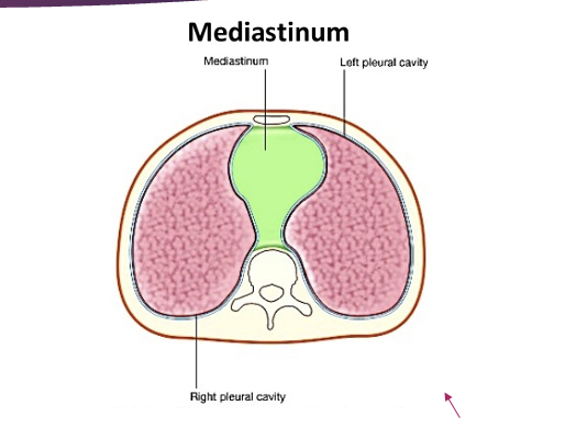
Mediasteinum
lies between the right and left plerura of the lungs
contains all the thoracic viscera except the lungs
(heart, esophagus, trachea, phrenic nerve, cardiac nerve, thoracic duct, thymus and lymph nodes of the central chest
Heart (where does it lie, superior part and where, inferior part and where)
lies within the mediastinum, with 2/3 lying left of the midsagittal plane
the superior portion of the heart, formed by the two atria, is considered the base
located at 2nd intercostal space
the inferior portion of the heart, apex, is defined by the top of the left venticle
located at the 5th intercostal space
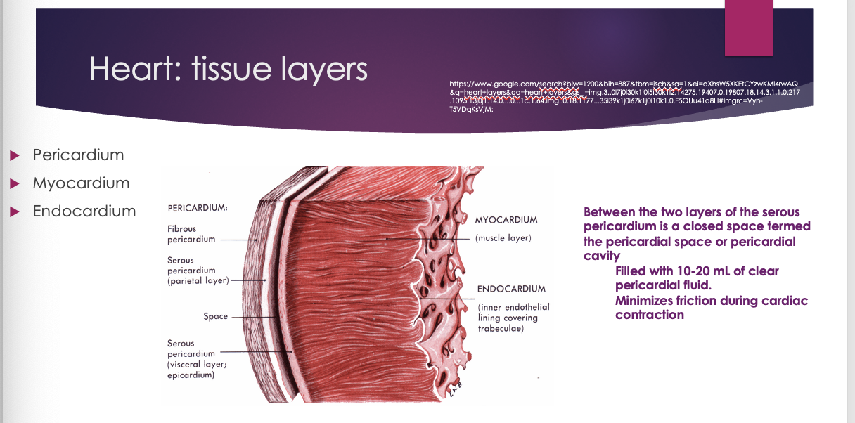
heart tissue layers
pericardium
myocardium
endocardium
between the two layers of the serous pericardium is a closed space termed the pericardial space or pericardial cavity
filled with 10-20 mL of clear pericardial fluid. minimizes friction during cardiac contraction
The space just like the lungs has fluid to prevent pericardial friction rub which we can hear with ausclultation
If there is too much fluid it causes increased pressure on the heart and it gets irritated, pissed off, and causes a skipped beat
Tissue Layers: Myocardium (three traits, categorized into two groups based on function)
Automaticity: ability to contract in the absence of a stimuli
Rhythmicity: ability to contract in a rhythmic manner
Conductivity: ability to transmit nerve impulse
Categorized into two groups based on function
Mechanical cells: (Myocytes) contraction properties
Conductive cells: electrical conduction properties. Cells joined by intercalated disks forming a structure known as syncytium
Intercalated disks contain two junctions: (1) Desmosomes, attaching one cell to another and (2) Connexins, allow the electrical flow to spread from one cell to another
______________________________________________
Automaticity: heart cell can beat on its self without being told: con-can have an arrythmia (they can beat abnormally at any time – they can do this when they are deprived of oxygen); pro- escape beats (if dr. C drops one scape beat may restart his heart)
What causes a lack of oxygen to the heart muscle – coronary artery is blocked –drinking caffeine, cocaine (any stimulant))
Rhythmacity: as one ecll gets the signal they talk to other cells and they get on same page – what we don’t want is them to beat at different rates
Conductivity: able to transmit and communicate to neighboring cells
These three traits allow myocardium to perform ideally in the perfect world. The problem with that is that if one cell decided to beat on its own unrhythmicaly, and you tell the cell next to you to do the same and because of conductivity they spread the word; then there is a problem and all the muscle cells are beating at different times which may not give a single contraction which leads to having no output
_______
MC and CC work together besed on the three traits – they talk to one another through the intercalated discs and a ____
clinical: Myocardium death
injured myocardial cells cannot be replaced, as the myocardium is unable to undergo mitotic activity
death of cells from an infarction or a cardiomyopathy may result in a significant reduction in contractile function
#1 cause of necrosis is lack of blood flow – once heart muscle is dead it does not come back
Clinical: endocardium
Endocardial infections (endocarditis) can spread into valvular tissue, developing active infections of the valve
Valve dysfunction
Bronchopulmonary hygiene procedures, including percussions and vibrations, are CONTRAINDICATED for patients with unstable infections, as they may dislodge, move as emboli, and cause an embolic stroke
Most common dysfunction is valvular dysfunction from endocarditis
We have to be careful – if they need vibration or percussion because of edema in their lungs – if they have an acute infection in endocardium its contraindicated – if its chronic we get medical clearance (after 4 weeks)
Its contraindicated because they may form a blood clot(emboli) which can cause embolic stroke
Chronic – NEED clearance from doctor
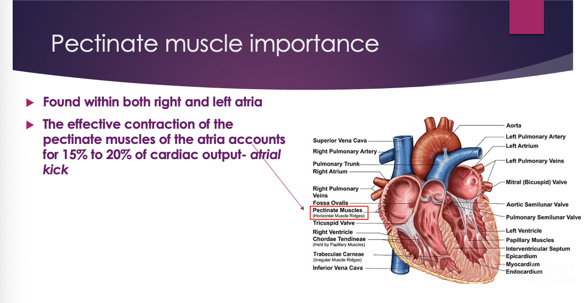
Pectinate muscle importance
Found within both right and left atria
The effective contraction of the pectinate muscles of the atria accounts for 15% to 20% of cardiac output- atrial kick
Main job – perform an atrial kick
Atrial kick – when atrium contract the pectinate muscles also contract to probide an extra umph – without that we lose 15-20% of the cardiac output
Atrial kick
Phenomenon of increased force generated by the atria during contraction
Assists with movement of blood from atria to ventricles
Young healthy adults do not rely on atrial kick as much, as 80% of the blood in young healthy adults moves from the atria to the ventricles
Overall, AK accounts for ~20% of volumetric blood towards ventricular pre-load
clinical: atrial fibrillation
abnormal electrical conduction causing a quivering of the atria
compromised cardiac output
if its quivering you dont get a full contraction leading to decrease CO
Clinical: Cor Pulmonale
Patients with chronic lung pathologies (COPD, pulmonary fibrosis) often present with hypoxemia and increased pressure within the pulmonary vasculature, pulmonary artery hypertension.
What is hypoxemia?
Due to the compromised perfusion capacity of the lung, increases in the pulmonary artery occur which increase the workload on the right ventricle
Causes right ventricular hypertrophy and resultant right ventricular failure called Cor Pulmonale
Cor Pulmonale refers to R sided heart failure only
When people say heart failure they typically refer to the entire L side failing – without L ventricle you cant live
Typical Cor P. is caused by pulmonary hypertension
Cause of R sided: Someone develops a lung pathology (means lungs have problem getting blood and o2 to themselves – if someone has COPD they have issues getting o2 to the lungs) – the only way the lungs can compensate is through the pulmonary artery (causes vasoconstriction of the pulmonary artery) - naturally arteries have smooth muscles, and the body realized theres a problem so they take the pulmonary artery a constrict and increases the pressure in the artery (this is called hypertension (pulmonary hypertension)) – so as a result from a lung problem we now have another lung problem – problem is that it makes it more narrow for the heart to pump into the lungs to the heart has to pump harder (r side works overtime) and eventually it will fail where you see cora pulmonale (so not the R side has failed so the L side has to work overtime (it has to help it) so now the L works overtime and now you have congestive heart failure and if the L side fails the blood backs up into the system and now you have renal hypertension/increase pressure in systemic circulation
So now we have hypoxia (decreased O2 saturation)
clinical: valvular dysfunction
tricuspid, bicuspid, pulmonic, aortic
valvular dysfunction can be discovered via auscultation
what to do next
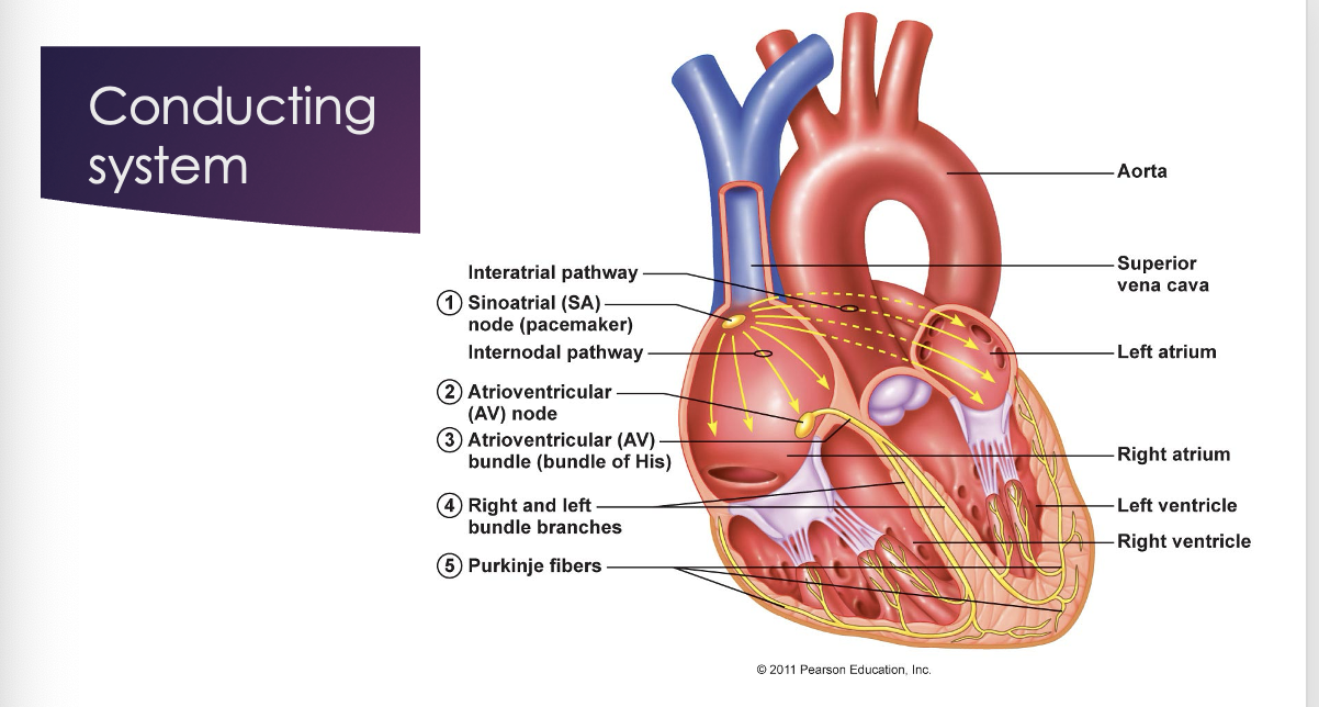
Conduction system through heart
SA node
AV node
AV bundle of his
bundle branches
purkinjie fibers
Innervation to heart
The ANS (autonomic nervous system) influences the rate of impulse generation, contraction, relaxation and strength of contraction
the ANS cause cause adjustments in CO to meet metabolic demands
consists of sympathetics and parasympathetic innervation
need a balance between the two in the heart
Heart: innervation, cardiac plexus (sympathetic)
Sympathetic input to the cardiac plexus arises from the sympathetic trunk in the neck
Sympathetic stimulation releases the neurohormone, catecholamines (epinephrine and norepinephrine)
Causes excitement of the cardiovascular system
Increases heart rate and blood pressure
Sympathetic = “fight or flight response”
Balance between SNS and PNS to make heart work
Sympathetic = epinephrine and norepinephrine
Heart: innervation, cardiac plexus (parasympathetic)
The cardiac plexus receives parasympathetic input from the right and left vagus nerves
Acetylcholine: neurohormone involved in parasympathetic stimulation
Vagal stimulation is inhibitory on the cardiovascular system, and therefore decreases heart rate and blood pressure
PNS decreased HR/BP
Hormone for PNS is Ach
angiology: systemic circulation (note the order, arteries are made of, elastic vs muscular arteries, veins have ___)
Arteries -> arterioles -> capillary beds <- venules <- veins
Arteries
Wall is composed of elastin, fibrous connective tissue and smooth muscle
Elastic arteries: have more elastic fibers than smooth muscle (aorta, pulmonary trunk) to allow for greater stretching as blood is ejected out of the heart
Muscular arteries: medium and smaller arteries. Contain more smooth muscle. These arteries can vasodilate and vasoconstrict (smooth muscle) to control the amount of blood to the periphery
Smooth muscle innervated by ANS, presence of a-receptors (alpha- receptors)
Veins
Compared to arteries, have thinner walls and a larger diameter.
In the LE, one way valves inhibit back flow of blood to the heart
clinical: varicosities, edema, DVT’s
Patients with incompetent valves in their veins may develop varicosities along with excess amounts of edema
Patients on prolong bed rest are at risk for developing DVT’s due to a lack of muscle activity resulting in the pooling of blood and clot formation
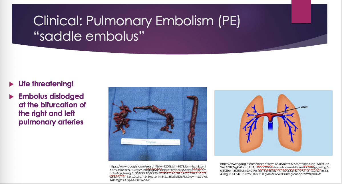
Clinical: Pulmonary Embolism (PE) “saddle embolus”
