histology- bone CT
 Call Kai
 Learn
Learn Practice Test
 Spaced Repetition
 Match
Match Flashcards
 Knowt Play
1/35

Earn XP
Description and Tags
foundations 1a- notes, lab notes, self-quiz
Name | Mastery | Learn | Test | Matching | Spaced | Call with Kai | Chat |
|---|
No analytics yet
Send a link to your students to track their progress
36 Terms
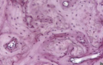
ID the bone type
woven bone
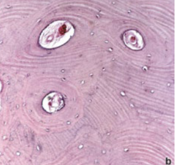
ID the bone type
lamellar bone
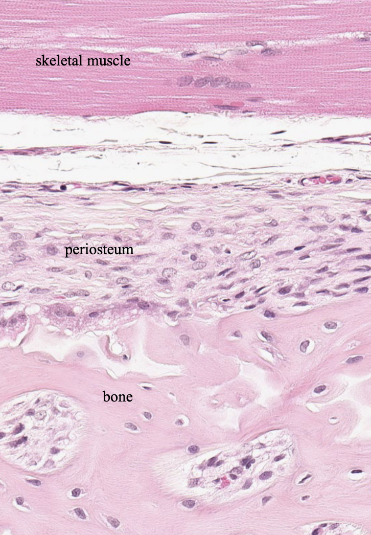
what type of CT is the periosteum made of?
dense irregular CT
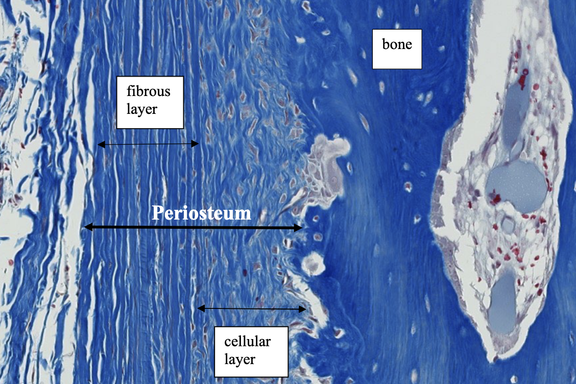
what type of CT is the periosteum made of?
dense irregular CT
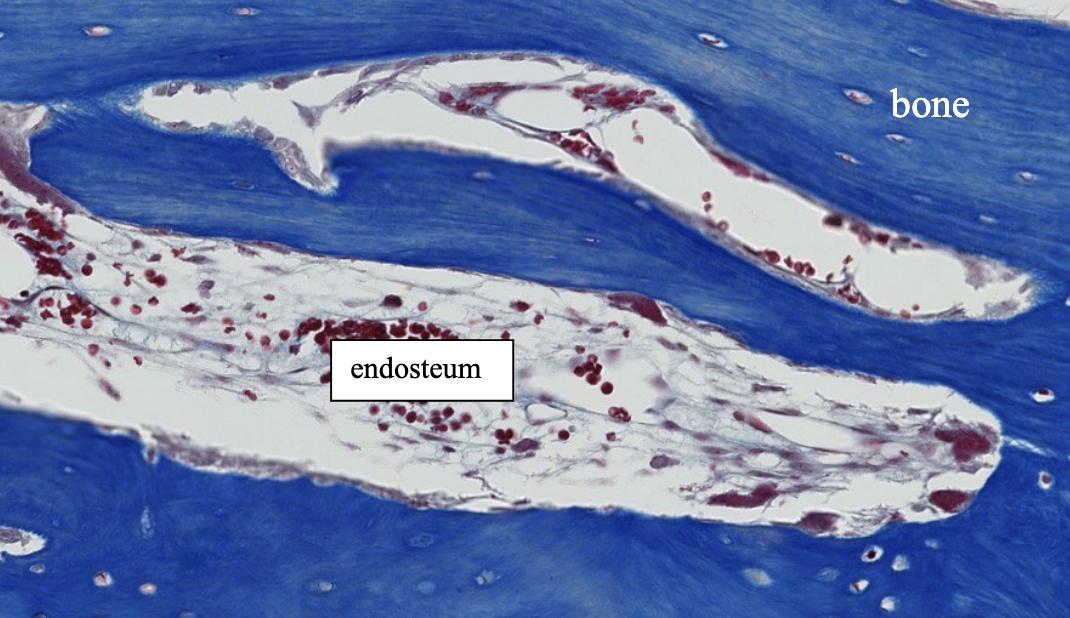
what type of CT do you see in the endosteum (note: bone marrow not very cellular makes it visible, usually not)?
loose irregular CT
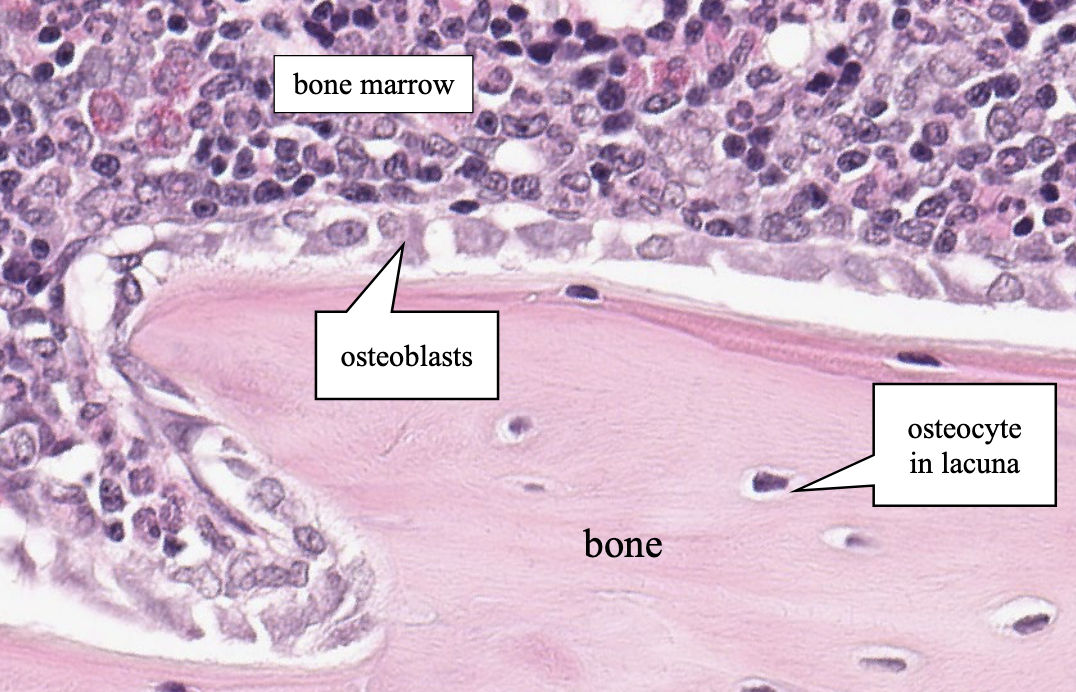
what is the term for the area covered by bone marrow in this picture?
endosteum
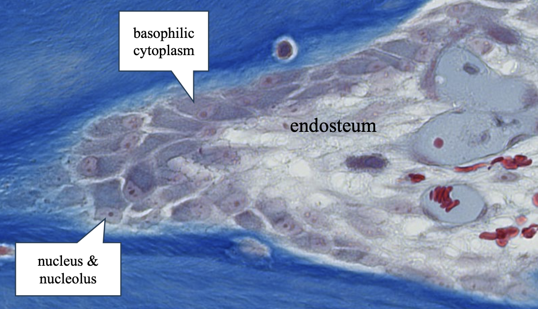
this is a cluster of _________ within the endosteum on the edge of bone.
osteoblasts
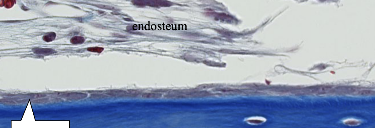
the white arrow is pointing to the less active ________ forming flatteded rows along the endosteal bone surface.
osteoblasts
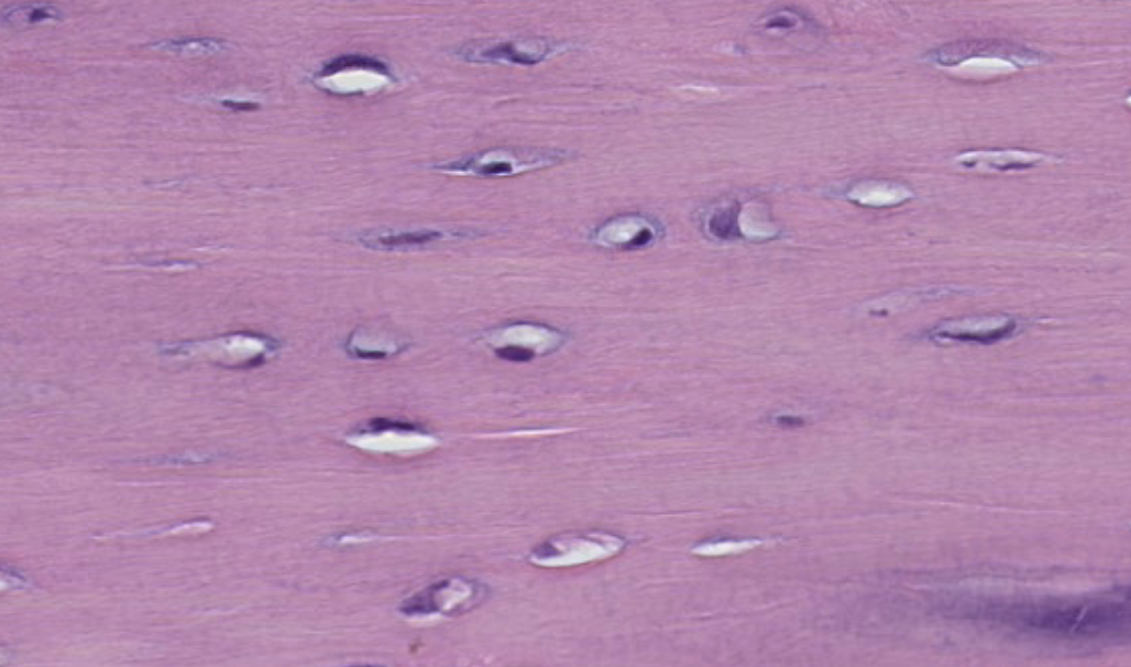
what sructures are located within the lacunae?
osteocytes
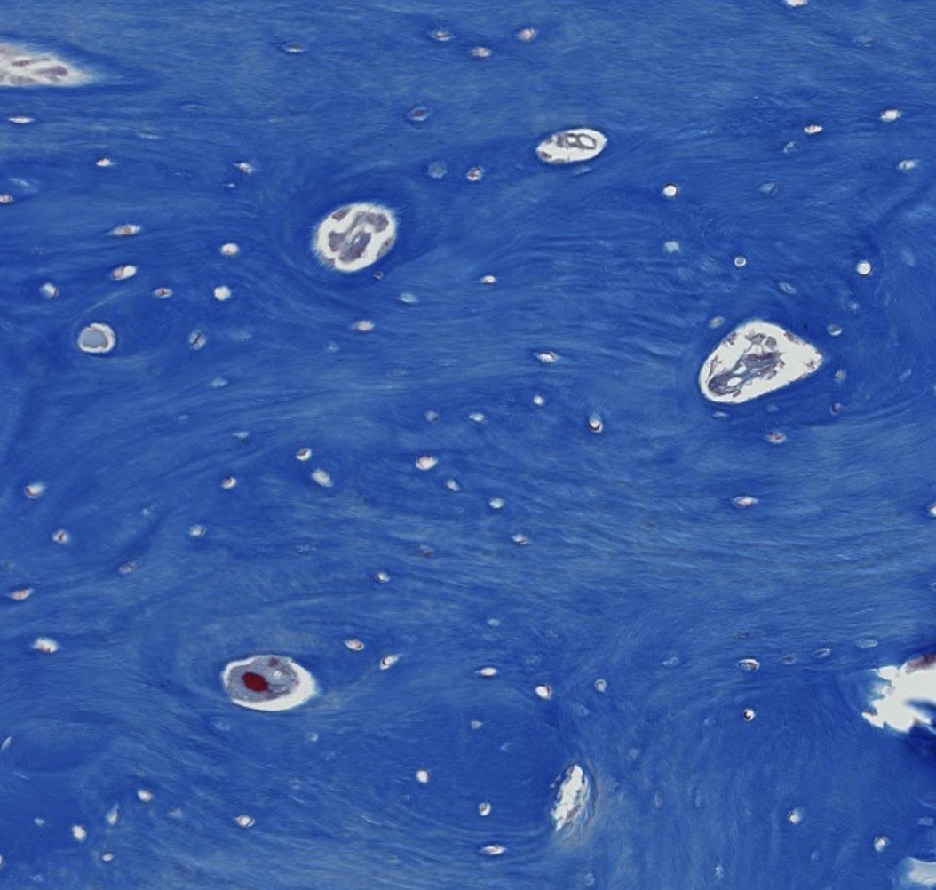
ID the bone type shown in this trichrome stain
woven bone
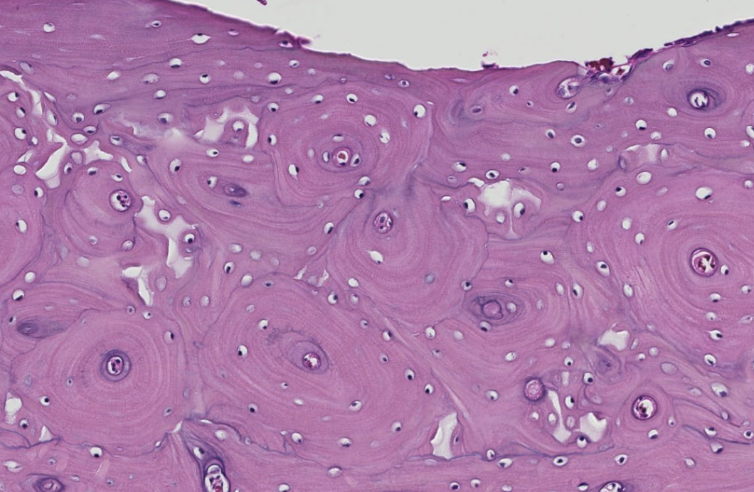
ID the bone type shown in this H&E stain (transverse tissue section)
lamellar bone
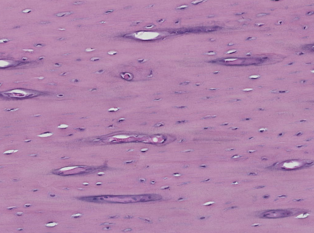
ID the bone type shown in this H&E stain (longitudinal tissue section)
lamellar bone
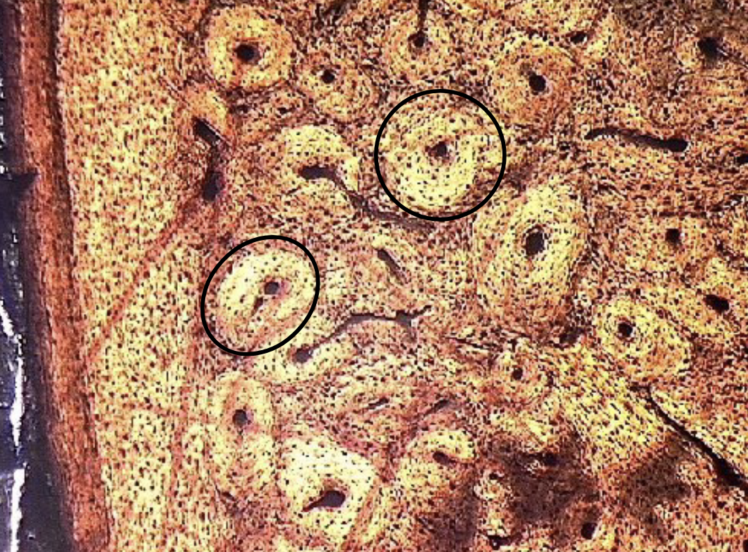
in this transverse section, what structures are circled?
osteons
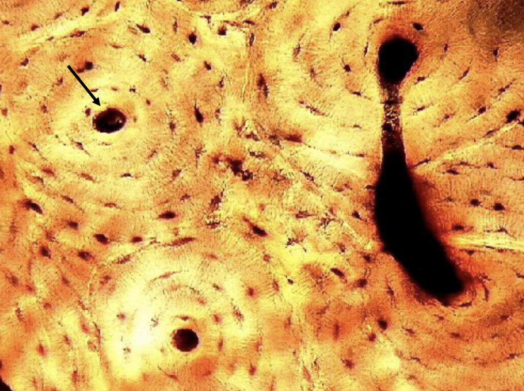
what structure is the arrow pointing to at the center of the osteon?
central (haversian) canal
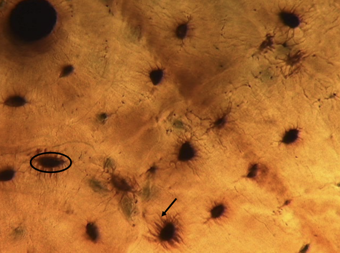
Concentric rings of ________ (black circle) are arranged in successive rings around the central canals.
Osteocytes live in these lacunae, and they interconnect with surrounding osteocytes via small channels called ________ (black arrowed line), which microscopically resemble fine hairs.
lacunae
canaliculi
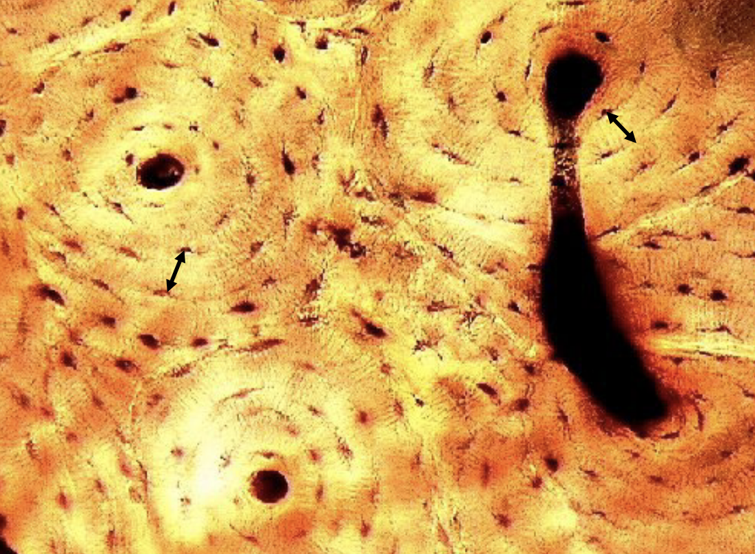
ID the bands between the rings of lacunae
lamellae
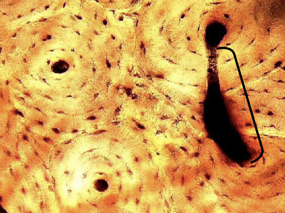
The vessels and nerves traveling in the central canal of an osteon are interconnected to the central canal in an adjacent osteon by travelling in ________.
perforating (Volkmann’s) canals
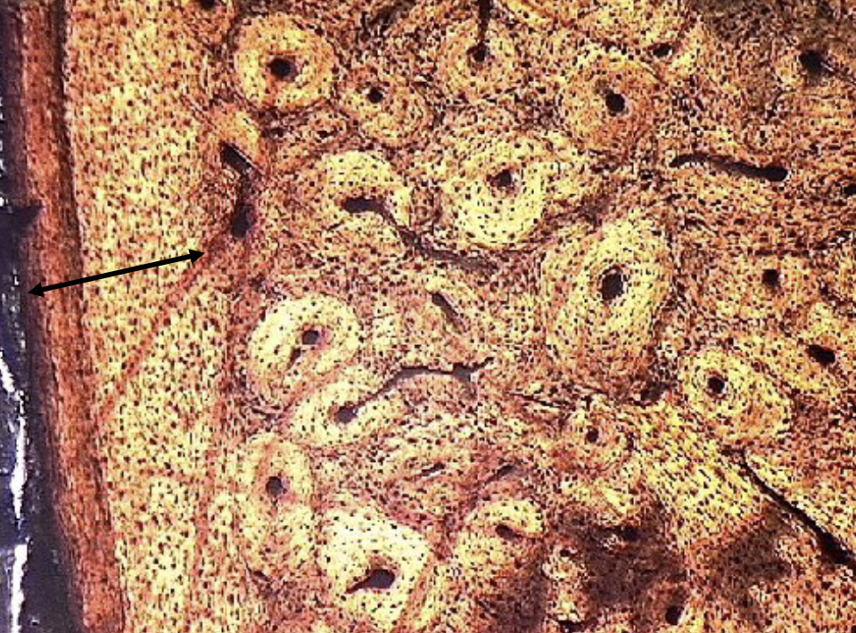
The width of the ____ ______ ______ on this ground bone
section is indicated by the black double-arrowed line.
outer circumferential lamellae
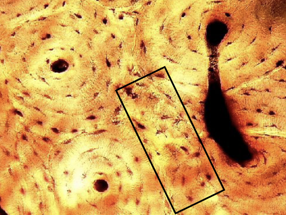
What is enclosed in the black rectangle
interstitial lamellae
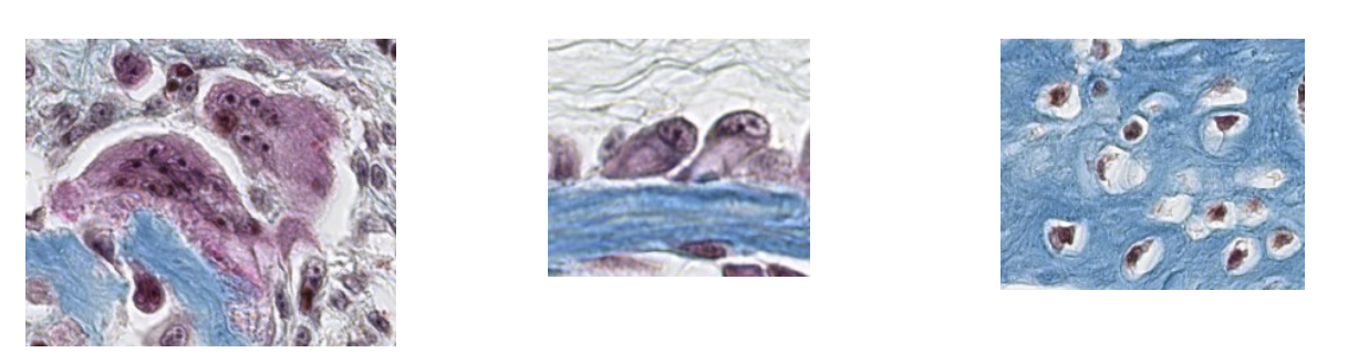
ID the bone cells, from left to right.
osteoclasts, osteoblasts, osteocytes
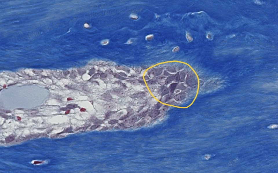
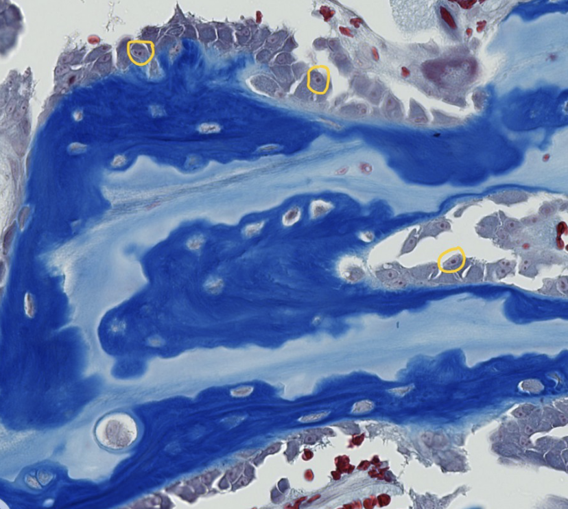
ID the cells encircled in yellow
what CT layer are these cells located within
osteoblasts
endosteum
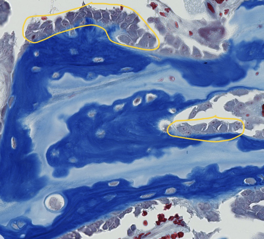
ID the encircled bone cells (hint: they are on he bone surface)
osteoblasts
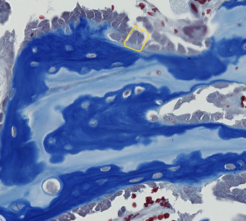
what is the name of the unmineralized matrix of collagen type I and proteoglycans secreted bu the encircled bone cell?
osteoid
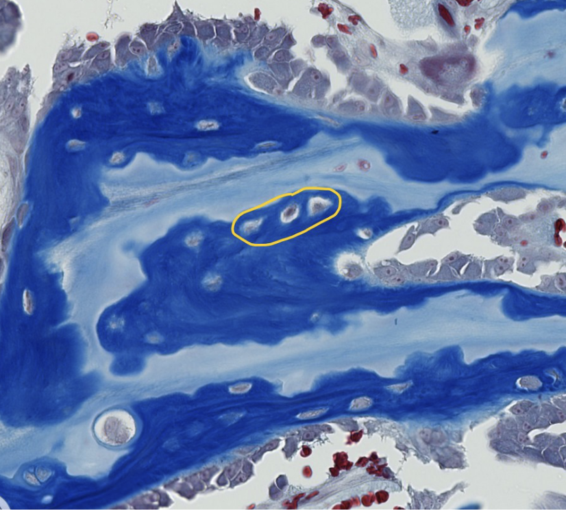
ID the encircled bone cells
osteocytes
which organelles are encircled with yellow?
what are the small dark circles visible inside the encircled cell organelles?
nuclei
nucleoli
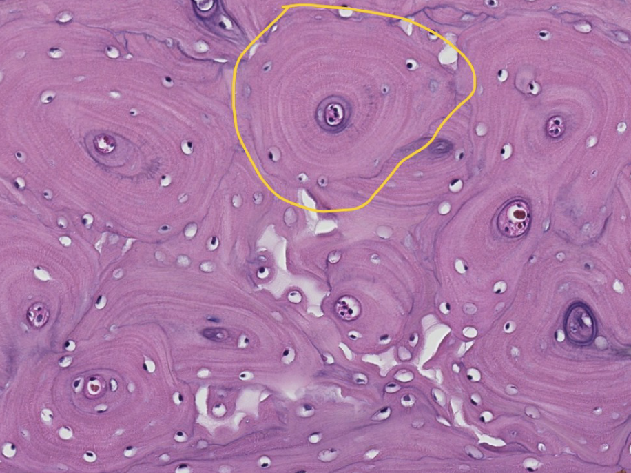
This is the diaphysis (shaft) of a long bone in transverse (cross) section. ID the encircled structural unit of mature bone.
Osteon (haversian system)
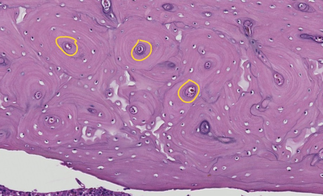
this is the diaphysis (shaft) of a long bone in a transverse (cross) section. ID the encircled region of the osteon
central canal
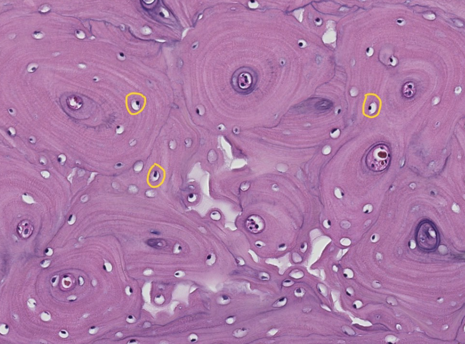
this is the diaphysis (shaft) of a long bone in a transverse (cross) section. ID the encircled region of the osteon
lacunae with osteocytes
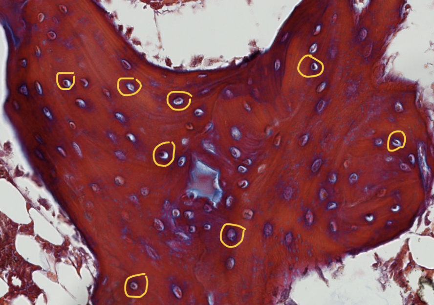
ID the encircled structures in this boen spicule
lacunae with osteocytes
The encircled tissue is bone marrow, surrounded by bone spicules. What do you think the clear spaces are? Hint: it is a type of adult CT proper.
adipocytes (white fat)
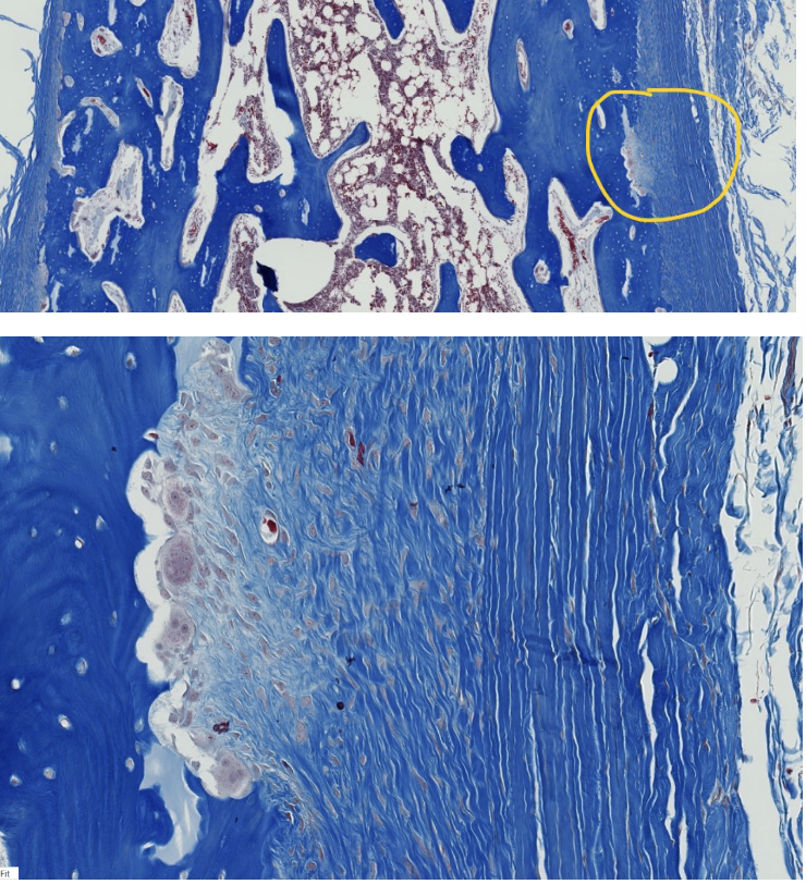
The upper image is fill thickness through the shaft of a long bone, with the external surfaces on the left and right sides of the image.
The encircled area in the upper image is enlarged in the lower image.
What do we call the encircled external fibrous membrane (outer CT covering) of bone?
What does fibrous refer to in this context?
periosteum
collagen fibers
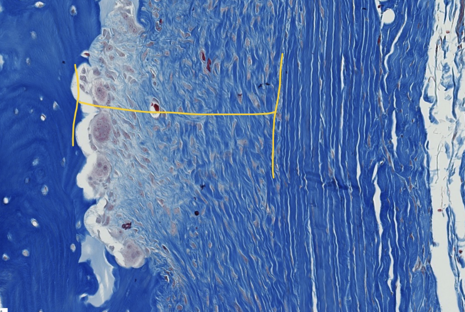
in actively growing bone, the periosteum has 2 visible layers. ID the layer within the yellow bracketed portion.
inner cellular layer
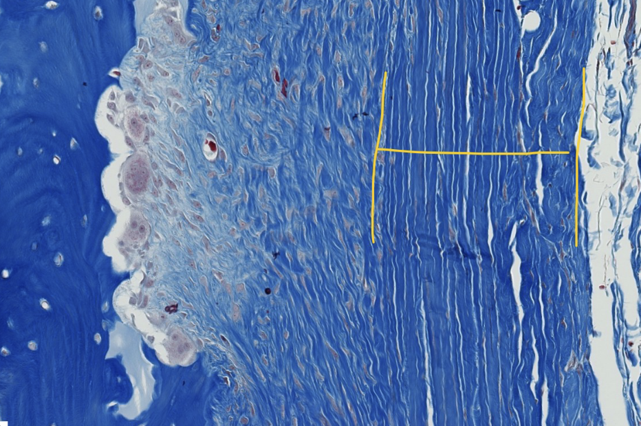
in actively growing bone, the periosteum has 2 visible layers. ID the yellow bracketed layer
outer fibrous layer
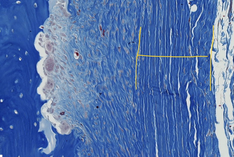
what type of collagen make up the bulk of the indicated layer?
type I collagen
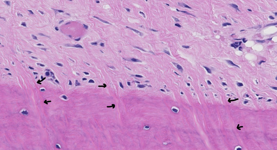
bundles of collagen fibers from tendons, ligaments, and the periosteum are embedded within the underlying bone as a means of anchoring these structures. What do we call these fibrous insertions?
sharpey’s (perforating) fibers
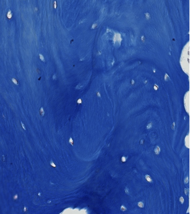
what is the microscopic architecture of woven bone (how are the collagen fibers arranged)?
randomly arranged in whorl-like layers (like fingerprints)