CH. 13 | Viruses, Viroids, and Prions
 Call Kai
 Learn
Learn Practice Test
 Spaced Repetition
 Match
Match Flashcards
 Knowt Play
1/16

There's no tags or description
Looks like no tags are added yet.
Name | Mastery | Learn | Test | Matching | Spaced | Call with Kai | Chat |
|---|
No analytics yet
Send a link to your students to track their progress
17 Terms
13 - 1 Differentiate a virus from a bacterium
Virus
Host specific
Attachment happens through receptor sites
Obligate intracellular parasites (However, some bacteria have this as well)
Requires living host cells / Needs to be inside a host in order to multiply
Acellular organisms (Do not contain cellular membranes, cellular structure, etc)
Contain a single type of nucleic acid; DNA or RNA (Cannot have both)
Can either be single-stranded or double-stranded, but never both
Can either be linear or circular, but never both
Contains a protein coat
Lack enzymes for protein synthesis and ATP generation
Bacteriophages / Phages
Viruses that infect bacteria
Host Range
A variety of cell types/host cells that the virus can infect
Two factors determine host range:
Attachment compatibility — The virus's outer surface must chemically match specific receptor sites on the target cell's surface. This is essentially a lock-and-key mechanism held together by weak bonds (like hydrogen bonds). The more attachment points, the stronger the association.
Internal cellular machinery — Even if a virus successfully attaches, it also needs the host cell to have the right internal factors to support viral replication.
What sets viruses apart from bacteria?
Viruses are acellular organisms that contain either DNA or RNA, but never both
13 - 2 Describe the chemical and physical structure of both an enveloped and a nonenveloped virus
Virion
A complete, infectious viral particle made of nucleic acid surrounded by a protein coat
Nucleic Acid
Encoded by either DNA or RNA (Never both)
Can be single or double stranded, linear, or circular
Capsid
Used for protection, protects the nucleic acid from nuclease enzymes and promotes attachment to host cells
The protein coat that protects the nucleic acid
Made of protein subunits called capsomeres
Determines most of the virus’s mass
Enveloped Virus
Has nucleic acid + capsid + an outer envelope (membrane)
The envelope is composed of lipids, proteins, and carbohydrates
Derived from the host cell’s plasma membrane during extrusion
May be covered by spikes — carbohydrate-protein complexes that:
Helps the virus attach to host cells
Can cause hemagglutination
Are used as identification markers
Makes the virus more versatile in attaching to host cells, but makes it more vulnerable to destruction
Nonenveloped Virus
Consists of only nucleic acid + capsid (Acts as the outermost layer)
Capsid serves 2 key functions:
Protects the nucleic acid from nuclease enzymes in biological fluids
Promotes attachment to susceptible host cells
More resistant to destruction
13 - 3 Define viral species
A group of viruses sharing the same genetic information and host range (ecological niche)
An example of this is SIV vs. HIV
SIV affects apes. Whereas HIV affects humans
13 - 4 Give an example of a family, genus, and common name for a virus
Viruses don’t follow scientific nomenclature for naming. Instead, they use common names. An example of this is HIV: Human immunodeficiency Virus
Examples
Family: Coronoavindae
Genus: Betacoronavirus
Common name: Severe acute respiratory syndrome coronavirus 2
SARS-CoV 2
Family: Herpesviridae
Genus: Simplexvirus
Species: Human herpesvirus-2
Viral Taxonomy
Family, Genus, Common Name
Family names end in -viridae
Genus names end in -virus
13 - 5 Describe how bacteriophages are cultured
Bacteriophages can be grown either in liquid or solid media
A bacteriophage sample is mixed with host bacteria and melted agar
Poured into a Petri plate
Virus-bacteria mixture solidifies into a thin top layer of bacteria
Virus multiplication occurs
Plaque
Lawn inoculation
Corresponds to a single virus in the suspension
13 - 6 Describe how animal viruses are cultured
3 Methods
Using living animals
Embryonated eggs
Cell cultures
In Living Animals
Viruses are grown in living animals (mice, rabbits, guinea pigs). The animal is inoculated with the specimen, then observed for signs of disease or sacrificed so infected tissues can be examined. Used when a virus cannot be grown any other way.
Embryonated Eggs
A fertilized egg with a developing chick embryo. A hole is drilled in the shell, and the virus is injected near the appropriate membrane. Viral growth is indicated by embryo death, cell damage, or pock/lesion formation on the membranes. Still used today to grow viruses for some vaccines, which is why you may be asked about egg allergies before vaccination.
Cell Cultures
ells grown in culture media in the lab — more convenient than living animals or eggs. Started by treating animal tissue with enzymes to separate individual cells, which are then suspended in nutrient solution and grow into a monolayer
Viral infection of the monolayer causes cell deterioration called cytopathic effect (CPE).
Two types of cell lines:
Primary cell lines — derived from tissue slices; die after a few generations
Continuous cell lines — transformed (cancerous) cells that survive indefinitely; called "immortal cell lines" (e.g., HeLa cells from Henrietta Lacks)
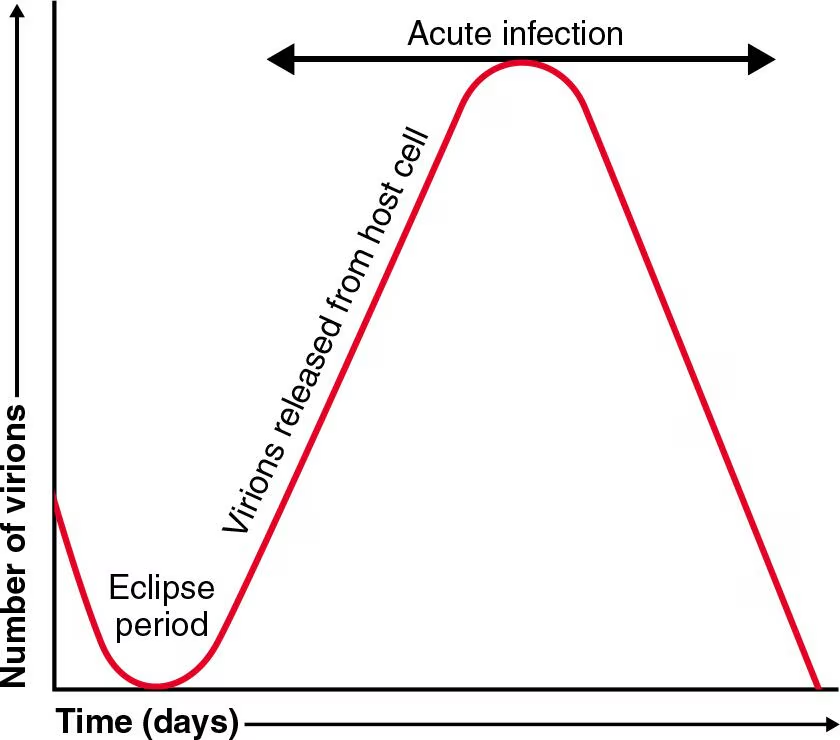
Virus multiplication | One-step growth curve
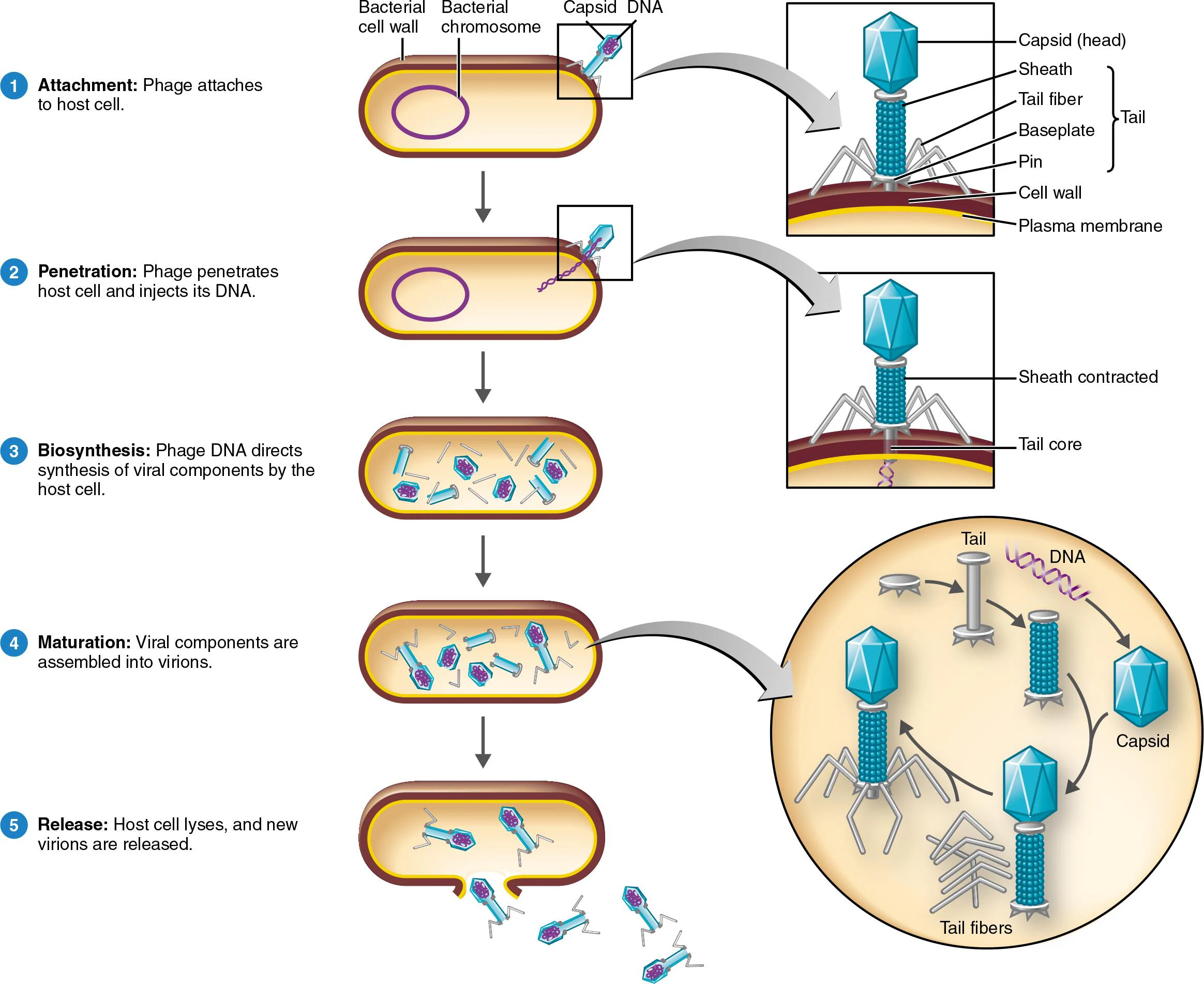
13 - 8 Describe the lytic cycle of T-even bacteriophages (infects E. coli)
Lytic Cycle
A mechanism of phage multiplication that ends with lysis and death of the host cell
—
5 Stages
Attachment
The phage collides with the bacterium
Attach to receptor sites on the bacterial cell wall
Penetration
Phage releases phage lysozyme to break down part of the cell wall
Phage injects its DNA into the host — capsid stays outside
Biosynthesis
Phage DNA takes over the host cell
Host DNA is degraded; protein synthesis is stopped
Host cellular components are hijacked to make viral components
Eclipse period
Maturation
Viral components self-assemble into complete virions spontaneously
Release
Lysozyme (encoded by Phage DNA) breaks down the bacterial cell wall
Plasma membrane bursts open → host cell dies
New virions are released and go on to infect neighboring cells
Key Details to Remember
The host cell always dies in the lytic cycle (unlike the lysogenic cycle where it survives)
The virus contributes DNA only — all machinery (ribosomes, enzymes, energy) comes from the host
The eclipse period = viral components present, but no detectable complete virions yet
Lysozyme is used twice — once to enter (penetration) and once to exit (release)
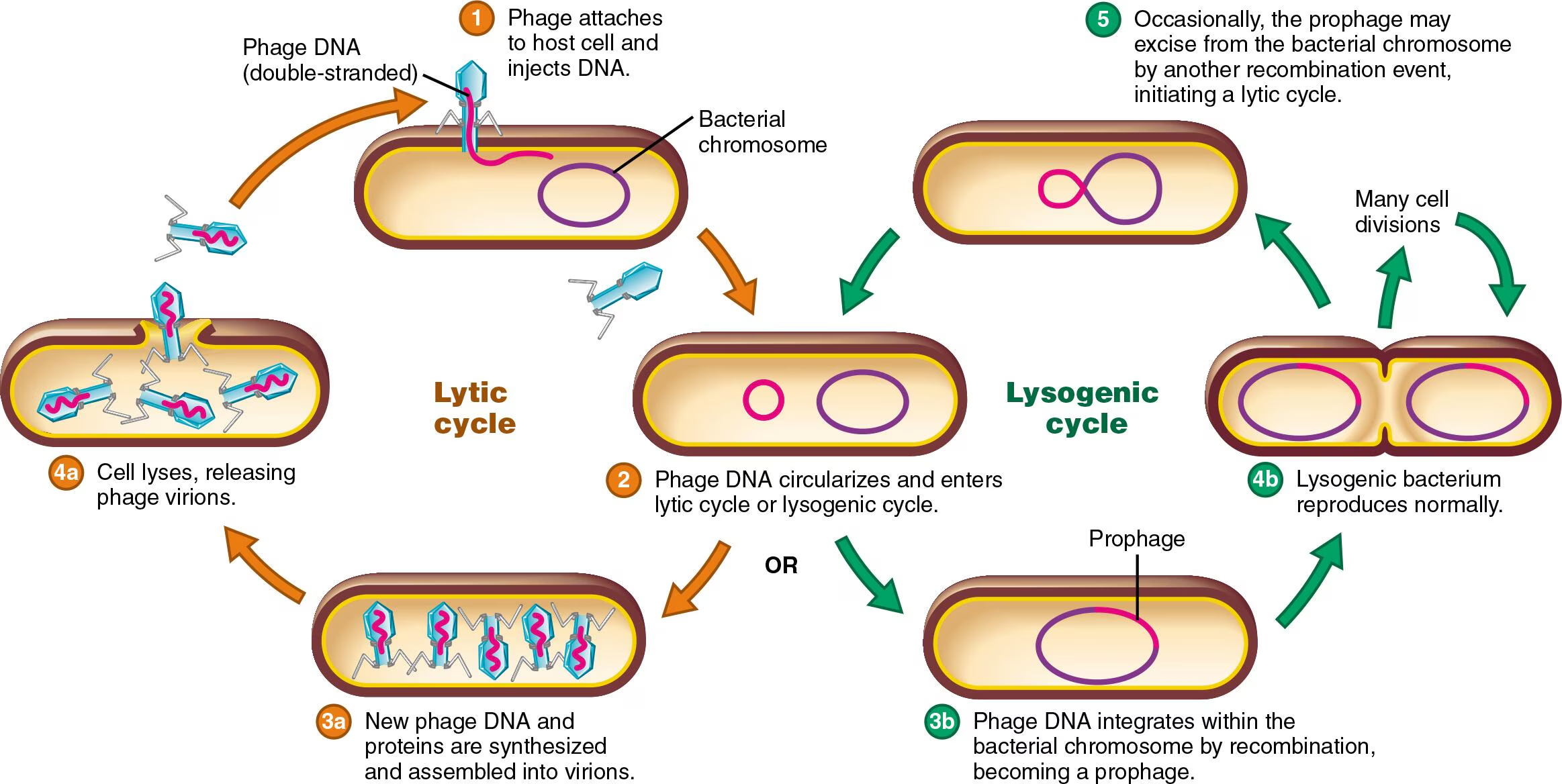
13 - 9 Describe the lysogenic cycle of bacterophage Lambdavirus
Lysogenic Cycle
Does not immediately kill the host cell. Instead, it is the incorporation of viral DNA into host DNA, which remains dormant — sometimes for many generations
—
5 Stages
Attachment & Penetration
Same as the lytic cycle: Phage attaches to the host cell and injects its DNA
Integration
Instead of taking over and destroying the host, the phage DNA integrates directly into the host’s chromosome, now called a prophage
Repressor proteins encoded by the phage silence all other phage genes, keeping them latent and inactive
The host cell has no idea that the extra DNA is there
Replication
Every time the host cell divides, it copies the prophage as well → all daughter cells carry the prophage (lysogenic cells)
Excision (Trigger required)
A spontaneous event.
UV light, or certain chemicals, can cause the prophage to pop out of the host chromosome → this initiates the lytic cycle
3 Important Consequences of Lysogeny
Phage Conversion
The prophage carries extra genes that give the host cell new properties — most importantly, toxin production
Examples:
Corynebacterium diphtheriae → causes diphtheria only when carrying a lysogenic phage (the prophage encodes the toxin)
Streptococci → can cause toxic shock syndrome only when lysogenic
Clostridium botulinum → botulism toxin is also prophage-encoded
This is clinically significant — a normally harmless bacterium can become dangerous through lysogeny
Immunity to Reinfection
Lysogenic cells are immune to reinfection by the same phage
The repressor proteins that keep the prophage silent also block any new incoming phage of the same type
Note: the cell is NOT immune to other phage types
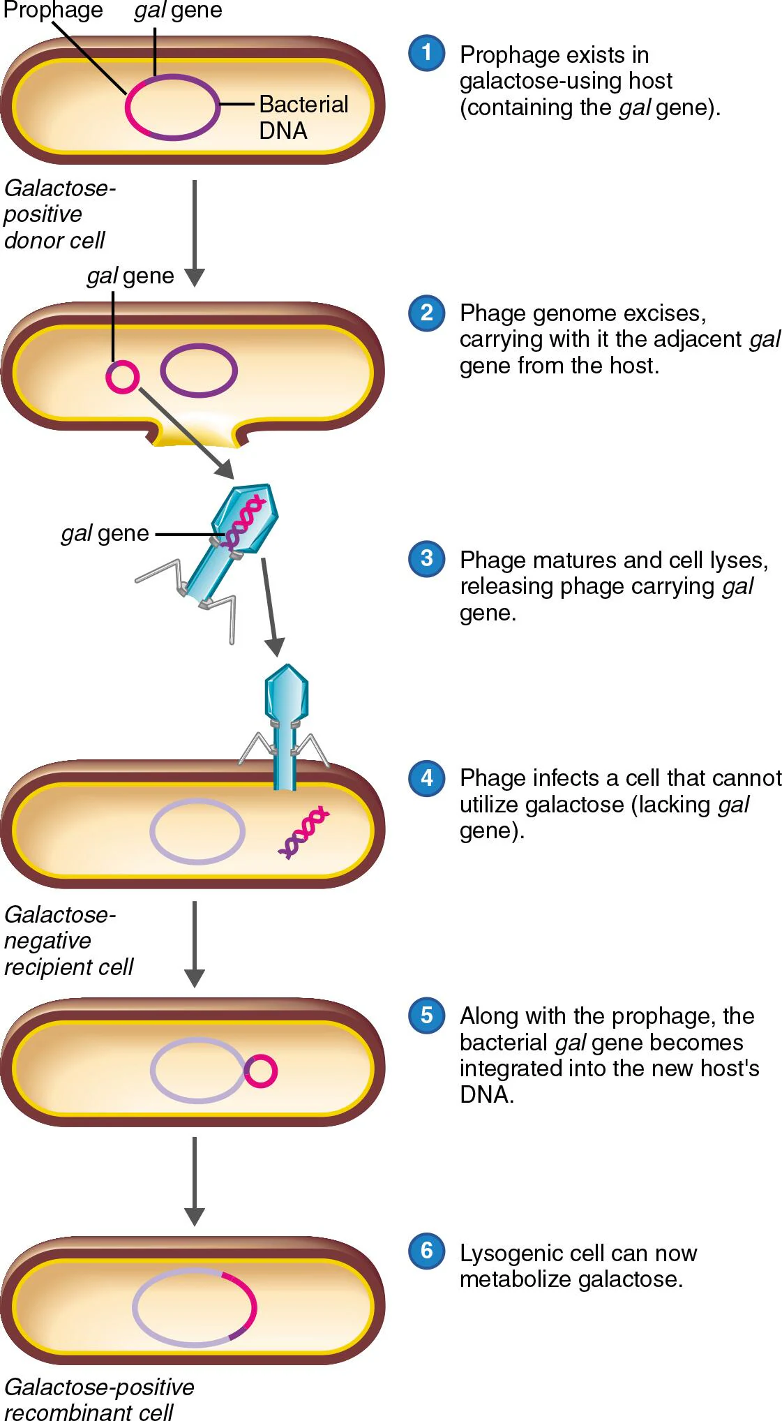
Specialized Transduction
When the prophage excises, it sometimes makes an unclean cut and accidentally carries a piece of the host's bacterial DNA with it
When this phage infects a new host cell, it delivers both its own DNA and that bacterial DNA fragment
The new host now has genes from a completely different bacterium — this is how traits can transfer between bacterial cells via a virus
Specialized Transduction
13 - 11 Define oncogene and transformed cell
Oncogene
A mutated proto-oncogene that triggers abnormal, uncontrolled cell growth that can lead to cancer
Can be activated, and when activated, it can lead to cancer
Proto-oncogenes
Normal genes in a healthy cell that encode proteins involved in stimulating normal cell growth
When mutated → become oncogenes
Mutations can be triggered by mutagenic chemicals, high-energy radiation, or viruses (oncoviruses)
Oncoviruses
Viruses that cause cancer by activating oncogenes → activation makes the cell abnormal → these abnormal cells are called transformed cells
Transformed Cell
A tumor cell that has acquired new properties distinct from normal cells, including uncontrolled growth, lack of apoptosis, irregular shape, and a virus-specific surface marker called a tumor-specific transplantation antigen (TSTA)
An abnormal cell that results from oncogene activation
13 - 14 Differentiate between latent and persistent viral infections
Latent Viral Infection
The virus remains in the host for a long period without producing disease — it is inactive/dormant until triggered
Examples include:
Simplexirus (cold sores)
Varicellovirus (chickenpox)
Persistent (Chronic) Viral Infection
The virus stays in the host but symptoms gradually increase in severity over a long period. Virions slowly build up over time rather than spiking suddenly
Typically fatal
Latent and Persistent Viral Infections
Red curve (Acute infection) — measured in days. Virions spike fast and high, then drop quickly. This is your typical infection where you get sick, your immune system fights it off, and it's over. Not latent or persistent — just included for comparison.
Green curve (Latent infection) — measured in months/years. Virion levels are flat/undetectable for a long time (the dormant period), then spike suddenly when a trigger reactivates the virus. That sudden spike is the key visual — it mirrors the acute curve but happens much later.
Blue curve (Persistent infection) — measured in months/years. Virion levels never fully drop to zero after the initial infection — they slowly and gradually build up over a long period. No sudden spike, just a slow creeping rise.
![<p><strong>Red curve (Acute infection)</strong> — measured in days. Virions spike fast and high, then drop quickly. This is your typical infection where you get sick, your immune system fights it off, and it's over. Not latent or persistent — just included for comparison.</p><p class="font-claude-response-body break-words whitespace-normal leading-[1.7]"><strong>Green curve (Latent infection)</strong> — measured in months/years. Virion levels are flat/undetectable for a long time (the dormant period), then spike suddenly when a trigger reactivates the virus. That sudden spike is the key visual — it mirrors the acute curve but happens much later.</p><p class="font-claude-response-body break-words whitespace-normal leading-[1.7]"><strong>Blue curve (Persistent infection)</strong> — measured in months/years. Virion levels never fully drop to zero after the initial infection — they slowly and gradually build up over a long period. No sudden spike, just a slow creeping rise.</p>](https://assets.knowt.com/user-attachments/14cb70a2-21e0-42a9-873a-c0c5a0f4e032.png)
13 - 15 Differentiate virus, viroid, and prion
Virus
Contains either DNA or RNA enclosed in a protein coat (capsid)
Requires a host cell in order to replicate
Can infect animals, plants, bacteria
Viroid
Infectious RNA
Short pieces of naked circular RNA only, with no protein coat
Smaller and simpler than a virus
Causes disease in plants by gene silencing — does not code for any proteins
Virusoid
A type of viroid that does not have a protein coat, but can only cause disease when the plant cell is already infected by a virus
Hepatitis D is a suspected virusoid in humans
13 - 17 Discuss how a protein can be infectious