Exam 1 Review (1-3)
 Call Kai
 Learn
Learn Practice Test
 Spaced Repetition
 Match
Match Flashcards
 Knowt Play
1/68

Earn XP
Description and Tags
Focuses on the learning objectives
Name | Mastery | Learn | Test | Matching | Spaced | Call with Kai | Chat |
|---|
No analytics yet
Send a link to your students to track their progress
69 Terms
Identify the most abundant functional groups found
in biomolecules
Amino (-NH3)
Hydroxyl (-OH)
Sulfhydryl (-SH)
Carboxyl (-C=OO-)
Phosphoryl (P=OOO-)
Methyl (-CH)
Polar
molecules that have a partial positive charge on one side and a partial negative charge on the other, leading to an unequal distribution of electrons
ex) H2O
Nonpolar
molecules that have an equal distribution of electrical charge, resulting in no partial positive or negative charges. ex) lipids
Name the four major classes of biomolecules,
Carbohydrates
Lipids
Proteins
Nucleic acids
What biomolecules form polymers?
Carbohydrates
Proteins
Nucleic Acids
Primary cellular function of carbohydrates
Energy source/storage
Cell recognition
Provide structural support
Primary cellular function of lipids
Forms cell membranes
Energy conversion/storage
Cell signaling
Primary cellular function of proteins
Act as catalysts
Structural support
Store & transmit genetic info
Primary cellular function of nucleic acids
Store & transmit genetic information
Enzyme catalysis
Signal transduction
Energy conversion
Explain the role of pathways in living systems
Coordinate & control complex processes in response to environment
Series of linked biochemical rxns where product of 1 rxn = reactant for the next
Identify the three components of a nucleotide
5C sugar
Phosphoryl group
Nitrogenous base
Compare and contrast the overall functional role of DNA and RNA
DNA → permanent storage for an organisms genetic info
RNA → temporary working copy that executes the instructions to build; transcriptome (mRNA), Translation (tRNA & rRNA)
Both → store, encode, or transmit the genetic instructions required to build and maintain living organisms
Define the Central Dogma of molecular biology
Describes the flow of genetic information within a biological system
DNA → RNA → Protein
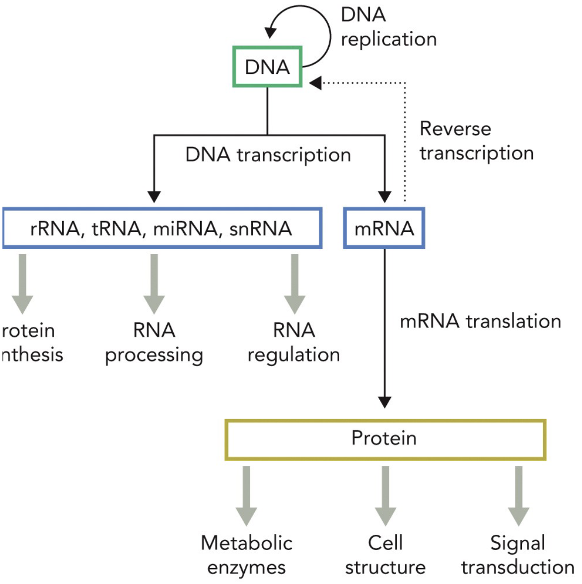
Transcriptome
Collection of DNA transcripts (RNA) generated by DNA transcription
Proteome
Set of proteins produced by mRNA translation either in entire organism or tissue
Identify the relationship between protein structure and function
Proteins 3D shape dictates its ability to recognize, bind, & interact with other molecules
Endothermic
A reaction absorbs heat from the surroundings
+ΔH (unfavorable/nonspont.)
Breaking of noncovalent interactions
Exothermic
Reaction releases heat
-ΔH (favorable/spont.)
Formation of noncovalent interaction
0th Law of Thermodynamics
Thermal equilibrium- if 2 bodies are in thermal equilibrium with a third body, they are also in equilibrium with each other
1st Law of Thermodynamics
Conservation of energy- Energy is conserved, but can be converted between types
Energy (E) can be neither created nor destroyed.
• Energy can only be converted from one form to another
• ΔE=q-W
2nd Law of Thermodynamics
Entropy- In all spontaneous processes, the entropy of the universe increases
3rd Law of Thermodynamics
Absolute zero- the entropy of a system approaches a constant value
as the system approaches absolute zero
Enthalpy
Total heat content/bond energy of a system
-ΔH = favorable/spontaneous
Entropy
ΔS
Number of ways energy can be distributed (resonance in Ochem); disorder
+ΔS = favorable/spontaneous
Gibbs Free Energy
ΔG
Energy available for work
-ΔG = favorable/spontaneous
Gibbs Free Energy Relation to ΔH & ΔS
ΔG = ΔH - TΔS
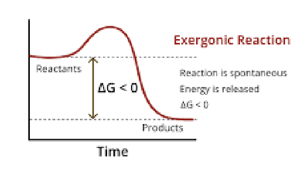
ΔG < 0
Rxn favorable
Equilibrium favors products (forward rxn)
Energy released
Exergonic
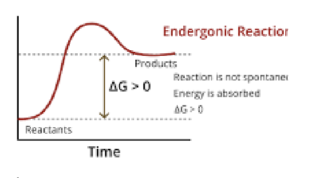
ΔG > 0
Rxn unfavorable
Equilibrium favors reactants (reverse rxn) Energy absorbed Endergonic
Exergonic
-ΔG
Rxn spontaneous
Energy released to surroundings
Endergonic & Coupled in Biological Systems
ΔG
Rxn non-spontaneous
Energy absorbed/required
Can be coupled to an exergonic rxn to become overall favorable
Common Coupling Rxn
ATP Hyrdolysis → ΔG = -30.5 kj/mol
Explain why ATP hydrolysis produces a large standard free energy change, in terms of electrostatic charge repulsion and product stabilization.
Due to the repulsion between negatively charged phosphate groups in ATP (-ΔH).
When ATP is hydrolyzed, the release of one phosphate group decreases this repulsion and allows for better stabilization of the products → more possible resonance forms , leading to a more favorable reaction.
Hydrogen Bonds
Responsible fro unique properties of water
H “shared” bt 2 eneg atoms
ΔH is - & favorable, attractive
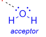
H Bond Donor
Atom that has H bonded

H Bond Acceptor
Has lone pair
Electrostatic Interactions
Attractive or repulsive forces
Strength of bond depends on charge, distance bt charges, and environment bt them
Dipole-dipole
Ionic interactions
Salt bridge → ion-ion bt + and - charged atoms
Van der Waals
Temporarily occur between the dipoles of nearby electrically neutral molecules
Depend strongly on distance bt 2 atoms
Very weak individual attraction
Hydrophobic Effect
Occur bt nonpolar molecules
Do not for H bonds w/ water
Play important role in protein folding rxns
Driven by water’s tendency to maximize its own internal H bonding and entropy
Strength of 4 IMF
Strongest: Electrostatic (salt-bridge #1, dipole #2) → 0.25nm
Moderate Strong: Hydrogen Bonding → 0.30nm
Weak: van der Waals → 0.3-0.6nm
Variable: Hydrophobic Effect
Relate pH to the concentration of H+ or OH–
pH = -log[H+]
pOH = -log[OH-]
[H+] increases = pH decreases
[OH-] increases = pH increases
Interpret titration curve for a weak acid

Use the Henderson-Hasselbalch equation to determine pH or the ionization state of a weak acid
pH = pKa + log [A-]/[HA]
pKa = -logKa
Relate ionization state to a group's capacity for electrostatic and hydrogen-bonding interactions.
The ionization state of a group affects its ability to participate in electrostatic interactions and hydrogen bonding. Higher ionization states generally enhance a group's capacity to form such interactions due to the presence of charged species.
Key structural differences bt DNA & RNA an their link to function
DNA: dbl stranded w/ deoxyribose sugar, N base, and uses Thymine, longer
RNA: single stranded w/ ribose sugar, N base, and uses Uracil, shorter
1° structure of DNA
Sequence of polymers (polarity 5’→3’)
Unique arrangement of deoxyribonucleotides/ribonucleotides arranged in a single chain
Usually depicted as single letters in a row

2° structure of DNA
Complementary H bonding interactions
2 comp. strands of DNA bind (anneal)
Double helix
Causes backbone (sugar phosphate) to adopt a particular shape
Restate Chargaff’s rule and explain typical base pairing in DNA structure.
The amount of adenine = amount of thymine, and the amount of cytosine = amount of guanine, leading to complementary base pairing (A-T and C-G) that stabilizes the DNA double helix.
Called Watson-Crick base pairs
A-DNA
Right handed helix
Wider turn
Compact and compressed
Areas of active transcription

B-DNA
Physiological form
Right handed helix

Z-DNA
Left handed helix
Narrower turn
Areas of active transcription
Zigzag backbone

Helical twist
Optimizes H bonding

Propeller twist
Optimizes stacking
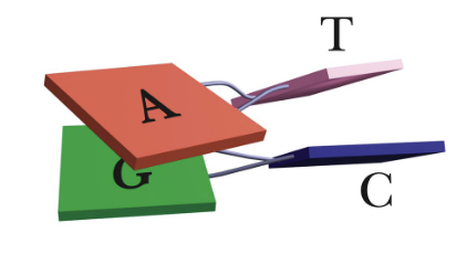
Base stacking
Most important stabilizing interaction in nucleic acids

Impact of strand length on Tm
Longer the strand = higher the Tm
More H bonds
More stacking
More Van der Waals
IMF are additive

Impact of ionic strength on Tm
Higher ionic strength = higher Tm
The backbone carries - charges → + ions in solution neutralize the - charges, reducing the repulsion between the strands and stabilizing the double helix structure.
Higher [salt] = higher Tm = higher stability
Higher heat required to denature strands
Counter ion Condensation
Impact of A-T vs G-C content on Tm
Higher G-C content increases Tm due to more hydrogen bonds and stronger base stacking interactions → more stable compared to A-T pairs.
Positive supercoiling
Right strand twists over left
DNA overwound in same direction as natural right handed helix
Increase physical stress of DNA
Negative supercoiling
Left strand twists over right, causing DNA to be underwound, which relieves physical stress and facilitates strand separation during processes like replication and transcription.
Linking number
Number of times a strand of DNA winds in the right-handed direction around the helix in an imaginary plane
Lk = Tw + Wr
Twist
Measures the winding of DNA strands around each other
Writhe
Measures crossing of DNA strands
Explain how linking number, twist, and writhe are related
The linking number (Lk) is the sum of the twist (Tw) and writhe (Wr) of a DNA molecule, showing how the DNA strands are intertwined.
Identify the histone proteins and the structures they form.
Histone proteins are the core components of nucleosomes, which are structures formed by DNA wrapped around histone octomers.
These proteins include H2A, H2B, H3, and H4, and they play a critical role in the compaction and organization of chromatin.

Key elements in DNA condensation
Structural proteins
Chemical/ionic factors
Enzymatic macheinery
Explain how a plasmid can transfer genetic material from one cell to another
Conjugation
Transformation
Transduction
List the elements of a plasmid that make it useful for producing
recombinant DNA
Origin of Replication
Selectable marker
Multiple cloning sites
Promoter region
Explain the role played by restriction enzymes in the production of recombinant DNA
Act as molecular scissors that cut DNA at a specific sequence, allowing the isolation of genes and splicing them into other DNA molecules
Add a vector and insert
Explain the cut-and-paste cloning and identify what is happening in each step
1st → restriction enzymes cut the DNA at specific sites
2nd (cut)→ the desired DNA fragment is isolated and a vector (like a plasmid) is prepared by cutting it with the same restriction enzyme.
3rd (paste) → the DNA fragment is inserted into the vector, which is then introduced into a host cell for replication.
Predict the expected size of a colony PCR product based on primer binding sites relative to the insert within a plasmid and interpret agarose gel results
Equals the exact number of base pairs from the 5′ start of the forward primer to the 5′ start (or 3′ end) of the reverse primer on the plasmid map, spanning the distance between their binding sites