Prokaryotic (bacterial ) cell biology -Ch. 4.1 - 4.3 - Microbiology
1/8
There's no tags or description
Looks like no tags are added yet.
Name | Mastery | Learn | Test | Matching | Spaced |
|---|
No study sessions yet.
9 Terms
Be able to provide similarities between Prok and Euk microbes (other than they are both living cells).
PROKARYOTIC external cell structures – know the structure and function of each item below – (**contrast these structures w/ what we see in eukaryotic
Cell membrane – concentrate on function –NOTE energy production (ATP production) across membrane**
Prokaryotes
Function: energy reactions (ATP creation), nutrient processing, synthesis (from embedded proteins that synthesize structural macromolecules to build cell envelope and appendages), and regulate transport (nutrients into the cell and discharge waste out of cell)
Structure: selectively permeable, embedded proteins, protein anchor
Site of ATP: across the membrane
Eukaryotes
Function: controlling the movement of substances into and out of the cell by regulating which molecules can pass through, and keep the internal organelles safe
Site of ATP: Mitochondria
Cell Wall **
Peptidoglycan structure – 3 components –know well
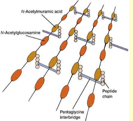
Polysaccharide backbone (NAG and NAM) [repeating disaccharide]
tetrapeptide side chain - [consist of 4 amino acids hanging from the saccharide]
peptide cross-links - [connect the side chains together - give peptidoglycan its strength]
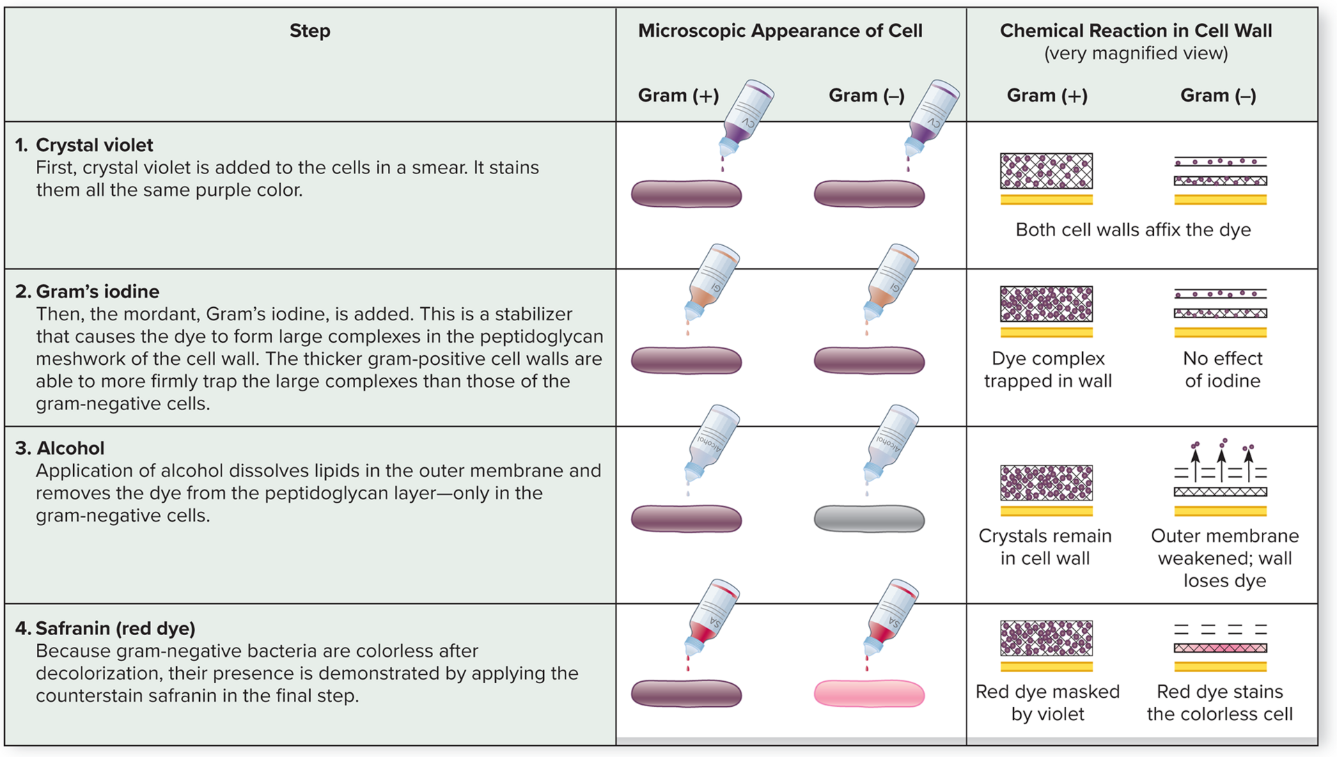
Difference between gram + and gram – cell wall (be able to describe the cell wall structure in detail)
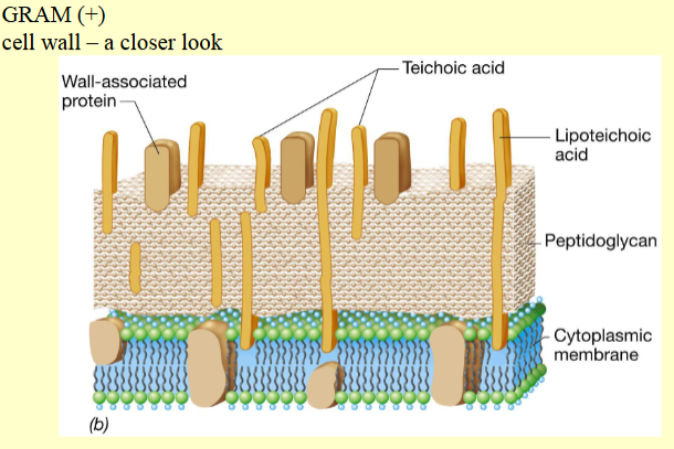
Has 2 layer (peptidoglycan and cell membrane)
Thicker peptidoglycan —> causing it to stay stain purple when gram staining
Contains both teichoic acid [Anionic polymers containing glycerol that appear in the walls of gram-positive bacteria.] and Lipoteichoic acids [Anionic polymers containing glycerol that are anchored in the cytoplasmic membranes of gram-positive bacteria.]—> function in cell wall maintenance (cell shape), enlargement during cell division, and acidic charge on the cell surface
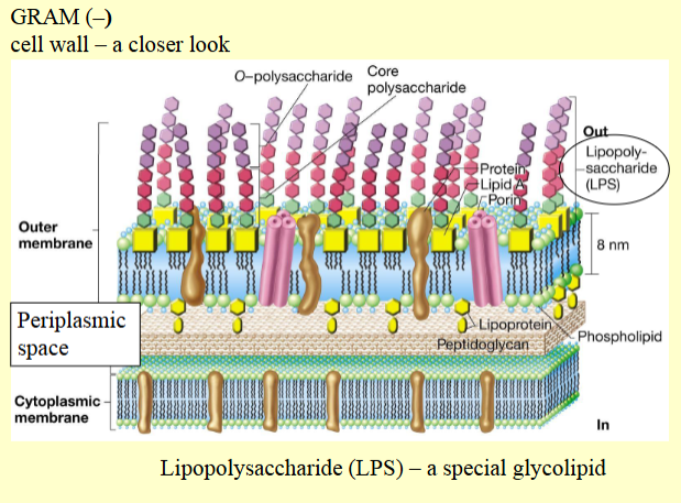
Has 3 layer (peptidoglycan, cell membrane, and outer membrane)
lipopolysaccharide: a special glycolipid - important for bacteria cells —> allows for cell-to-cell communication by extending off as cell markers and receptors
endotoxin: lipid portion of the lipopolysaccharide - a toxin (toxic shock) - (skeptic shock and dropping of blood pressure)
infection of meningitis and typhoid fever
main molecule that makes up the outer membrane; major structural component → hybrid molecule with lipid component inside and polysaccirde outside
Periplasmic space: area between the inner and outer membrane where enzymes are that can help builds peptidoglycan —> providing a space to segregate potentially harmful enzymes from the cytoplasm
Understand principles behind how the Gram stain works and why it works the way it does (make sure to read the 'insight' box that talks about this- important for lab also!)
Fimbriae, Pili (pilus), Capsule and S-layer – structure and function –Also be able to talk about what advantage this structure would give to the bacterial cell that has it.
Fimbriae:
Function: Stick to each other and to surfaces, responsible for the mutual clinging of cells that leads to biofilms and other thick aggregates of cell on the surface of liquids and for the microbial colonization of inanimate solids such as rocks and glass
Structure: small, bristle like fibers sprouting off the surface of bacteria cells (made up of proteins)
Advantage:
Pili (pilus):
Function: assist with attachment, by connecting two different cell in conjugation, where partial transfer of DNA from one cell to another
Structure: A long, rigid, tubular structure made of special proteins called pilin.
Advantage: only occurs between gram-negative cells
Capsule:
Function: This layer is protective and can be associated with virulence. —> protect the bacteria against white blood cell (phagocytes)
Structure: the loose, denser, thicker, more tightly bound, gel-like covering or slime made chiefly of polysaccharides.
Advantage
S-layer:
Function: can aid in attachment
Structure: single layers of thousands of copies of a single protein linked together like a tiny chain-link fence - “armor”
Advantage
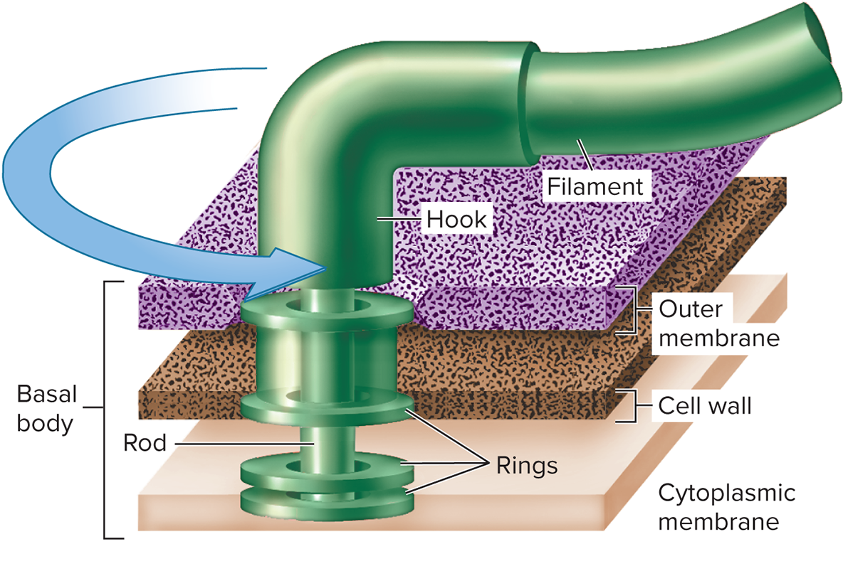
Bacterial Flagella:
Function: To provide motility, or self-propulsion so that the cell can swim freely throughout an aqueous habitat
Structure: 3 distinct part: filament, hook, and the basal body
filament: helical structure composed of proteins (20nm diameter + 1-70 microns in length), is inserted into the hook
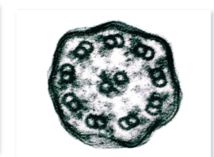
hook: is anchored to the cell by the basal body and is anchored by stacked rings that are through the cell wall, allowing for 360 rotation (with the filament attached)
basal body: the collective outer membrane, cell wall, and cell membrane (cytoplasmic membrane)
Advantage: helps with identification in the lab to diagnosis pathogens via their motility—> also perform chemotaxis which is movement in response to chemical signals
Positive chemotaxis: cells moves towards the direction of a favorable chemical stimulus (nutrient)
Negative chemotaxis: cells move away from repellent (potentially harmful) compound
Phototaxis: movement in response to light than chemicals
Be able to describe the structure of the prokaryotic flagella and how the prokaryotic flagella moves (Also be able to compare this to the structure and movement of the Eukaryotic flagella - see 5.2)
Prokaryotic Flagella:
Function: To provide motility, or self-propulsion so that the cell can swim freely throughout an aqueous habitat
Structure: 3 distinct part: filament, hook, and the basal body
filament: helical structure composed of proteins (20nm diameter + 1-70 microns in length), is inserted into the hook
hook: is anchored to the cell by the basal body and is anchored by stacked rings through a rod that are through the cell wall, allowing for 360 rotation (with the filament attached)
basal body: the collective outer membrane, cell wall, and cell membrane (cytoplasmic membrane)
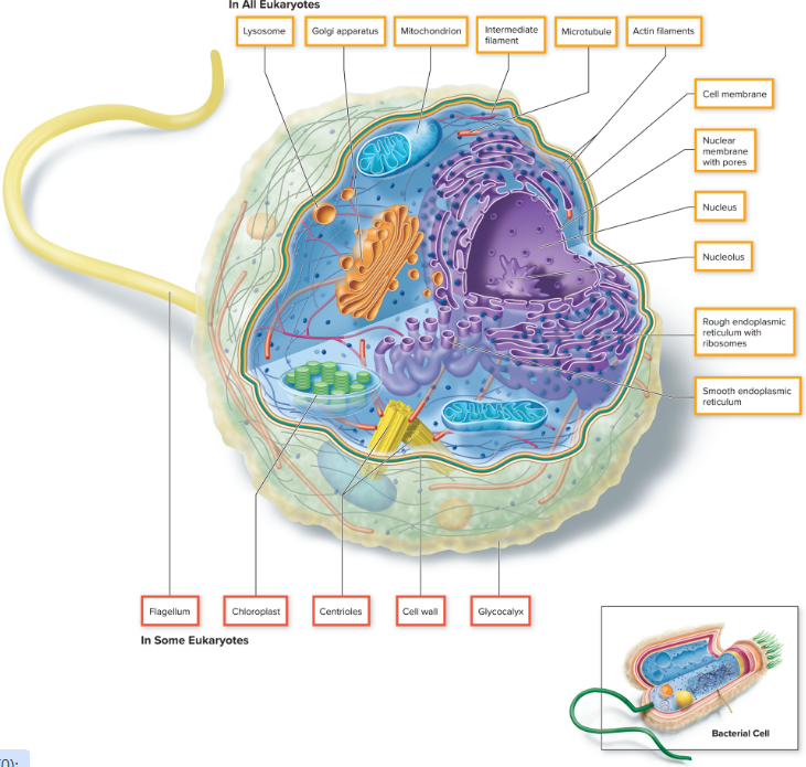
Eukaryotic flagella
Differences between the two
Euk: 10x thicker, structurally more complex, and covered by an extension of the cell membrane
structure: long, sheathed cylinder containing regularly spaced, hollow tubules (microtubules) that extend along its entire length. there are 9 closely attached microtubules inside a flagella
Movement: adjacent microtubules slide past each other , whipping the flagellum back and forth.
Understand the concept of chemotaxis and how the bacteria move while undergoing chemotaxis (be sure to be clear on whether you are being asked for the flagella movement OR the bacteria movement).
chemotaxis: The tendency of organisms to move in response to a chemical gradient (toward an attractant or to avoid adverse stimuli).
Positive chemotaxis: cells moves towards the direction of a favorable chemical stimulus (nutrient)
Negative chemotaxis: cells move away from repellent (potentially harmful) compound
Phototaxis: movement in response to light than chemicals
Mechanism of Chemotaxis:
Chemotaxis involves two key components:
Sensing the environment:
Bacteria have specialized proteins called chemoreceptors (or methyl-accepting chemotaxis proteins, MCPs) embedded in their cell membrane. These detect attractants or repellents in the environment.
Chemoreceptors transmit signals to a signaling pathway, which ultimately controls the movement of the bacterial flagella.
Movement of bacteria (Bacterial Motion):
Bacteria move by rotating their flagella.
Movement alternates between two types:
Run: Smooth, straight-line movement caused by counterclockwise (CCW) rotation of flagella, which bundle together.
Tumble: Random, reorienting movement caused by clockwise (CW) rotation of flagella, which fly apart and disrupt the bundle.
flagellum is effective due to its system for detecting chemical is linked to the mechanisms that drive the flagellum—> cluster of receptors in the cytoplasmic membrane that bind specific molecules that cause them to attach, transmitting signals to flagellum to set it into rotary motion
the movement is caused by a proton gradient (hydrogen ions)
generated by metabolism of the bacterium and bind to and detaches from parts of the flagellar motoe within the cytoplasmic membrane, causing the filament to roate
Flagella Movement:
Controlled by the direction of rotation:
Counterclockwise (CCW) rotation:
Causes the flagella to bundle and work together.
Results in a run (directed motion).
Clockwise (CW) rotation:
Causes the flagella to spread apart.
Results in a tumble (random reorientation).
Bacterial Movement:
Bacteria exhibit biased random walks during chemotaxis:
In the presence of an attractant:
Runs become longer, and tumbles become less frequent, directing the bacteria toward higher concentrations of the attractant.
In the presence of a repellent:
Runs become shorter, and tumbles become more frequent, helping bacteria reorient away from the repellent.