Cell Structure and Function Chapter 8
1/35
There's no tags or description
Looks like no tags are added yet.
Name | Mastery | Learn | Test | Matching | Spaced |
|---|
No study sessions yet.
36 Terms
3 Transport Mechanisms
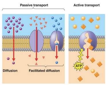
Simple Diffusion
Facilitated Diffusion
Active Transport
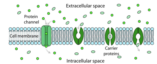
Transport Proteins
Assist most solutes across membranes
Some move solutes to regions of lower concentration (facilitated diffusion) which uses no energy.
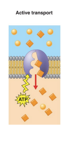
Active Transport
Transport proteins move solutes against the concentration gradient
Requires energy
Hydrolysis of ATP
Simultaneous transport of another solute down an energy gradient (diffusion down one gradient drives diffusion up another gradient)
Concentration gradient
Determines the movement of a molecule that has no net charge
Passive Transport - exergonic movement “down”
Negative free energy
Active Transport - endergonic movement “up”
positive free energy
Electrochemical potential
Determines movement of ion
Concentration gradient and charge gradient across membrane
Charged Gradient/Membrane Potential (Vm)
Active Transport of ions across a membrane creates it
It is a charge separation across a membrane (i.e. voltage)
Membrane voltage can be used for nerve impulse conduction and driving transport of solutes
Excess of negatively charged solutes inside the cell
Simple Diffusion
the unassisted net movement of a solute from high to lower concentration
Only Possible for:
Gases
nonpolar molecules
small polar molecules (water, ethanol)
Diffusion is always movement toward equilibrium
Free energy minimized
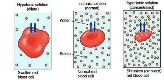
Osmosis
Water moves towards the region of higher solute concentration
For most cells water tends to move inward
Osmolarity
Total solute concentrations inside versus outside the cell
Hypertonic - solute concentration is higher outside the cell (concentrated)
Hypotonic - solute concentration is lower outside the cell (diluted)
Isotonic Solution - Same solute concentration inside and outside the cell
Cell Response
Shrink in hypertonic
Swell/burst in hypotonic
Cell walls prevent swelling and bursting and instead become very firm from turgor pressure
Cells without cell walls pump out inorganic ions to reduce intracellular osmolarity
Solute Size
lipid bilayers are more permeable to small molecules
without a transporter however they most more slowly
molecular weight less than 100 Da it can diffuse across the membrane.
Solute Polarity
Lipid bilayers are more permeable to nonpolar substances than to polar ones
Dissolve readily into the hydrophobic region of the bilayer
Even large nonpolar molecules cross easily
Solute Charge
Charged Solutes do not spontaneously cross the bilayer
Cells use this property to create electrochemical gradients
IT IS NECESSARY FOR PROPER FUNCTION
ATP generation, signaling etc.
Could be gradient of either sodium ions of protons and are established by active transporter
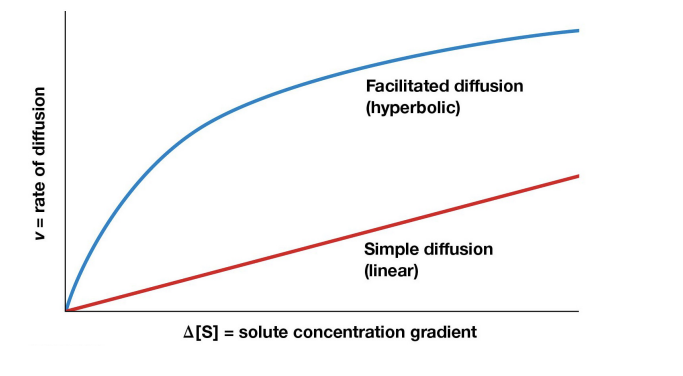
Diffusion Kinetics
Rate of simple diffusion is directly proportional to the concentration gradient
Facilitated Diffusion
Molecules move down their concentration gradient (from high to low) across a cell membrane with the help of membrane proteins
Limited # of transport proteins so saturation occurs
Types of Transporters
Carrier Proteins - bind solute molecules on onside of a membrane, undergo a conformation change, and release the solute on the other side of the membrane
Channel Proteins - form hydrophilic channels through the membrane to provide a passage route for solutes
Alternating Conformation Model
a Carrier protein is allosteric protein and alternates between 2 conformation states
1 - Solute binding site is accessible on one side of membrane
2 - Shifts to the alternate conformation, with the solute binding site on the other side of the membrane, triggering solute release.
Specificity of Carrier Proteins
Carrier proteins are analogous to enzymes
High specificity
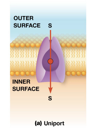
Uniport
When a carrier protein (uniporter) transports a single solute across the membrane
Coupled Transport
2 solutes transported simultaneously and their transport is coupled
Symport
2 solutes moves across a membrane in the same direction
Antiport
2 solutes moves in opposite directions
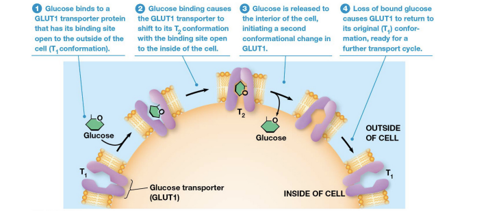
Glucose Transporter
Uniport carrier
Glucose is transported inward by a glucose transported (GLUT; GLUT1 in erythrocytes)
Integral membrane Protein with 12 transmembrane segments which form a cavity with hydrophilic side chains
Process of GLUT1
Glucose collides with and binds to GLUT1 in T1 conformation
GLUT1 shifts to T2 conformation
Conformation change releases glucose
GLUT1 returns to original T1 conformation
Can be reversed.
Glucose concentration kept low inside most animal cells.
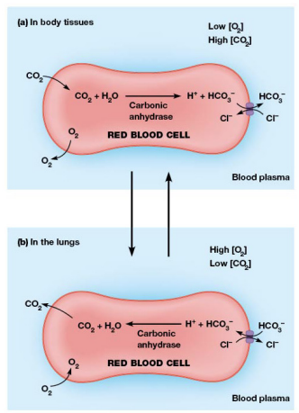
Anion Exchange Protein
Chloride bicarbonate exchanger
Facilities reciprocal exchange of Cl- and HCO3- ions only
Strict 1:1 ratio and can only occur if both anions are present
“Ping Pong” Mechanism
2 conformation states
1st - Protein binds a chloride ion on one side of the membrane which causes a change to the second state
2nd - chloride moved across the membrane and released
This release causes the protein to bind bicarbonate cause a shift back to the first conformation
Bicarbonate moves out of the cell allowing the carrier to bind chloride again.
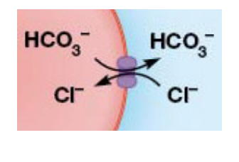
Significance of Anion Exchange Protein
Waste co2 diffuses into erythrocytes where it is converted into HCO3-
Moves out to prevent net charge imbalance
In lungs this is reversed
Channel Proteins
Form hydrophilic transmembrane channels that allow specific solutes to cross the membrane directly
Types of Channels
Ion
porins
aquaporins
Ion Channels
tiny pores lined with hydrophilic atoms
remarkably selective
Gated Channels
Most ion channels are gated
Voltage Gated Channels - open and close in response to changes in membrane potential
Ligand Gated Channels -triggered by the binding of certain substances to the channel protein
Mechanosensitive channels - responds to mechanic forces on membrane
Functions of Ion Channels
cellular communication
eg. muscle contraction and electrical signaling of nerve cells
Maintain salt balance in cells and airways linking the lungs
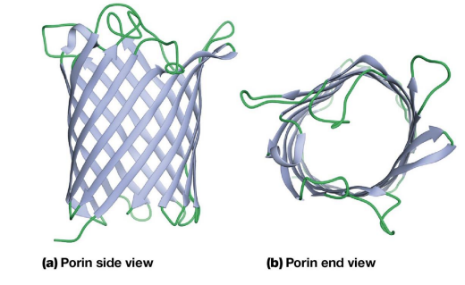
Porins
Pores on outer membranes of bacteria, mitochondria, and chloroplasts
Larger and less specific
Formed by multipass transmembrane proteins aka Porins
Structure of Porins
Beta Barrel has a water filled pore at its center
Polar side chains line the inside of the pore, allowing passage of many hydrophilic solutes
The outside of the barrel contains many nonpolar side chains that interact with the hydrophobic interior of the membrane
Aquaporins (AQP)
Water channel proteins found in cell membranes that selectively facilitate the rapid passage of water, and sometimes small solutes like glycerol or urea, across the membrane.
erythrocytes and kidney cells
root cells and vacuolar membranes
AQP structure
Tetrameric integral membrane proteins
4 central channels
Just large enough for water molecules to pass through one at a time
