anatomy of female pelvis PENNY chapt. 16
1/59
There's no tags or description
Looks like no tags are added yet.
Name | Mastery | Learn | Test | Matching | Spaced | Call with Kai | Chat |
|---|
No analytics yet
Send a link to your students to track their progress
60 Terms
1. Which of the following is not part of the colon that is located within the female pelvis?
a. Ascending
b. Descending
c. Sigmoid
d. Transverse
d. Transverse
2. Which of the following is a pelvic ligament that extends from the lateral surface of the cervix to the lateral fornix of the vagina?
a. Common iliac ligament
b. Cardinal ligament
c. Iliopsoas ligament
d. Broad ligament
b. Cardinal ligament
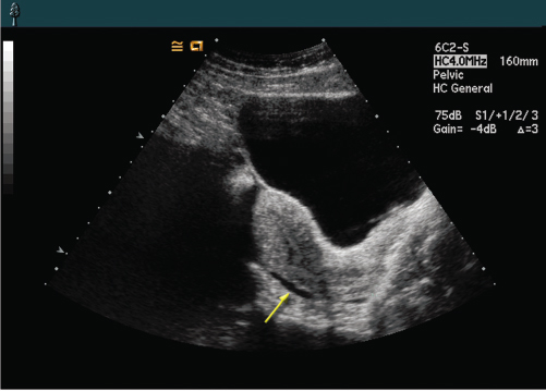
3. In Figure 16-15, what does the arrow indicate?
a. Fluid within the rectouterine pouch
b. Fluid within the vesicouterine pouch
c. Fluid within the space of Retzius
d. Fluid within the anterior cul-de-sac
a. Fluid within the rectouterine pouch
4. The uterine vasculature is located within the:
a. broad ligaments.
b. cardinal ligaments.
c. suspensory ligament of the ovary.
d. obturator internus ligament.
b. cardinal ligaments.
5. What do the arrows in Figure 16-16 indicate?
a. Radial arteries
b. Uterine arteries
c. Spiral arteries
d. Arcuate arteries
d. Arcuate arteries
6. What is another name for the true pelvis?
a. Greater pelvis
b. Lesser pelvis
c. Superior pelvis
d. Major pelvis
b. Lesser pelvis
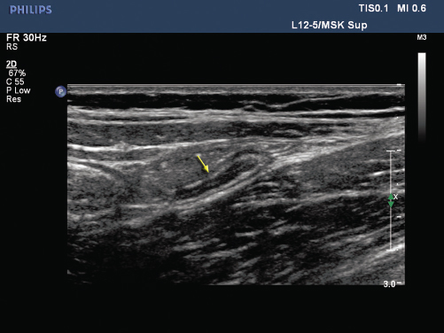
7. The patient in Figure 16-17 complained of right lower quadrant pain and presented for a transabdominal pelvic sonogram. Upon closer presentation with a linear transducer, you noted the structure identified by the arrow. What is the most likely etiology of this structure?
a. Cecum
b. Rectum
c. Appendix
d. Sigmoid colon
c. Appendix
8. Which of the following muscles is located posteriorly within the pelvis and helps support the sacrum?
a. Iliopsoas
b. Coccygeus
c. Obturator internus
d. Piriformis
b. Coccygeus
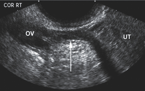
9. The arrow in Figure 16-18 indicates the:
a. ovarian ligament.
b. round ligament.
c. uterosacral ligament.
d. broad ligament.
d. broad ligament.
10. Which of the following is not supported by the broad ligaments?
a. Pelvic diaphragm
b. Uterus
c. Uterine tubes
d. Ovaries
a. Pelvic diaphragm
11. Which of the following is not a levator ani muscle?
a. Pubourethralis
b. Pubovaginalis
c. Puboileacus
d. Iliococcygeus
c. Puboileacus
12. What artery directly supplies blood to the basal layer of the endometrium?
a. Spiral
b. Arcuate
c. Straight
d. Radial
c. Straight
13. Which of the following is not true of the uterine plexus?
a. They are tortuous like the arteries.
b. They supply blood to the uterine tubes.
c. They anastomose with each other and the ovarian vein.
d. They are located along the sides of the cervix and the cornua.
b. They supply blood to the uterine tubes.
14. What is the fibrous structure located along the midline of the abdomen that separates the rectus abdominis muscles?
a. Linea terminalis
b. Linea rectus
c. Linea alba
d. Linea aspera
c. Linea alba
15. What is the relationship of the lesser pelvis to the greater pelvis?
a. It is located more inferiorly.
b. It is located more laterally.
c. It is located more anteriorly.
d. It is located more superiorly.
a. It is located more inferiorly.
16. What part of the cervix is closest to the vagina?
a. Internal os
b. Internal canal
c. Adventitia
d. External os
d. External os
17. What midline, anterior pelvic structure may produce an acoustic shadow when scanning the female pelvis?
a. Iliac crest
b. Anterior superior iliac spine
c. Pubic symphysis
d. Ischial ramus
c. Pubic symphysis
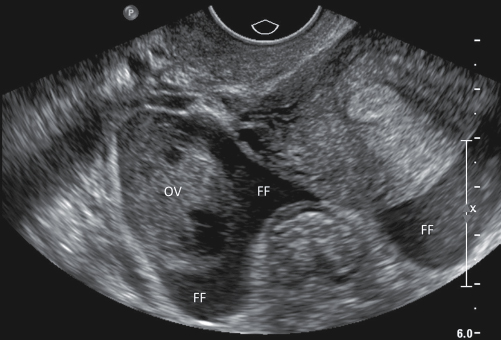
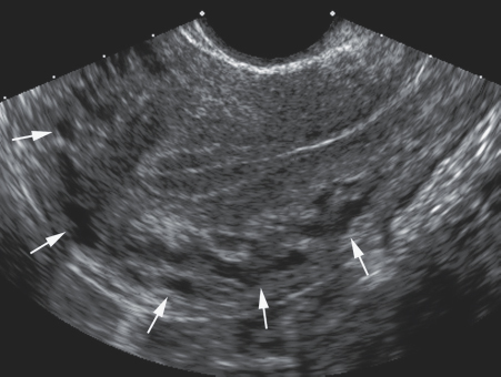
18. Figure 16-19 is a transverse sonogram of the female pelvis. Which of the following statements is not true of this image?
a. The free fluid is noted within the right adnexa.
b. The free fluid is noted within the rectouterine pouch.
c. The free fluid is noted within the posterior cul-de-sac.
d. The free fluid appears to be anechoic and simple.
d. The free fluid appears to be anechoic and simple.
19. The common iliac veins combine to create the:
a. inferior vena cava.
b. external iliac vein.
c. internal iliac vein.
d. abdominal aorta.
a. inferior vena cava.
20. You are performing a female pelvic sonogram and identify a solid mass adjacent to the right ovary, just right lateral to the uterus. What is the most likely location of this mass?
a. Within the right adnexa
b. Within the pouch of Douglas
c. Within the pelvic diaphragm
d. Within the space of Retzius
a. Within the right adnexa
21. What structure within the female pelvis lies posterior to the urinary bladder and anterior to the rectum?
a. Broad ligament
b. Rectus abdominis muscle
c. Space of Retzius
d. Uterus
d. Uterus
22. Fluid noted posterior to the uterus would most likely be located within the:
a. space of Retzius.
b. pouch of Douglas.
c. anterior cul-de-sac.
d. adnexa.
b. pouch of Douglas.
23. Both the straight and spiral arteries are branches of the:
a. common iliac artery.
b. radial artery.
c. arcuate artery.
d. external iliac artery.
b. radial artery.
24. The left ovarian vein drains directly into the:
a. right renal vein.
b. inferior vena cava.
c. aorta.
d. left renal vein.
d. left renal vein.
25. Pelvic bones, when visualized on sonography, will produce:
a. posterior shadowing.
b. posterior enhancement.
c. mirror image artifact.
d. minimal enhancement.
a. posterior shadowing.
26. The uterine arteries supply blood to all of the following except:
a. fallopian tubes.
b. rectum.
c. ovaries.
d. uterus.
b. rectum.
27. The anterior cul-de-sac is also referred to as the:
a. space of Retzius.
b. rectouterine pouch.
c. pouch of Douglas.
d. vesicouterine pouch.
d. vesicouterine pouch.
28. What is considered the most dependent part of the peritoneal cavity?
a. Space of Retzius
b. Anterior cul-de-sac
c. Pouch of Douglas
d. Rectovesical pouch
c. Pouch of Douglas
29. The right ovarian vein drains directly into the:
a. right renal vein.
b. aorta.
c. inferior vena cava.
d. common iliac vein.
c. inferior vena cava.
30. The innominate bones of the pelvis consist of the:
a. ischium, ilium, and pubic bones.
b. ilium, sacrum, and coccyx bones.
c. sacrum, coccyx, and pubic bones.
d. sacrum, ischium, and ilium bones.
a. ischium, ilium, and pubic bones.
31. What other term is used to describe the space of Retzius?
a. Posterior cul-de-sac
b. Anterior cul-de-sac
c. Murphy pouch
d. Retropubic space
d. Retropubic space
32. The true pelvis is delineated from the false pelvis by the:
a. space of Retzius.
b. adnexa.
c. linea terminalis.
d. linea alba.
c. linea terminalis.
33. The vagina is located __ to the uterus:
a. anterior
b. posterior
c. inferior
d. medial
c. inferior
34. The muscles that may be confused with the ovaries on a pelvic sonogram include the:
a. rectus abdominis and obturator internus muscles.
b. levator ani and coccygeus muscles.
c. obturator internus and levator ani muscles.
d. piriformis and iliopsoas muscles.
d. piriformis and iliopsoas muscles.
35. Which vessels supply blood to the deeper layers of the myometrium?
a. Radial arteries
b. Spiral arteries
c. Straight arteries
d. Arcuate arteries
a. Radial arteries
36. Pelvic muscles appear:
a. echogenic.
b. anechoic.
c. hypoechoic.
d. complex.
c. hypoechoic.
37. The abdominal aorta bifurcates into the:
a. internal iliac arteries.
b. common iliac arteries.
c. ovarian arteries.
d. external iliac arteries.
b. common iliac arteries.
38. Which of the following are the paired anterior abdominal muscles that extend from the xiphoid process of the sternum to the pubic bone?
a. Iliopsoas muscles
b. Rectus abdominis muscles
c. Obturator internus muscles
d. Piriformis muscles
b. Rectus abdominis muscles
39. Peritoneal spaces located posterior to the broad ligament are referred to as the:
a. rectouterine spaces.
b. anterior cul-de-sacs.
c. lateral cul-de-sacs.
d. adnexa.
d. adnexa.
40. The paired muscles that are located lateral to the uterus and anterior to the iliac crest are the:
a. iliopsoas muscles.
b. rectus abdominis muscles.
c. obturator internus muscles.
d. piriformis muscles.
a. iliopsoas muscles.
41. Fluid noted anterior to the uterus would most likely be located within the:
a. pouch of Douglas.
b. vesicouterine pouch.
c. space of Retzius.
d. rectouterine pouch.
b. vesicouterine pouch.
42. The bilateral muscles that are located posterior to and extend from the sacrum to the femoral greater trochanter are the:
a. levator ani muscles.
b. rectus abdominis muscles.
c. obturator internus muscles.
d. piriformis muscles.
d. piriformis muscles.
43. The pelvic ligament that provides support to the ovary to the pelvic side wall is the:
a. cardinal ligament.
b. ovarian ligament.
c. broad ligament.
d. suspensory ligament of the ovary.
d. suspensory ligament of the ovary.
44. The pelvic muscle group that is located between the coccyx and the pubis is the:
a. levator ani muscles.
b. rectus abdominis muscles.
c. obturator internus muscles.
d. piriformis muscle.
a. levator ani muscles.
45. The sonographic pelvic examination of a female patient reveals an extensive amount of ascites. In the transverse plane, you visualize two echogenic structures extending from the side walls of uterus to the pelvic side walls bilaterally. These structures are most likely the:
a. broad ligaments.
b. cardinal ligaments.
c. ovarian ligaments.
d. uterosacral ligaments
a. broad ligaments.
46. The space of Retzius is located:
a. between the uterus and the bladder.
b. between the bladder and the ilium.
c. along the lateral aspect of the uterus.
d. between the bladder and the pubic bone.
d. between the bladder and the pubic bone.
47. The right ovarian artery branches off of the:
a. aorta.
b. right renal artery.
c. uterine artery.
d. internal iliac artery.
a. aorta.
48. The muscle located lateral to the ovaries is the:
a. iliopsoas muscle.
b. rectus abdominis muscle.
c. obturator internus muscle.
d. piriformis muscle.
c. obturator internus muscle.
49. The arteries that directly supply blood to the functional layer of the endometrium are the:
a. radial arteries.
b. spiral arteries.
c. straight arteries.
d. arcuate arteries.
b. spiral arteries.
50. Another name for the rectouterine pouch is the:
a. space of Retzius.
b. pouch of Retzius.
c. pouch of Douglas.
d. anterior cul-de-sac.
c. pouch of Douglas.
51. A patient presents to the sonography department with a history of uterine prolapse. Which of the following best describes this disorder?
a. A condition that results from the weakening of the pelvic diaphragm muscles and allows for the displacement of the uterus, often through the vagina.
b. A congenital anomaly that results in the duplication of the uterus.
c. A condition that results in the abnormal invasion of the myometrium through the bladder wall, leading to hematuria.
d. An abnormality that describes the inversion of the myometrium and endometrium.
a. A condition that results from the weakening of the pelvic diaphragm muscles and allows for the displacement of the uterus, often through the vagina.
52. The pelvic ligament that extends from the lateral aspect of the uterus to the side walls of the pelvis is the:
a. broad ligament.
b. ovarian ligament.
c. piriformis ligament.
d. round ligament.
a. broad ligament.
53. The uterine artery branches off of the:
a. abdominal aorta.
b. uterine plexus.
c. internal iliac artery.
d. external iliac artery.
c. internal iliac artery.
54. The peripheral arteries of the uterus are the:
a. radial arteries.
b. spiral arteries.
c. straight arteries.
d. arcuate arteries.
d. arcuate arteries.
55. The urinary bladder, uterus, and ovaries are located within the:
a. true pelvis.
b. false pelvis.
a. true pelvis.
56. The pelvic ligament that provides support to the ovary and extends from the ovary to the lateral surface of the uterus is the:
a. cardinal ligament.
b. ovarian ligament.
c. broad ligament.
d. suspensory ligament of the ovary.
b. ovarian ligament.
57. The surface of the pelvic bones, when visualized on sonography, will appear:
a. anechoic.
b. hypoechoic.
c. dark.
d. hyperechoic.
d. hyperechoic.
58. Which vessel is the longest?
a. left ovarian vein
b. left ovarian artery
c. right ovarian vein
d. right ovarian artery
a. left ovarian vein
59. The ovary is supplied blood by the:
a. ovarian artery.
b. ovarian artery and uterine artery.
c. uterine artery.
d. arcuate artery.
b. ovarian artery and uterine artery.
60. Prolapse of the pelvic organs most often involves the:
a. rectus abdominis and obturator internus muscles.
b. levator ani and coccygeus muscles.
c. obturator internus and levator ani muscles.
d. piriformis and iliopsoas muscles.
b. levator ani and coccygeus muscles.