Normal Sonographic Findings During the First Trimester
 Call Kai
 Learn
Learn Practice Test
 Spaced Repetition
 Match
Match Flashcards
 Knowt Play
1/85

There's no tags or description
Looks like no tags are added yet.
Name | Mastery | Learn | Test | Matching | Spaced | Call with Kai | Chat |
|---|
No analytics yet
Send a link to your students to track their progress
86 Terms
— of the endometrium is essentially the first sonographically identifiable sign of a pregnancy
The decidual reaction
The decidualized endometrium will appear — as a result of the continued production of progesterone by the corpus luteum
thick and echogenic
A decidual reaction is considered to be a — of pregnancy because the endometrium can also appear thick and echogenic during the secretory phase of the endometrial cycle and in the presence of an ectopic pregnancy
nonspecific sonographic finding
The first definitive sonographic sign of an IUP is identification of the —
gestational sac within the decidualized endometrium.
— is the developmental stage of the conceptus that implants into the uterine cavity
The blastocyst
The blastocyst gives rise to the —
gestational sac, or chorionic sac
The early gestational sac, which is first seen at —, appears as a small, anechoic sphere within the decidualized endometrium
5 weeks
the gestational sac will grow at a rate of — in early pregnancy
1mm per day
— denotes the appearance of the small gestational sac in the uterine cavity surrounded by the thickened, echogenic endometrium
The intradecidual sign
The intradecidual sign can be misdiagnosed because it may resemble the —
pseudogestational sac of an ectopic pregnancy
To differentiate an intrauterine gestational sac from the pseudogestational sac, sonographers can assess the endometrium for evidence of the —
double sac sign or double decidual sign
— denotes the typical appearance of the two distinct layers of decidua, the decidua capsularis (inner layer) and decidua parietalis (outer layer), separated by the anechoic fluid-filled uterine cavity.
The double sac sign

Gestational sac at 5.0 weeks’ gestation. The gestational sac (*) is seen as a round fluid collection within the endometrium, with no structures yet seen within it. The gestational sac is surrounded by two echogenic rings, an inner ring (short arrows) and an outer ring (long arrows), corresponding to the two layers of the decidua. This is referred to as the double sac sign or double decidual sign.
chorionic cavity
The space between the gestational sac and the amniotic sac.
The location of the secondary yolk sac.
chorion frondosum
The decidualized tissue at the implantation site containing the chorionic villi.
The fetal contribution of the placenta.
chorion laeve
The portion of the chorion that does not contain chorionic villi
chorionic villi
Fingerlike extension of trophoblastic tissue that invades the decidualized endometrium.
decidua basalis
The endometrial tissue at the implantation site.
The maternal contribution of the placenta.
decidua capsularis
The portion of the decidua opposite the uterine cavity, across from the decidua basalis.
decidua parietalis (vera)
The decidualized tissue along the uterine cavity adjacent to the decidua basalis.
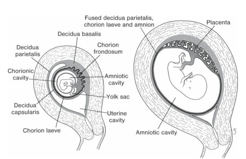
Relation of fetal membranes to wall of the uterus.
— is the earliest sonographic measurement that can be obtained to date the pregnancy
The measurement of the gestational sac
A mean sac diameter (MSD) is achieved by adding the measurements of the —
length, width, and height of the gestational sac and dividing by 3.
By — (measurement in millimeters). sonographers can obtain an estimate for the gestational age in days.
adding 30 to the MSD
An irregularly shaped gestational sac and an MSD of — that does not contain a fetal pole are both signs potential pregnancy failure.
>25 mm
The first structure seen with sonography within the gestational sac is the —
secondary yolk sac
the secondary yolk sac appears —
within the gestational sac as a round, anechoic structure surrounded by a thin, echogenic rim
the secondary yolk sac is located —
within the chorionic cavity, between the amnion and the chorion
This cavity between the amnion and the chorion may also be referred to as the —
extraembryonic coelom or extracelomic space
The yolk sac produces — and plays an important role in angiogenesis and hematopoiesis during early embryologic development
alpha-fetoprotein
the secondary yolk sac is connected to the embryo by the —, which contains one artery and one vein
vitelline duct, also referred to as the omphalomesenteric duct

Gestational sac and secondary yolk sac at 5.5 weeks’ gestation. The gestational sac (arrowheads) contains the secondary yolk sac (arrow). No embryo is seen at this time.
The gestational sac consists of two cavities: —
the chorionic cavity and amniotic cavity
The chorionic cavity lies between the—
amnion and the chorion.
the chorionic cavity contains —
the yolk sac and fluid
The amniotic cavity contains
simple-appearing amniotic fluid and the developing embryo
The amniotic membrane, or amnion, can be seen within the gestational sac as a —
thin, echogenic line loosely surrounding the embryo
The amnion and chorion typically fuse around the middle of the first trimester, but may not be totally fused until —
16 weeks’ gestation
By —, the embryo can be seen located within the amniotic cavity adjacent to the yolk sac, with transvaginal sonography
6 weeks
The documentation of fetal heart activity is performed using —
motion mode (M-mode)
Occasionally, a tiny heartbeat is often seen before an embryo can be measured, with sonographic documentation of heart activity being present between —
5 and 6 weeks
Heart motion can be detected in a 4-mm embryo, with motion certainly evident within the —.
5-mm embryo
The embryo will grow at a rate of — in the first trimester
1 mm per day
Embryonic heart rate is considered normal at — between 5 and 6 weeks
100 to 110 bpm
The heart rate increases to — by 9 weeks
150 bpm
From —, the fetal heart rate is typically around 150 bpm although it will vary with gestation age.
second trimester to term
— is associated with a poor prognosis and is often the first sonographic sign of an eminent embryologic demise
Bradycardia
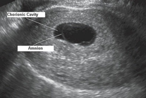
Chorionic cavity and amnion. The chorionic cavity and amnion are seen within this image of an early pregnancy.
The most accurate sonographic measurement of pregnancy is the —
crown rump length
The crown rump length (CRL) can be taken when a — is identified and should not include the yolk sac or fetal limb buds within the measurement
fetal pole

Crown rump length (CRL). A. CRL (between calipers) at 6.5 weeks. The yolk sac can be clearly identified (arrow) adjacent to the embryo. B. CRL (between calipers) at 10 weeks and 4 days.
Fetal limb buds are readily identified by —
7 weeks
Within the fetal head, a cystic structure may be noted. This most often represents the —
rhombencephalon, or hindbrain
The rhombencephalon will eventually develop into the —
fourth ventricle and several other essential brain structures
As early as —, the stomach may be visualized in the upper abdomen as well
8 weeks
— begins at 8 weeks, which marks the developmental stage when the midgut migrates into the base of the umbilical cord
Physiologic bowel herniation
Conversely, if physiologic bowel herniation does not resolve by —, a follow-up examination is often warraned
12 weeks

Rhombencephalon. The rhombencephalon (arrow) is seen within the head of this 8-week embryo, appearing as a cyst.

Physiologic bowel herniation. Transverse image of the fetal abdomen at 11.5 weeks’ gestation demonstrating the normal herniation of the bowel (arrow) into the base of the umbilical cord.
the fetal kidneys may be seen between —
13 and 14 weeks

Choroid plexus within the lateral ventricles. Choroid plexus (arrows) is seen within the lateral ventricle of this 13-week gestation fetus
The developing — may be noted at the end of first trimester as a welldefined, crescent-shaped homogeneous mass of tissue, along the margins of the gestational sac
placenta
The placenta is formed by the —
decidua basalis, the maternal contribution of the placenta, and the chorion frondosum, the fetal contribution.
The umbilical cord is visible during the latter half of the first trimester as a tortuous structure connecting the —
fetus to the developing placenta.
The evaluation of the — has become a vital part of early first-trimester screening.
nuchal translucency (NT)
The term nuchal refers to the —
neck
this translucency is represented by a thin membrane along the —, which can be measured sonographically
posterior aspect of the fetal neck
The most common abnormalities associated with increased NT are —
trisomy 21, trisomy 18, Turner syndrome, and congestive heart failure.
The NT is optimally measured between —, when the CRL measures between 45 and 84 mm based on recommendations by The Fetal Medicine Foundation, although laboratory specifications may vary
11 and 13 weeks’ 6 days gestation
The normal range of thickness of the NT is based on the gestational age, although most often a measurement — between 11 and 13 weeks 6 days is considered abnormal
greater than 3 mm
The cutoff measurement for NT screening for some institutions may be as high as —
3.5 mm

Placenta at 11 weeks. The developing placenta (arrowheads) is seen in this early pregnancy.
It has been recognized that with many Down syndrome fetuses, the — is either hypoplastic or absent between 11 and 13 weeks’ gestation
nasal bone
the sonographic assessment of the nasal bone can be performed as part of a screening —
first-trimester protocol
In a normal fetus, this will provide an — in the area of the nasal bone and overlying nasal skin.
“equal sign”

Nuchal translucency (NT). A. Normal NT. B. Abnormal NT. C. The first set of calipers is the correct placement for the NT measurement according to the American Institute of Ultrasound in Medicine.

Possible ectopic pregnancy locations.
The most common pelvic mass associated with pregnancy is the —
ovarian corpus luteum cyst
The corpus luteum of pregnancy is a functional cyst that is maintained during the first trimester by hCG, which is produced by the —
developing placenta
As a result, the corpus luteum secretes — and thereby maintains the thickness of the endometrium
progesterone
Typically, the corpus luteum measures between — and regresses near the end of the first trimester although it may continue to grow as large as 10 cm.
2 and 3 cm
sonographic guidelines for a nuchal translucency (NT) measurement
The margins of the NT edges must be clear enough for proper placement of the calipers.
The fetus must be in the midsagittal plane.
The image must be magnified so that it is filled by the fetal head, neck, and upper thorax.
The fetal neck must be in a neutral position, not flexed and not hyperextended.
The amnion must be seen as separate from the NT line.
The (+) calipers on the ultrasound must be used to perform the NT measurement.
Electronic calipers must be placed on the inner borders of the nuchal space with none of the horizontal crossbar itself protruding into the space.
The calipers must be placed perpendicular to the long axis of the fetus.
The measurement must be obtained at the widest space of the NT.
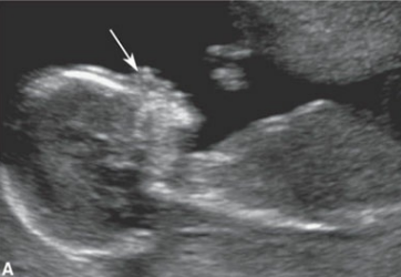
Absent nasal bone

Normal nasal bone
clinical findings of the corpus luteum of pregnancy
Asymptomatic
Pain associated with hemorrhage and enlargement of cyst
sonographic findings of the corpus luteum of pregnancy
Simple cyst appearance
A cysts with a thick, echogenic rim around it (may be difficult to differentiate from other solid and cystic adnexal masses)
Hemorrhagic cyst appearance, including complex components or entirely echogenic depending on the amount of blood and stage of lysis