Chest, Hernia, and Soft Tissue Ultrasound Diagram | Quizlet
 Call Kai
 Learn
Learn Practice Test
 Spaced Repetition
 Match
Match Flashcards
 Knowt Play
1/12

There's no tags or description
Looks like no tags are added yet.
Name | Mastery | Learn | Test | Matching | Spaced | Call with Kai | Chat |
|---|
No analytics yet
Send a link to your students to track their progress
13 Terms
Pleural Effusion
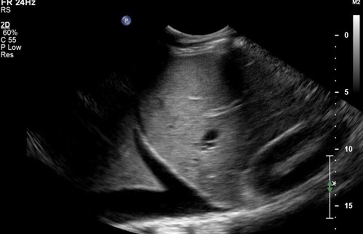
Pneumothorax
No sliding motion seen when patient breathes
Collapsed lung - air in chest cavity outside lung
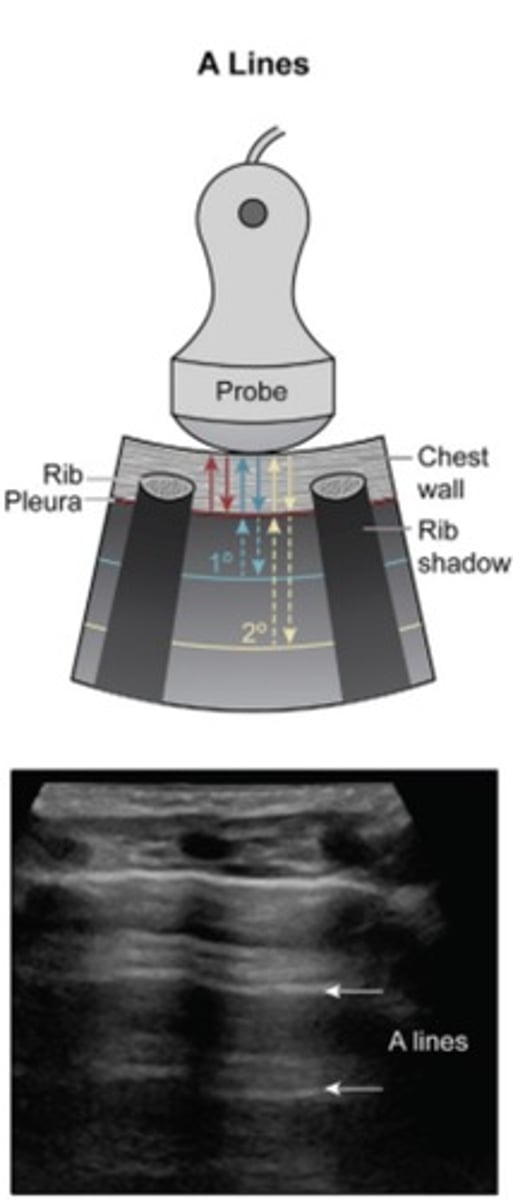
A-Lines
Horizontal
Seen in normal lung
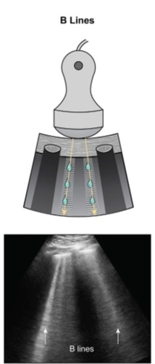
B-Lines
Vertical
Seen in pathological lungs
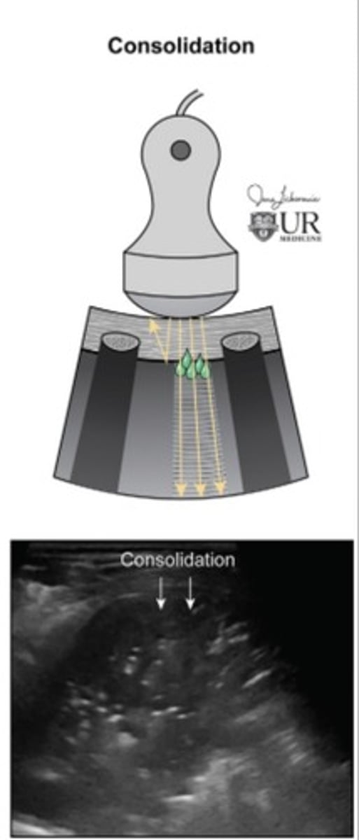
Consolidation
Fluid fills alveoli
Normal A-Lines and air in lungs are lost
Lung looks like liver tissue
Why it is important to scan patients while supine and while standing when evaluating for a hernia?
Gravitational and intra-abdominal pressures change
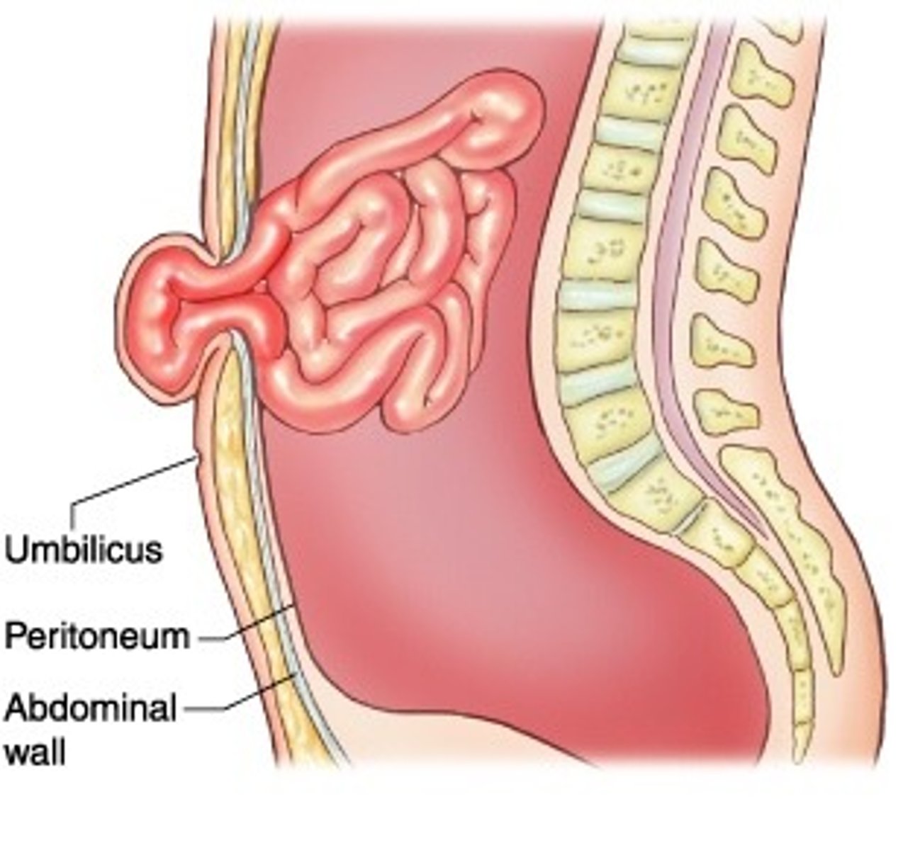
Umbilical Hernia
TERM
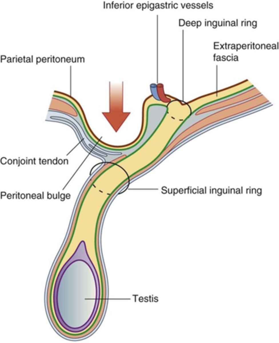
Direct Inguinal Hernia
DEFINITION
TERM
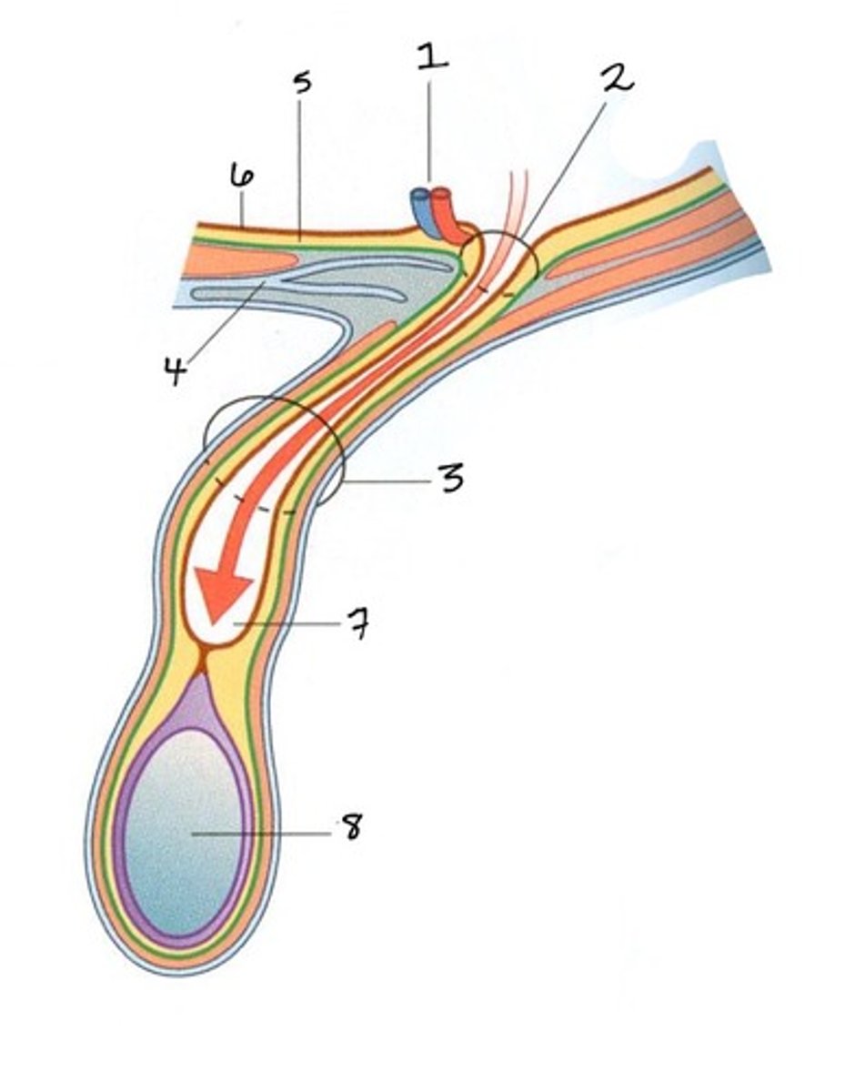
Indirect Inguinal Hernia
DEFINITION
Contents go through spermatic cord
TERM
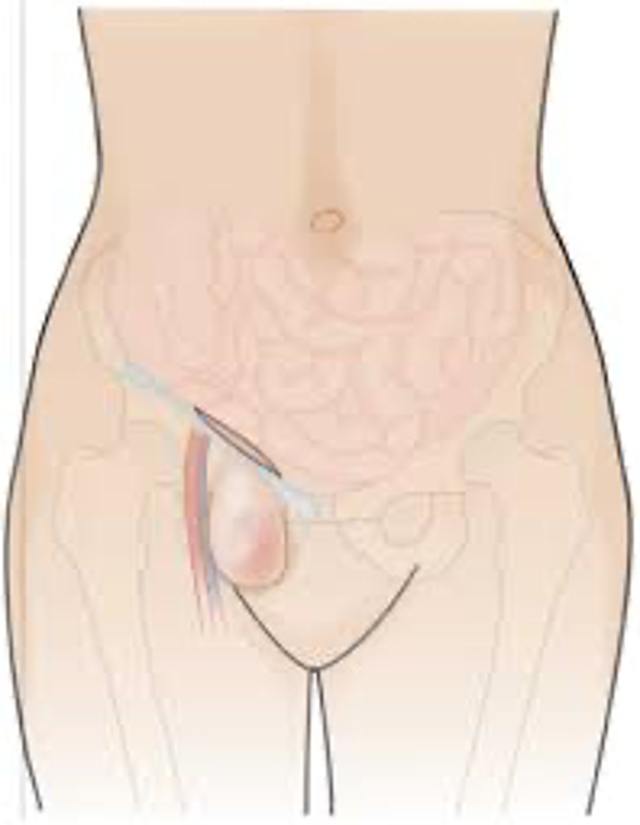
Femoral Hernia
DEFINITION
Below inguinal ligament
TERM
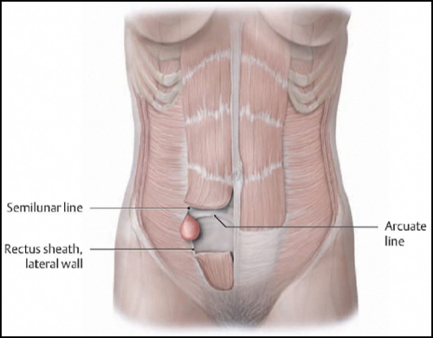
Spigelian Hernia
DEFINITION
Between transverse abdominus and rectus muscles
Scanning Technique for Soft Tissue Masses
Use lots of gel - light skin contact
Adjust settings for low flow (decrease scale, increase gain & frequency)
Use compression when needed
Look deep to the mass (don't miss something)
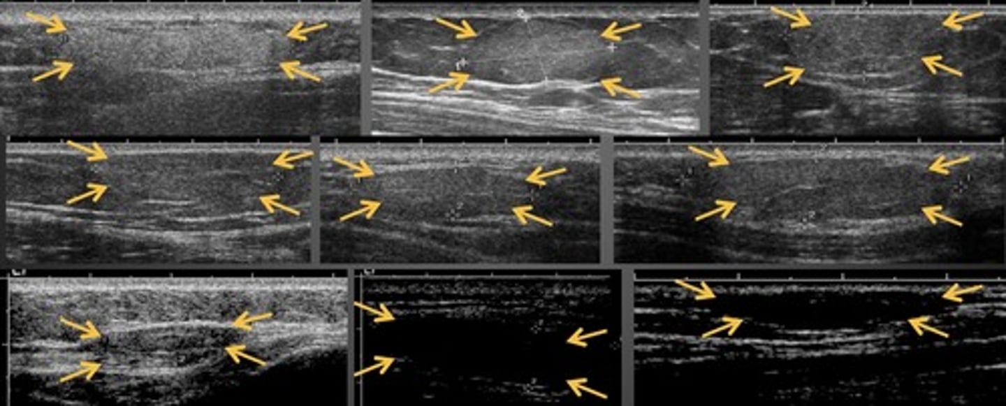
Lipomas
Most common soft tissue tumor
Subcutaneous
Smooth borders
No shadowing
Minimal/no color flow
Somewhat compressible
Range in echogenicity