Tissues: Epithelial and Connective
 Call Kai
 Learn
Learn Practice Test
 Spaced Repetition
 Match
Match Flashcards
 Knowt Play
1/81

Name | Mastery | Learn | Test | Matching | Spaced | Call with Kai | Chat |
|---|
No analytics yet
Send a link to your students to track their progress
82 Terms
Simple epithelia
contain a single layer of cells, with each cell attached to the basement membrane

Stratified epithelia
contain more than one layer of cell. The cells on the basal surface are attached to the basement membrane; those on the apical surface border an open space
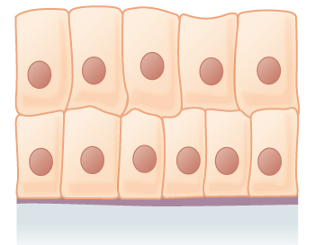
Squamous cells
flat cells with flat, disc-shaped nuclei

Cuboidal cells
cube-shaped cells with spherical, centrally located nuclei

Columnar cells
taller than they are wide, like columns. The nuclei of columnar cells are located near the basal surface and are commonly oval in shape, elongated from top to bottom

simple squamous epithelium
function: allows materials to pass by diffusion and filtration in sites where protection is not important; secretes lubricating substances in serosae (linings of ventral body cavity)
location: kidney glomeruli; air sacs of lungs; lining of heart, blood vessels, and lymphatic vessels; serosaeis

simple cuboidal epithelium
function: secretion and absorption
location: kindey tubules; ducts and secretory portions of small glands; ovary surface

simple columnar epithelium
function: absorption; secretion of mucus, enzymes, and other substances; ciliated type propels mucus (or reproductive cells) by ciliary action
location: conciliated type lines most of the digestive tract (stomach to rectum), gallbladder, and excretory ducts of some glands; cilated variety lines small bronchi uterine tubes, and some regions of the uterus
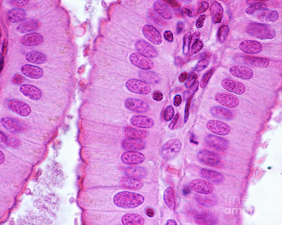
pseudostratified columnar epithelium
function: secrete substances, particularly mucus; propulsion of mucus by ciliary action
location: ciliated variety lines the trachea and most of the upper respiratory tract; nonciliated type in males’ sperm-carrying ducts and ducts of large glands

stratified squamous epithelium
function: protects underlying tissues in areas subjected to abrasion
location: nonkerantinized type forms the most linings of the esophagus, mouth, and vagina; keratinized variety forms the epidermis of the skin, a dry epithelium

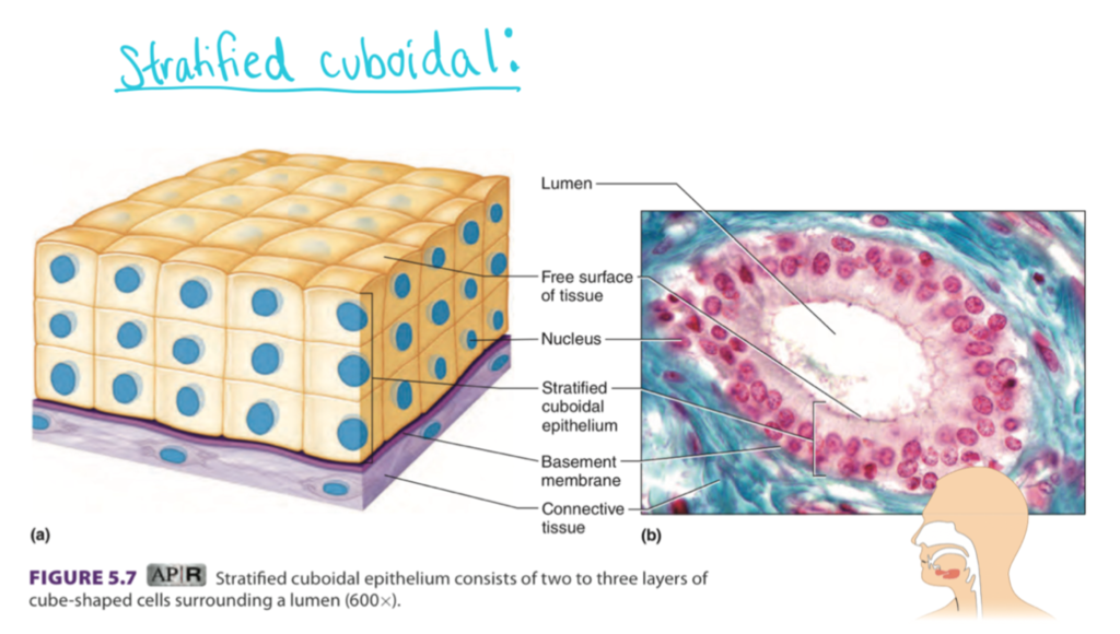
stratified cuboidal epithelium
function: protection
location: largest ducts of sweat glands, mammary glands, and salivary glands
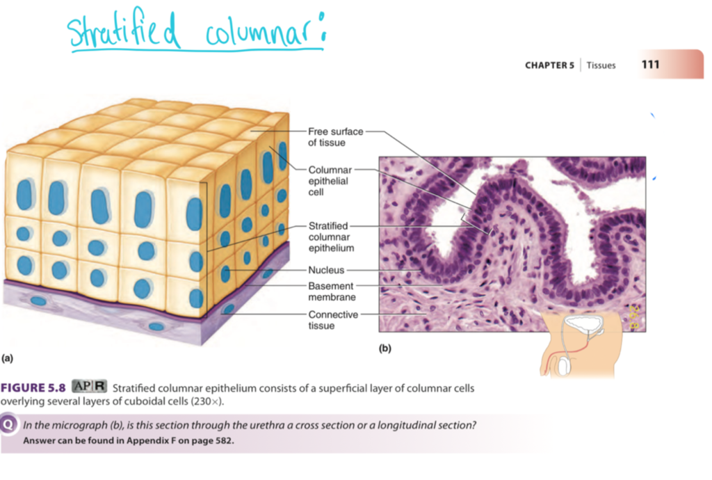
stratified columnar epithelium
function: protection; secretion
location: rare in the body; small amounts in male urethra and in large ducts of some glands
transitional epithelium
function: stretched readily, permits stored urine to distend urinary organ
location: lines the ureters, bladder, and part of the urethra

endocrine glands
internal secretion; ductless glands; produce hormones
exocrine glands
external secretion; numerous; secrete their products onto body surfaces (skin) or into body cavities (such as the digestive tube)
unicellular exocrine glands
goblet cell; shaped like a goble; scattered within the epithelial lining of the intestines and respiratory tubes
multicellular exocrine glands
an epithelium-walled duct and secretory unit (secretory epithelium)
cell junctions
specialized protein structures the connect neighboring cells to each other or to the extracellular matrix
tight junctions
belt-like junctions that encircle the apical region epithelial cells, near the free surface
adhesive belt junctions
type of anchoring cell junction found just below the tight junctions in epithelial tissues
desmosomes
binding cells together tightly; an anchoring junction
gap junction
a tunnel-like junction that can occur anywhere along the lateral membranes of adjacent cells
basal lamina
sheets; thin, noncellular sheet consists of proteins secreted by the epithelial cells
basement membrane
a layer between an epithelium and the under lying connective tissue; consists of both a basal lamina and a network of reticular fibers.
microvilli
fingerlike extensions of the plasma membrane of apical epithelial cells
cilia
whiplike, highly motile extensions of the apical surface membranes of certain epithelial cells
basal body
centriole at the base of each cilium
flagellum
an extremely long, isolated cilium
loose connective tissue
1) Areolar
2) Adipose
3) Reticular
dense connective tissue
1) regular
2) irregular
3) elastic
cartilage
1) hyaline cartilage
2) elastic cartilage
3) fibrocartilage
bone tissue
1) compact bone
2) spongy bone
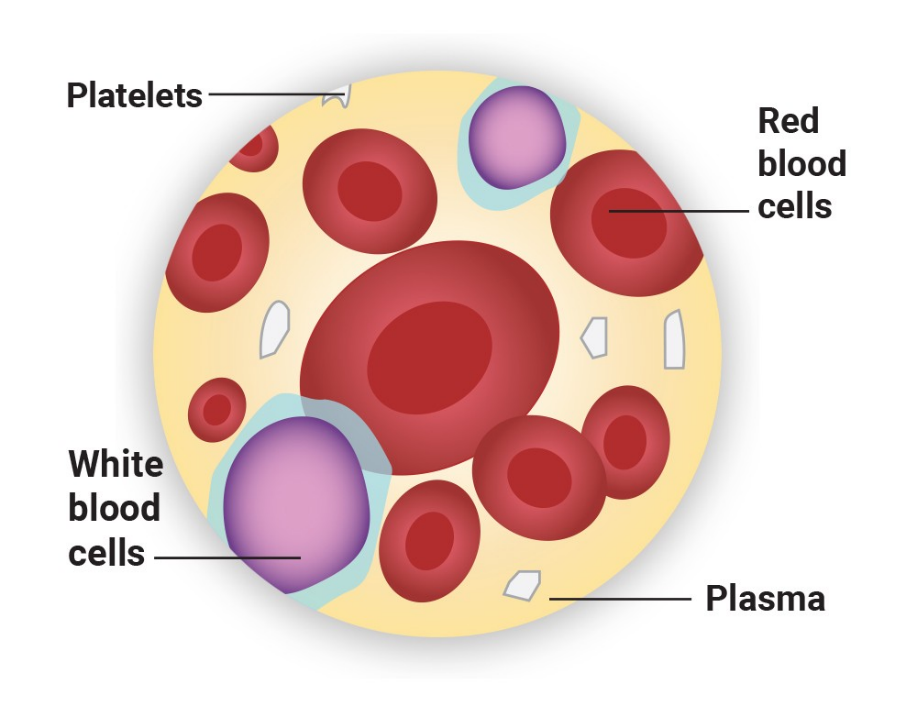
blood
blood cell formation and differentiation are quite complex (RBCs, WBCs, platelets)
extracellular matrix
cells of connective tissues are separated from one another by a large amount of extracellular material
ground substance
nonfiber part of the extracellular matrix of connective tissue
mesenchyme
another feature common to connective tissues is that they all originate from the embryonic tissue
connective tissue → embryonic → mesenchyme
function: gives rise to all other connective tissue
location: primarily in embryo

connective tissue → proper → loose → areolar
function: wraps and cushions organs; its macrophages phagocytize bacteria; plays important role in inflammation; holds and conveys tissue fluid
location: widely distributed under epithelia of body, e.g., forms laminated propria of mucous membranes; packages organs; surrounds capillaries

connective tissue → proper → loose → adipose
function: provides reserve food fuel; insulates against heat loss; supports and protects organs
location: under skin in subcutaneous tissue; around kidneys and eyeballs; within abdomen; in breasts

connective tissue → proper → loose → reticular
function: fibers form a soft internal skeleton (stroma) that supports other cell types including white blood cells, mast cells, and macrophages
location: lymphoid organs (lymph nodes, bone marrow, and spleen)

connective tissue → proper → dense → irregular
function: withstands tension exerted in may directions; provides structural strength
location: fibrous capsules of organs and joints; dermis of the skin; m submucosa of digestive tract
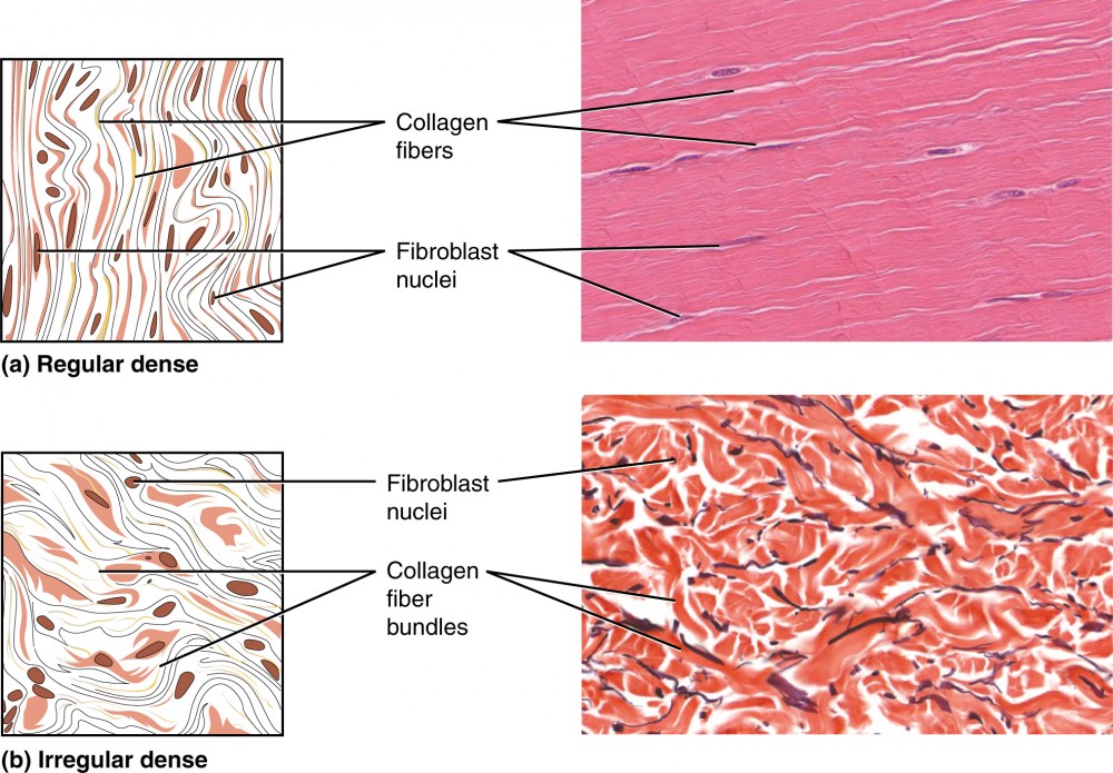
connective tissue → proper → dense → regular
function: attaches muscles to bones or muscles; attaches bones to bones; withstands great tensile stress when pulling force is applied in one direction
location: tendons, most ligaments, aponeuroses

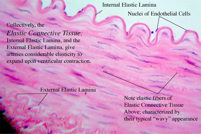
connective tissue → proper → dense → elastic
function: allows tissue to recoil after stretching; maintains pulsatile flow of blood through arteries; aids passive recoil of lungs following inspiration
location: walls of large arteries; within certain ligaments associated with the vertebral column; within the walls of the bronchial tubes
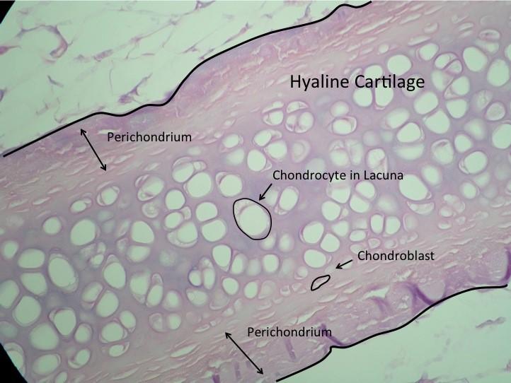
connective tissue → cartilage → hyaline
function: supports and reinforces; serves as resilient cushion; resists compressive stress
location: forms most of the embryonic skeleton; covers the ends of long bones in join cavities; forms costal cartilages of the ribs; cartilages of the nose, trachea, and larynx
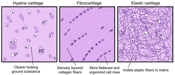
connective tissue → cartilage → elastic
function: maintains the shape of a structure while allowing great flexibility
location: supports the external ear (pinna); epiglottis

connective tissue → cartilage → fibrocartilage
function: tensile strength allows it to absorb compressive shock
location: intervertebral discs; pubic symphysis; disc of knee joint
connective tissue → bone
function: support and protects (by enclosing); provides levers for the muscles to act on; stores calcium and other minerals and fat; marrow inside bones is the site for blood cell formation (hematopoiesis)
location: bones

connective tissue → blood
function: transport respiratory gases, nutrients, wastes, and other substances
location: contained within blood vessels
fibroblasts
fiber buds or fiber formers; make the protein subunits of fibers
chondroblast
cells that secrete the matrix; cartilage formers
osteoblasts
cells that secrete in bone; bone formers
adipose
cells, store nutrients
white blood cells (WBCs)
respond to and protect against infectious agents (neutrophils, lymphocytes, eosinophils)
macrophages
specialize in phagocytosis; engulf and devour a wide variety of foreign materials
mast cells
mediate inflammation and promote healing
collagen fibers
the strongest and most abundant type of fiber in the connective tissue
reticular fibers
bundles of special type of collagen fibril; network
elastic fibers
contain rubberlike protein → function like rubber bands
loose areolar connective tissue
supporting and binding other tissues
holding body fluids
defending the body against infection
storing nutrients as fat
interstitial fluid
the cells of the body are bathed in tissue fluid
adipose tissue
similar to areolar connective tissue in structure and function; nutrient-storing function is much greater
subcutaneous tissue
much of the body’s adipose tissue occurs in the layer beneath the skin
visceral fat
a specific type of adipose (fat) tissue located inside the abdominal cavity
brown adipose tissue
produces heat and is a nutrient consumer
reticular connective tissue
resembles areolar tissue → only fibers in its matrix are reticular fibers
dense connective tissue
contains more collagen than areolar connective tissue does
dense irregular connective tissue
similar to areolar tissue; collagen fibers are much thicker
dense regular connective tissue
run in the same direction, parallel to the direction of pull; crowded between the collagen fibers are rows of fibroblasts
ligaments
bands or sheets that bind bones to one another
fascia
a fibrous membrane that wraps around muscles, muscle groups, large vessels and nerves
elastic connective tissue
a high proportion of elastic fibers; recoil after stretching
cartilage
a firm but flexible tissue, occurs in several parts of the skeleton
chondrocyte
mature form of a cartilage cell
lacuna
little depression or cavity; in bone and cartilage, each lacuna is occupied by a cell
bone tissue
a tremendous ability to support and protect body structures
osteocytes
mature bone cells
inhabit cavities:
lacunae
singular - lacuna
blood
the fluid in the blood vessels
cutaneous membranes
covers the body surface (skin)
mucous membranes (mucosae)
line body cavities that are open to the exterior
serous membranes (serosae)
line body cavities that are to the exterior
mesothelium
a serous membrane consists of simple squamous epithelium
serous fluid
lying on a thin layer of areolar connective tissue; filtrate of the blood in capillaries in the connective tissue