Week 26: Bacteria identification
5.0(1)
5.0(1)
Card Sorting
1/44
There's no tags or description
Looks like no tags are added yet.
Study Analytics
Name | Mastery | Learn | Test | Matching | Spaced |
|---|
No study sessions yet.
45 Terms
1
New cards
@@Cell Shape and Morphology@@
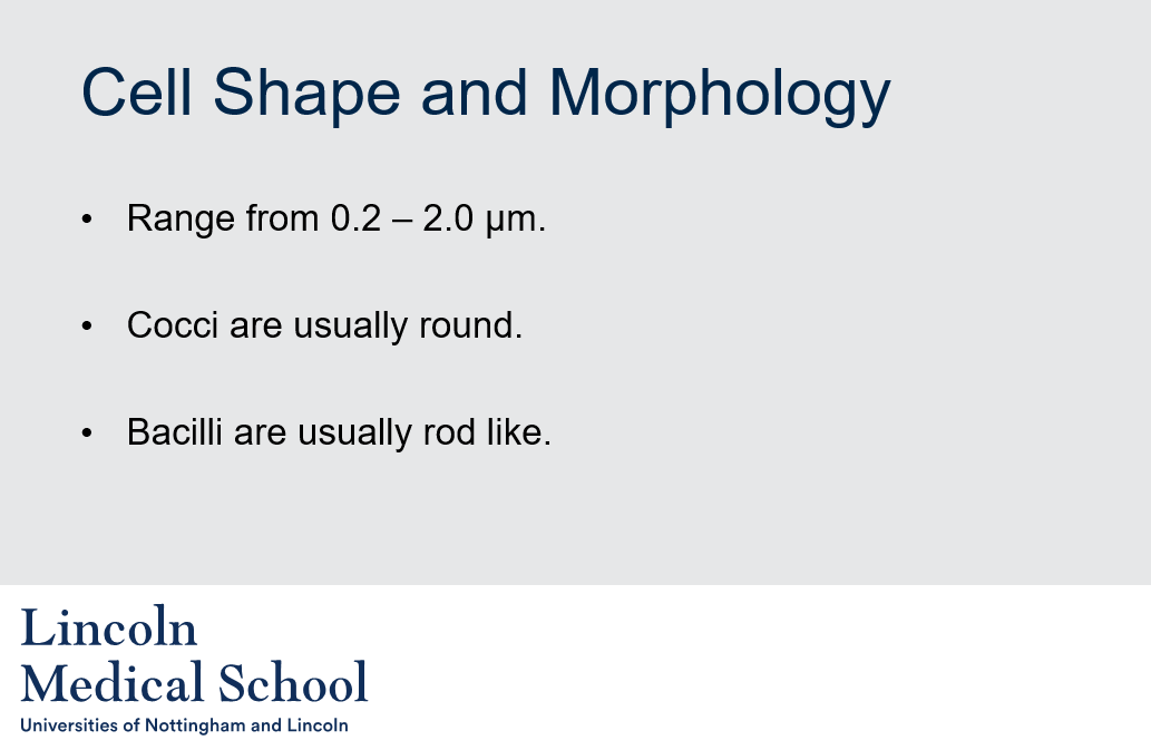
1. What are cocci and bacilli?
2. What is the size range of cocci and bacilli?
1. What are cocci and bacilli?
2. What is the size range of cocci and bacilli?
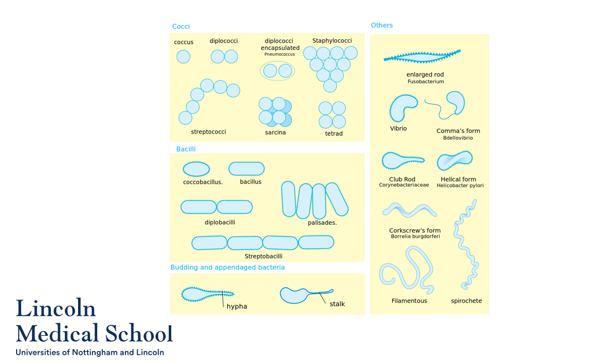
1. Cocci and bacilli are two different shapes of bacteria. Cocci are spherical or round in shape, while bacilli are rod-shaped.
2. The size range of cocci and bacilli can vary, but generally cocci have a diameter ranging from 0.2 to 2.0 micrometers (µm), while bacilli have a length ranging from 0.2 to 2.0 µm.
2
New cards
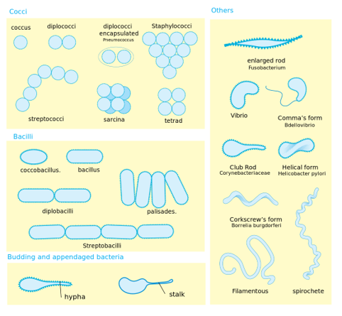
Can you label, describe and explain what this diagram is/shows?
3
New cards
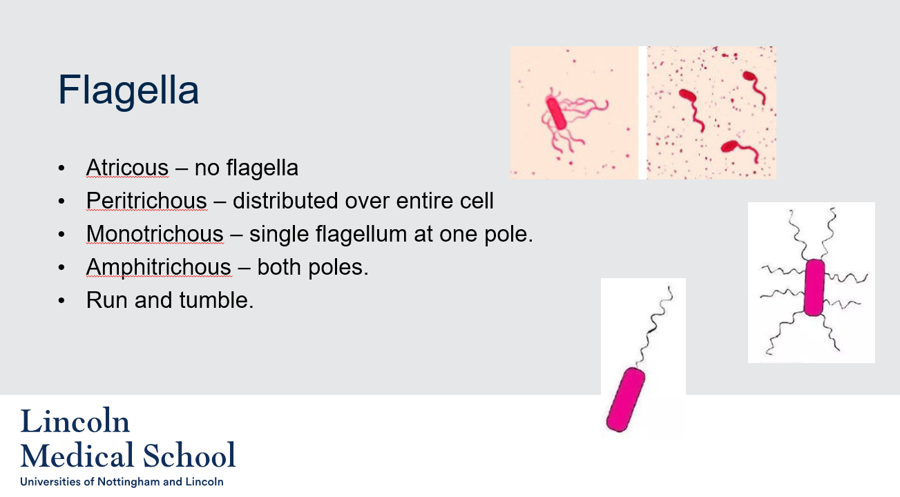
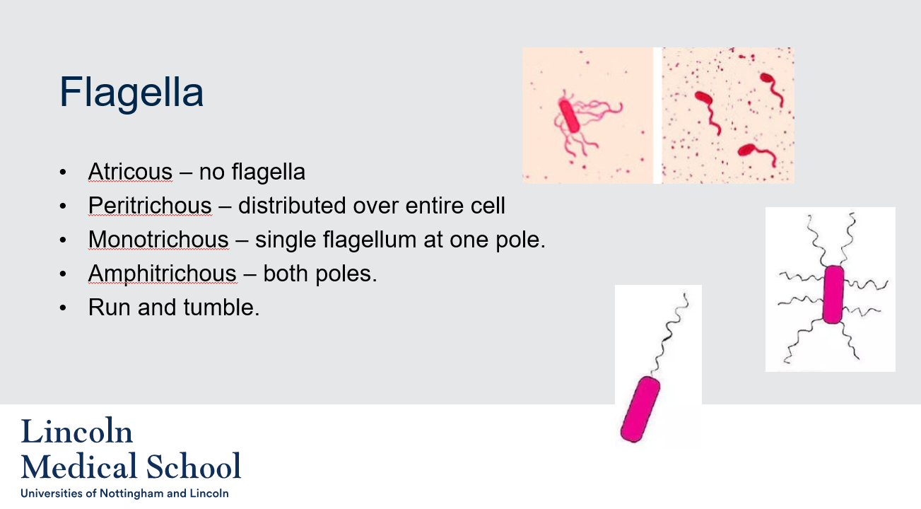
@@Flagella@@
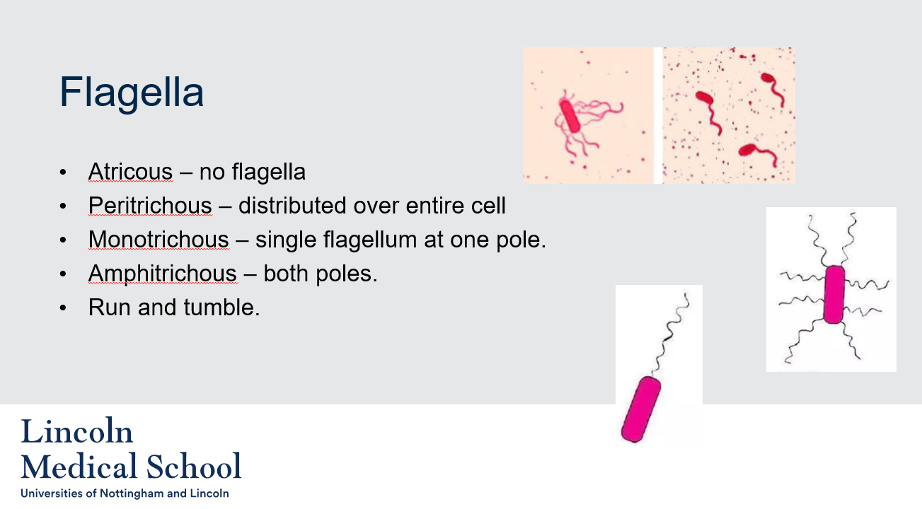
1. What is the meaning of the term "atricous"?
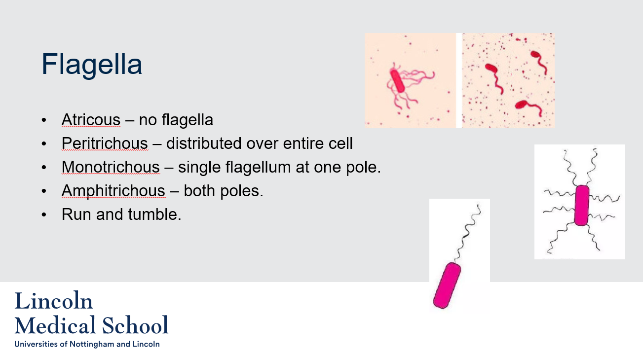
2. What is meant by the term "peritrichous"?
3. What is meant by the term "monotrichous"?
4. What is meant by the term "amphitrichous"?
5. What is "run and tumble" in bacteria?
1. What is the meaning of the term "atricous"?
2. What is meant by the term "peritrichous"?
3. What is meant by the term "monotrichous"?
4. What is meant by the term "amphitrichous"?
5. What is "run and tumble" in bacteria?
1. The term "atricous" refers to a type of bacteria that does not have any flagella.
2. The term "peritrichous" refers to the distribution of flagella on the surface of a bacterium. In peritrichous bacteria, the flagella are distributed over the entire cell surface, giving the bacteria the ability to move in any direction.
3. The term "monotrichous" refers to a type of bacteria that has a single flagellum at one pole, or end, of the cell. This allows the bacterium to move in a specific direction.
4. The term "amphitrichous" refers to a type of bacteria that has flagella located at both poles, or ends, of the cell. This allows the bacterium to move in two different directions.
5. "Run and tumble" is a type of movement used by some bacteria to navigate their environment. During a "run," the bacterium moves in a straight line due to the rotation of its flagella. However, when the bacterium encounters an obstacle or reaches the end of its run, it will stop and randomly change direction in a process called "tumble." This allows the bacterium to explore its environment and find nutrients or other resources.
4
New cards
@@Flagella@@
Can you label, describe and explain what this diagram is/shows?
Can you label, describe and explain what this diagram is/shows?
5
New cards
@@Flagella@@
Can you label, describe and explain what this diagram is/shows?
Can you label, describe and explain what this diagram is/shows?
6
New cards

@@Flagella@@
Can you label, describe and explain what this diagram is/shows?
Can you label, describe and explain what this diagram is/shows?
7
New cards
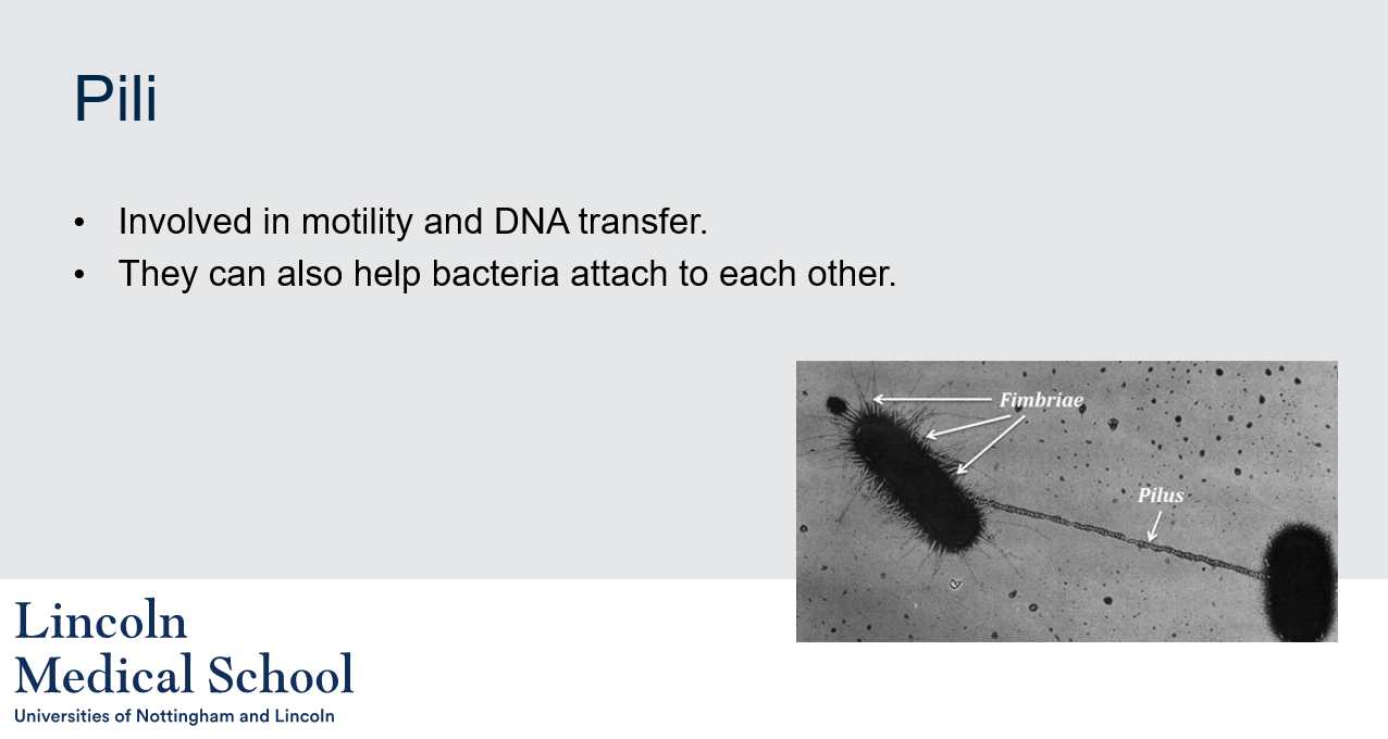
@@Pili@@
1. What are pili?
2. What is the role of pili in motility?
3. What is the role of pili in DNA transfer?
4. How do pili help bacteria attach to each other?
1. What are pili?
2. What is the role of pili in motility?
3. What is the role of pili in DNA transfer?
4. How do pili help bacteria attach to each other?
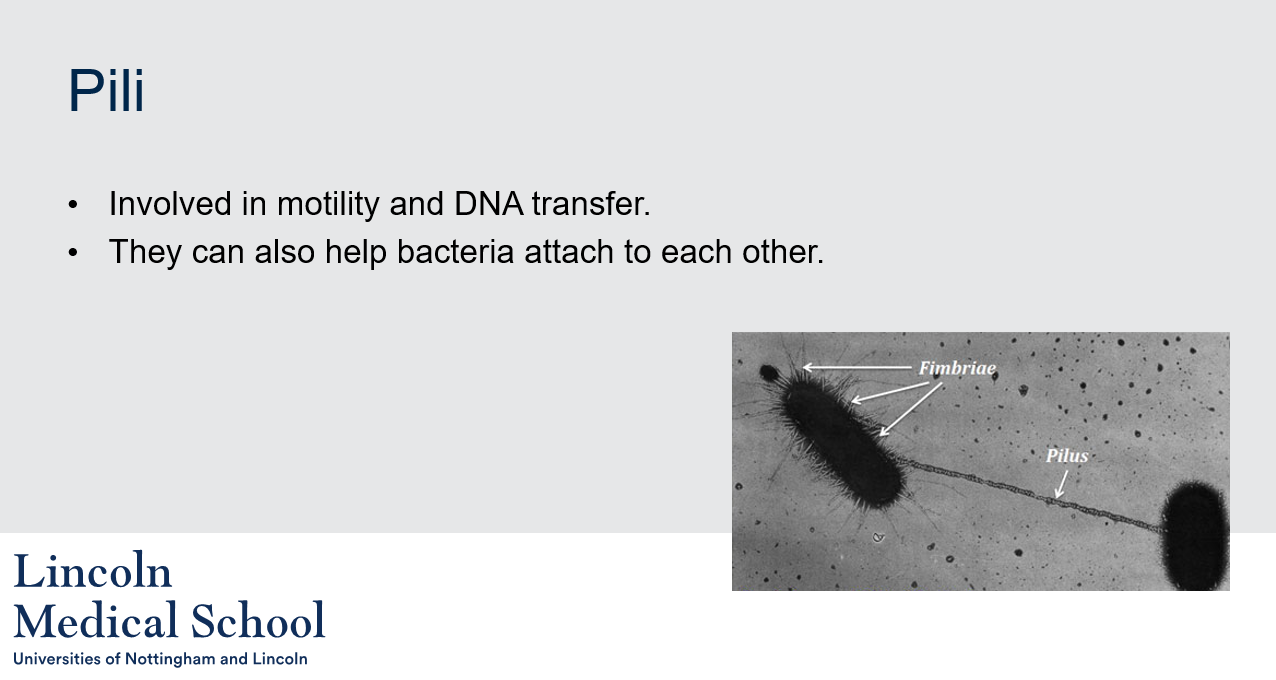
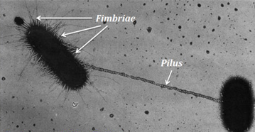
1. Pili are hair-like structures found on the surface of some bacteria. They are made up of protein subunits and can serve a variety of functions.
2. Pili can be involved in motility in some bacteria by allowing the bacterium to move over surfaces or by twitching. The twitching motility is accomplished by extending and retracting the pili to pull the bacterium along the surface.
3. Pili can play a role in DNA transfer between bacteria by allowing them to form a physical connection and transfer genetic material through a process called conjugation. During conjugation, a pilus is extended from one bacterium and contacts another bacterium, forming a temporary bridge that allows DNA to be transferred from one bacterium to the other.
4. Pili can help bacteria attach to each other and to surfaces by allowing them to adhere to specific molecules. For example, some bacteria can produce pili that bind to sugars or other molecules on the surface of host cells, allowing the bacteria to colonize the host and cause infection. In other cases, pili can help bacteria attach to surfaces such as rocks or other bacteria, forming biofilms.
8
New cards
@@Pili@@
Can you label, describe and explain what this diagram is/shows?
Can you label, describe and explain what this diagram is/shows?
9
New cards
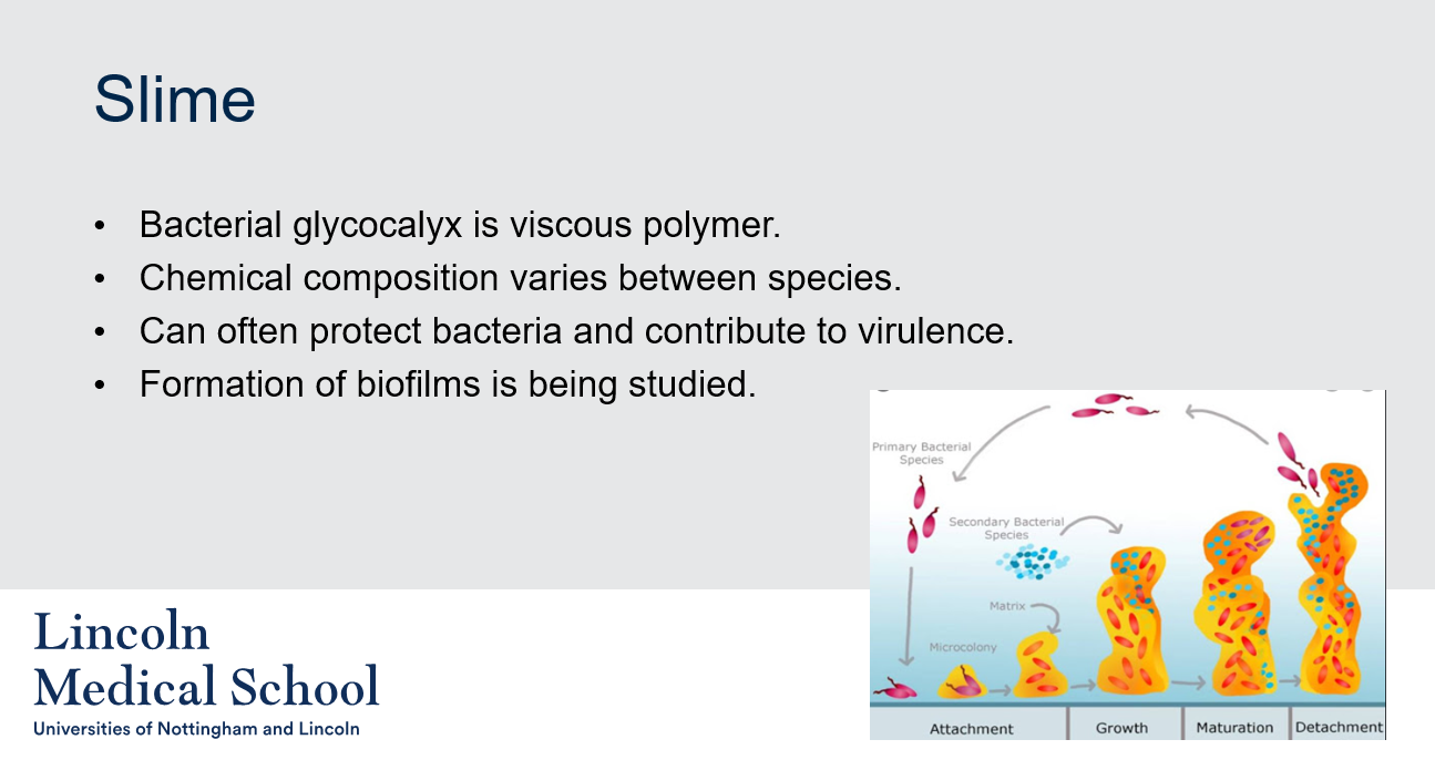
@@Slime@@
1. What is bacterial glycocalyx?
2. Does the chemical composition of bacterial glycocalyx vary between species?
3. Can bacterial glycocalyx protect bacteria and contribute to virulence?
4. Why is the formation of biofilms in bacteria being studied?
1. What is bacterial glycocalyx?
2. Does the chemical composition of bacterial glycocalyx vary between species?
3. Can bacterial glycocalyx protect bacteria and contribute to virulence?
4. Why is the formation of biofilms in bacteria being studied?
1. Bacterial glycocalyx is a viscous polymer that surrounds the surface of some bacteria. It is often referred to as slime.
2. Yes, the chemical composition of bacterial glycocalyx can vary depending on the species of bacteria.
3. Yes, bacterial glycocalyx can often protect bacteria from the immune system and from antibiotics by forming a physical barrier around the bacteria. It can also help bacteria adhere to surfaces, including host tissues, and evade detection by the immune system. Additionally, bacterial glycocalyx can contribute to the virulence of some bacterial species by promoting the formation of biofilms, which can help bacteria resist antimicrobial agents and host defenses.
4. The formation of biofilms in bacteria is being studied because it is believed to contribute to the persistence of bacterial infections and antibiotic resistance. Biofilms are communities of bacteria that adhere to surfaces and form a protective matrix of extracellular material, including glycocalyx. They can be difficult to treat with antibiotics because they can resist penetration and provide a physical barrier that protects bacteria from the immune system. Biofilm formation is being investigated as a potential target for new antimicrobial strategies.
10
New cards
@@Slime@@
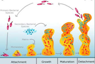
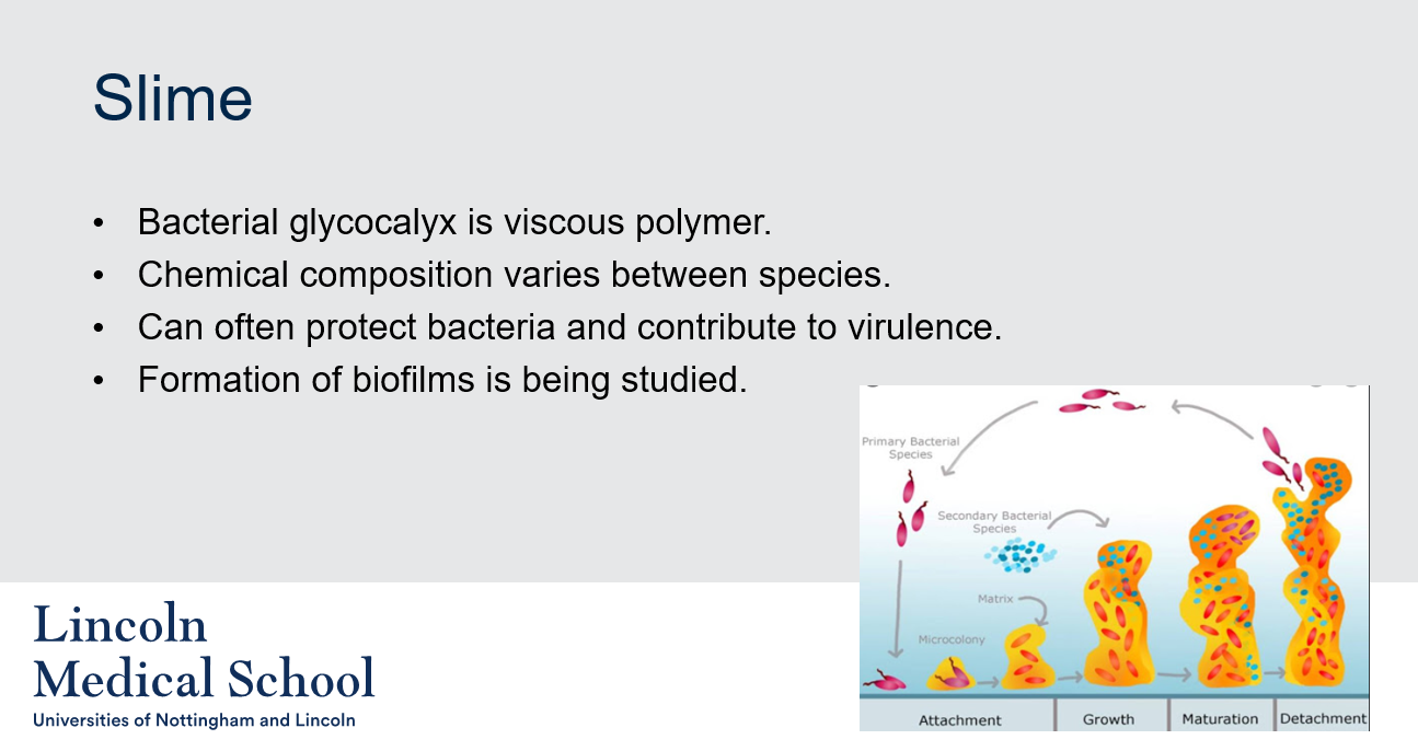
Can you label, describe and explain what this diagram is/shows?
Can you label, describe and explain what this diagram is/shows?
11
New cards
@@Colony Shape@@
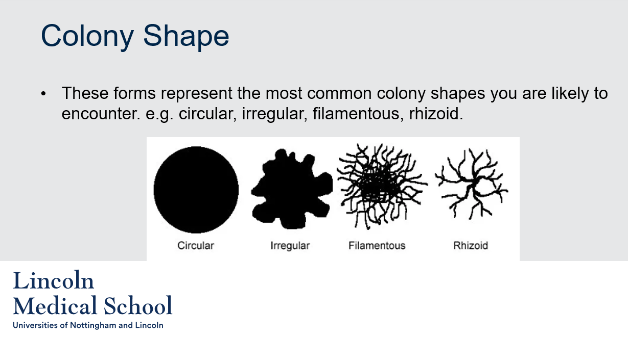
What are the most common colony shapes that one is likely to encounter in microbiology?
What are the most common colony shapes that one is likely to encounter in microbiology?
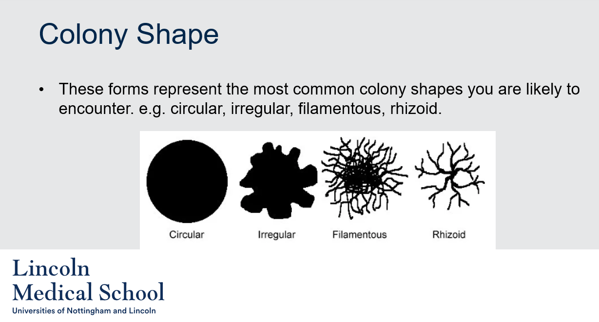
The most common colony shapes that one is likely to encounter in microbiology include circular, irregular, filamentous, and rhizoid. The circular shape is a common shape for bacterial colonies, while irregular shapes can be seen in fungal colonies. Filamentous colonies are composed of a tangled network of filaments or threads, while rhizoid colonies have a root-like appearance. These colony shapes can provide clues about the identity of the microorganisms present in a sample.
12
New cards

@@Colony Shape@@
Can you label, describe and explain what this diagram is/shows?
Can you label, describe and explain what this diagram is/shows?
13
New cards
@@Colony Margin@@
1. What is the colony margin in microbiology and why is it important?
2. What are some examples of colony margins in microbiology?
1. What is the colony margin in microbiology and why is it important?
2. What are some examples of colony margins in microbiology?
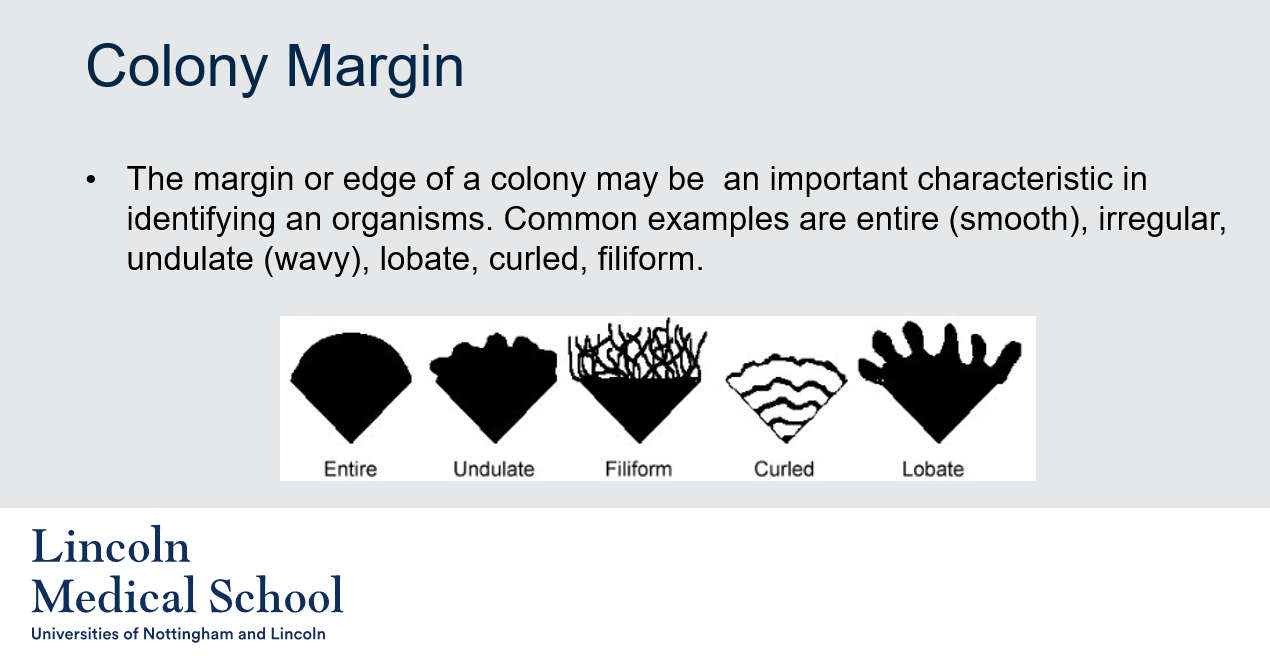
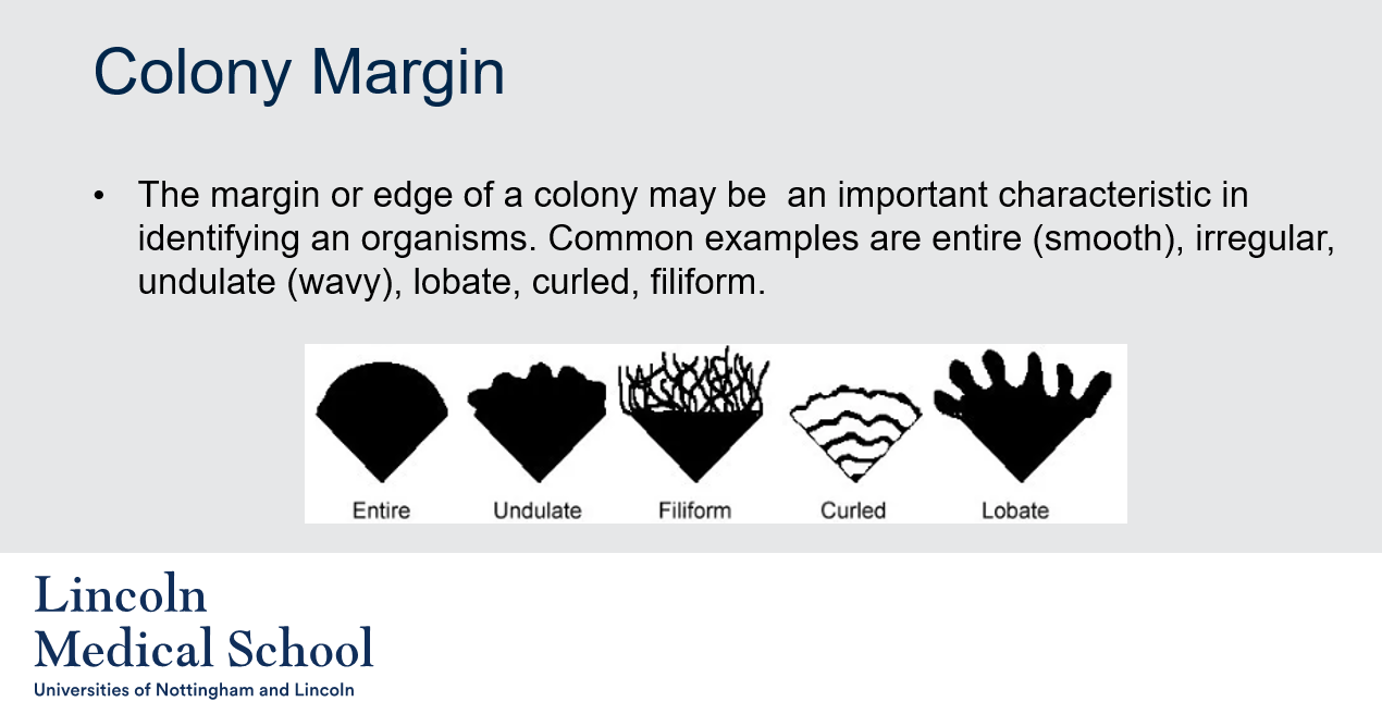
1. The colony margin in microbiology refers to the edge of a bacterial or fungal colony. It can be an important characteristic in identifying microorganisms, as different species often have distinct colony margins.
2. Some examples of colony margins in microbiology include:
* Entire (smooth): A colony with a smooth, circular edge.
* Irregular: A colony with an uneven or jagged edge.
* Undulate (wavy): A colony with a slightly wavy or curved edge.
* Lobate: A colony with a lobed or branched edge.
* Curled: A colony with a curled or rolled edge.
* Filiform: A colony with a thin, thread-like edge.
14
New cards

@@Colony Margin@@
Can you label, describe and explain what this diagram is/shows?
Can you label, describe and explain what this diagram is/shows?
15
New cards
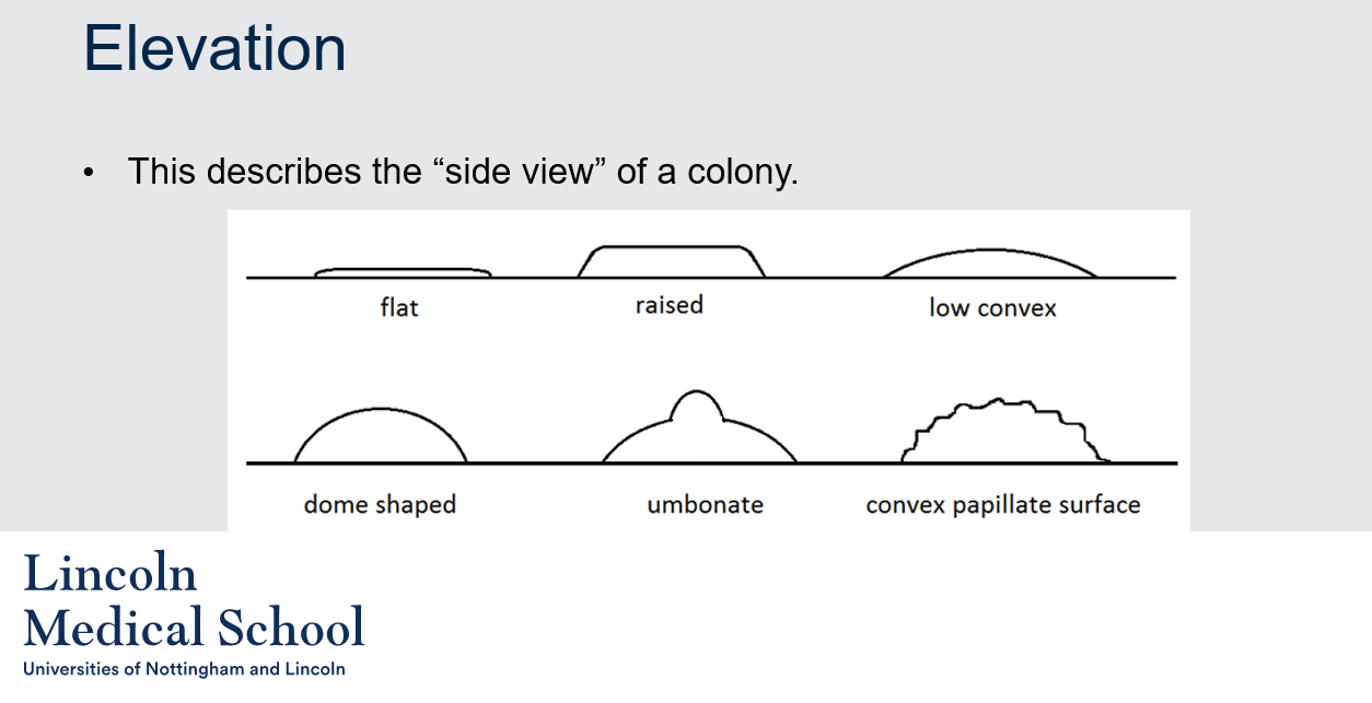
@@Elevation@@
1. What does the term "elevation" mean in microbiology?
2. What are some examples of colony elevations in microbiology?
1. What does the term "elevation" mean in microbiology?
2. What are some examples of colony elevations in microbiology?
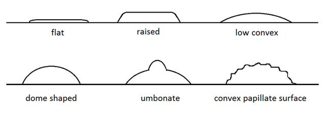
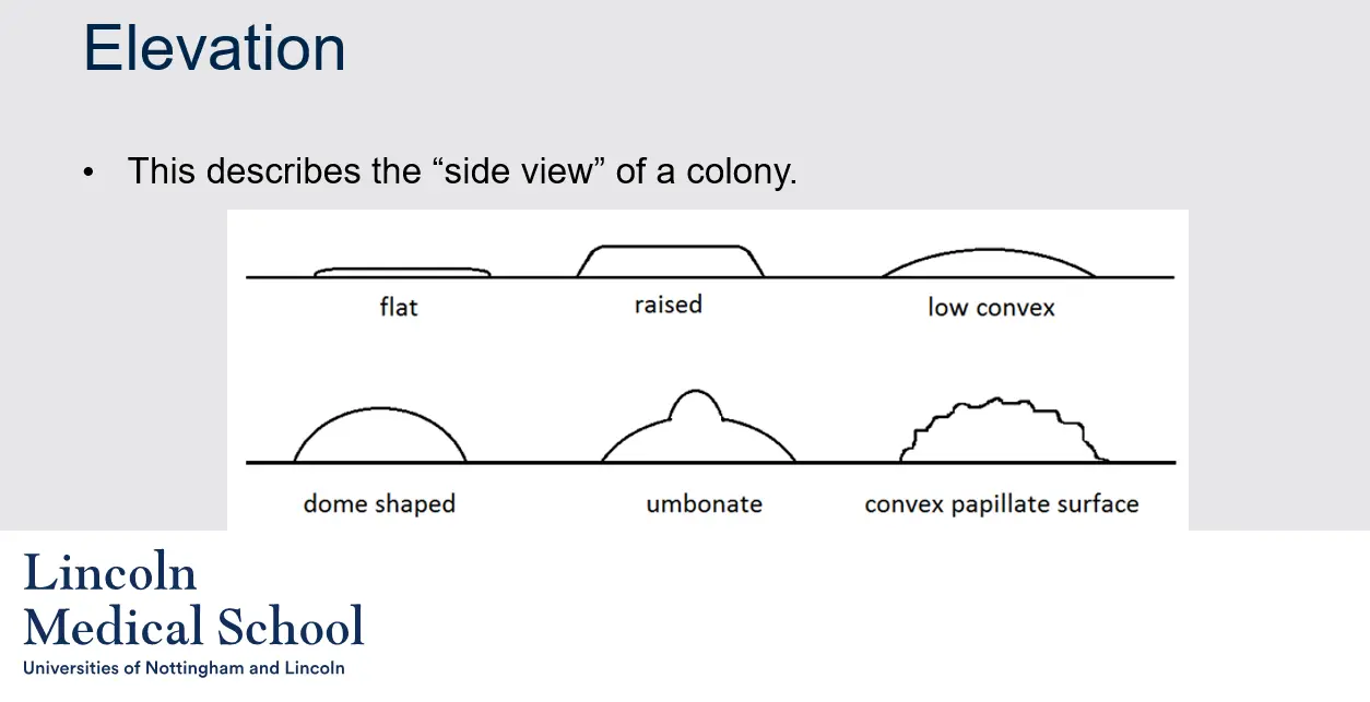
1. In microbiology, elevation refers to the side view of a bacterial or fungal colony. It describes the height or thickness of the colony relative to the surrounding medium.
2. Some common examples of colony elevations include:
* Flat: A colony that is at the same level as the surrounding growth medium.
* Raised: A colony that is elevated above the surface of the growth medium.
* Low convex: A colony that has a slight elevation in the center and slopes downward towards the edge.
* Dome-shaped: A colony that is highly convex and shaped like a dome.
* Umbonate: A colony that is raised in the center and has a distinct central bump or knob.
* Convex papillate surface: A colony that is raised and has a central protrusion that is taller than the rest of the colony.
16
New cards
@@Elevation@@
Can you label, describe and explain what this diagram is/shows?
Can you label, describe and explain what this diagram is/shows?
17
New cards
@@Colour@@
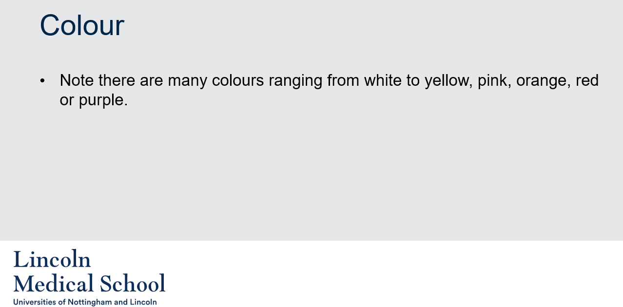
What is the significance of colony color in microbiology?
What is the significance of colony color in microbiology?
In microbiology, colony color is an important characteristic used in the identification and classification of microorganisms. Colony color can range from white to yellow, pink, orange, red, or purple, among other hues.
18
New cards
@@Texture@@
How is texture used to describe microbial colonies in microbiology?
How is texture used to describe microbial colonies in microbiology?
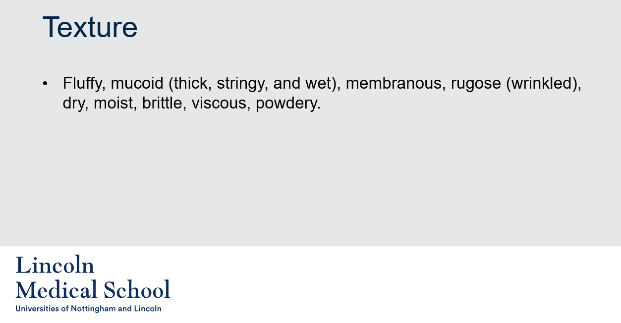
In microbiology, texture refers to the physical characteristics of the surface of a microbial colony. Texture can provide important information about the type of microorganism present, as well as the conditions under which it is growing. Some common examples of colony textures include:
* Fluffy: A colony that is soft and airy, with a feathery or cotton-like texture.
* Mucoid: A colony that is thick, stringy, and wet, with a slimy or sticky texture.
* Membranous: A colony that is thin and flat, with a smooth and shiny texture.
* Rugose: A colony that is wrinkled or folded, with a rough or bumpy texture.
* Dry: A colony that is rough or powdery, with a dry texture.
* Moist: A colony that is slightly damp or sticky, with a smooth texture.
* Brittle: A colony that is hard and easily broken, with a crunchy or crumbly texture.
* Viscous: A colony that is thick and gel-like, with a sticky or syrupy texture.
* Powdery: A colony that is dry and powdery, with a fine and dusty texture.
Texture is often used along with other colony characteristics, such as color, shape, margin, and elevation, to help identify and classify microorganisms.
19
New cards
@@Optical Quality@@
How is optical quality used to describe microbial colonies in microbiology?
How is optical quality used to describe microbial colonies in microbiology?
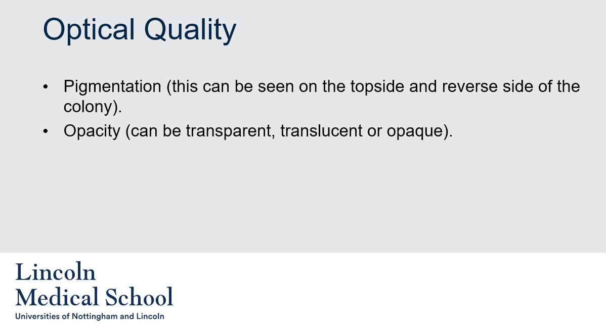
Optical quality is a term used in microbiology to describe the visual appearance of microbial colonies when viewed under different lighting conditions. Two important aspects of optical quality are pigmentation and opacity:
* Pigmentation: This refers to the color of the colony, which can vary depending on the type of microorganism present and the environmental conditions under which it is growing. Pigmentation can be seen on both the topside and reverse side of the colony.
* Opacity: This refers to how much light is able to pass through the colony. Opacity can range from transparent (allowing light to pass through easily) to translucent (allowing some light to pass through, but not enough to see through completely) to opaque (blocking all light).
20
New cards
@@Virulence@@
1. What does LD stand for in microbiology and how is it related to virulence?
2. What is LD50 and how is it used to measure acute toxicity of a material?
1. What does LD stand for in microbiology and how is it related to virulence?
2. What is LD50 and how is it used to measure acute toxicity of a material?
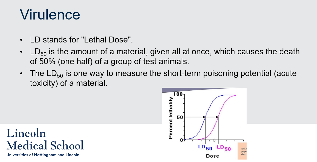
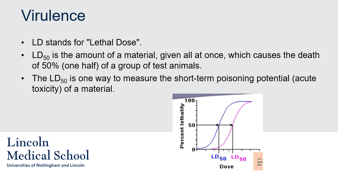
1. In microbiology, LD stands for "Lethal Dose". Virulence refers to the degree of pathogenicity, or the ability of a microorganism to cause disease in a host.
2. LD50 is the amount of a material, given all at once, which causes the death of 50% (one half) of a group of test animals. The LD50 is one way to measure the short-term poisoning potential (acute toxicity) of a material.
21
New cards
@@Virulence@@
Can you label, describe and explain what this diagram is/shows?
Can you label, describe and explain what this diagram is/shows?
22
New cards
@@Virulence@@
1. What does CFU stand for in microbiology?
2. What is the relationship between LD50 and CFU/ml in measuring the virulence of bacteria or viruses?
1. What does CFU stand for in microbiology?
2. What is the relationship between LD50 and CFU/ml in measuring the virulence of bacteria or viruses?
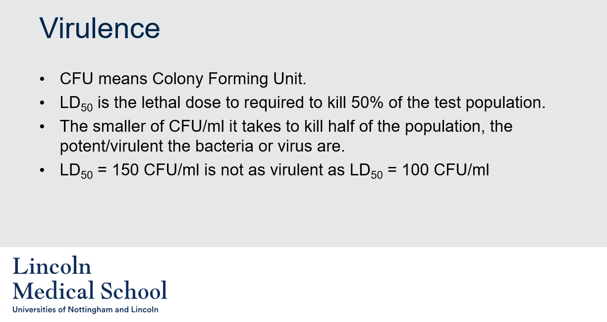
1. CFU stands for Colony Forming Unit, which is a measure of viable bacteria or fungal cells in a sample. It represents the number of cells that are capable of forming a visible colony on a growth medium under specific conditions.
2. LD50 stands for Lethal Dose 50, which is the amount of a material that causes the death of 50% of a test population. In microbiology, LD50 is often used to measure the potency or virulence of a pathogenic microorganism. CFU/ml, on the other hand, refers to the number of colony-forming units per milliliter of a sample, which represents the viable cells that are capable of growing into visible colonies on a growth medium. The smaller the CFU/ml it takes to cause death in half of the test population, the more potent/virulent the bacteria or virus are. For example, an LD50 of 100 CFU/ml is considered more virulent than an LD50 of 150 CFU/ml.
23
New cards
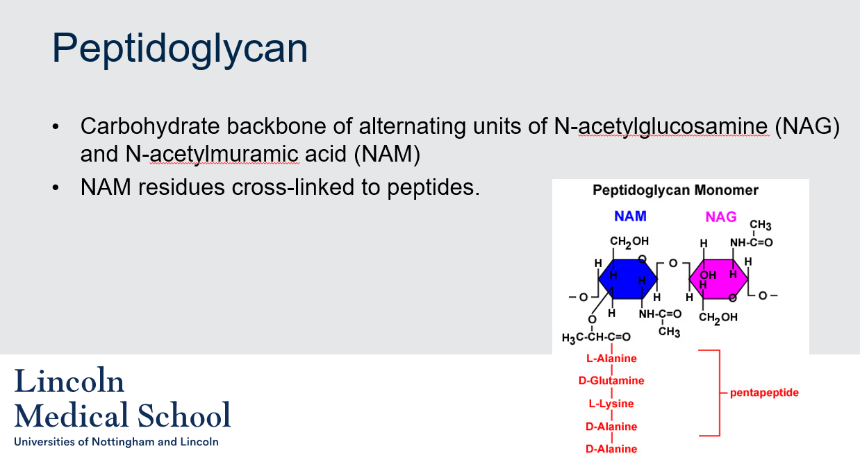
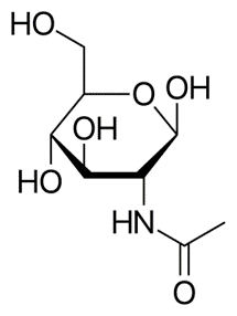
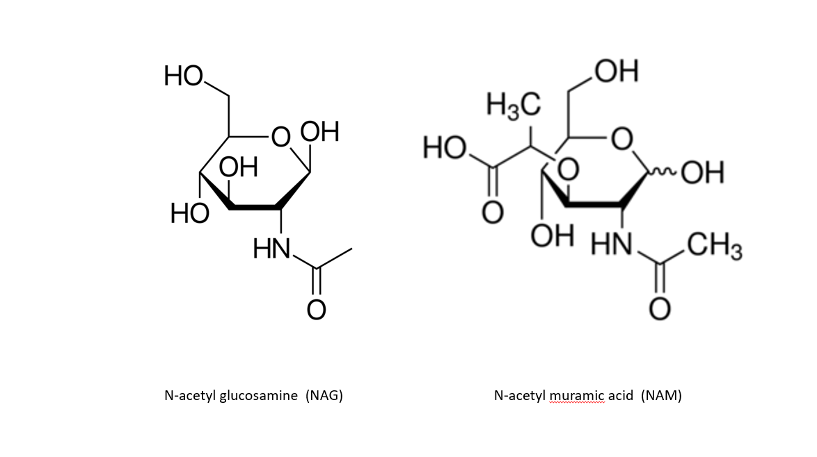
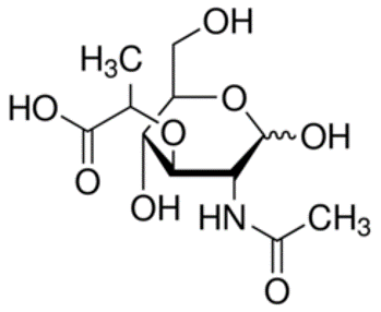
@@Peptidoglycan@@
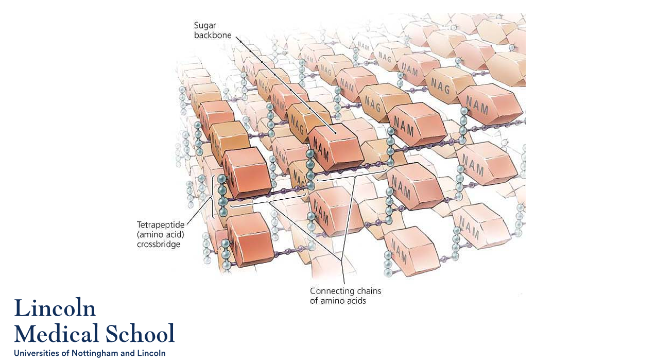
What is the basic structure of peptidoglycan in bacteria?
What is the basic structure of peptidoglycan in bacteria?
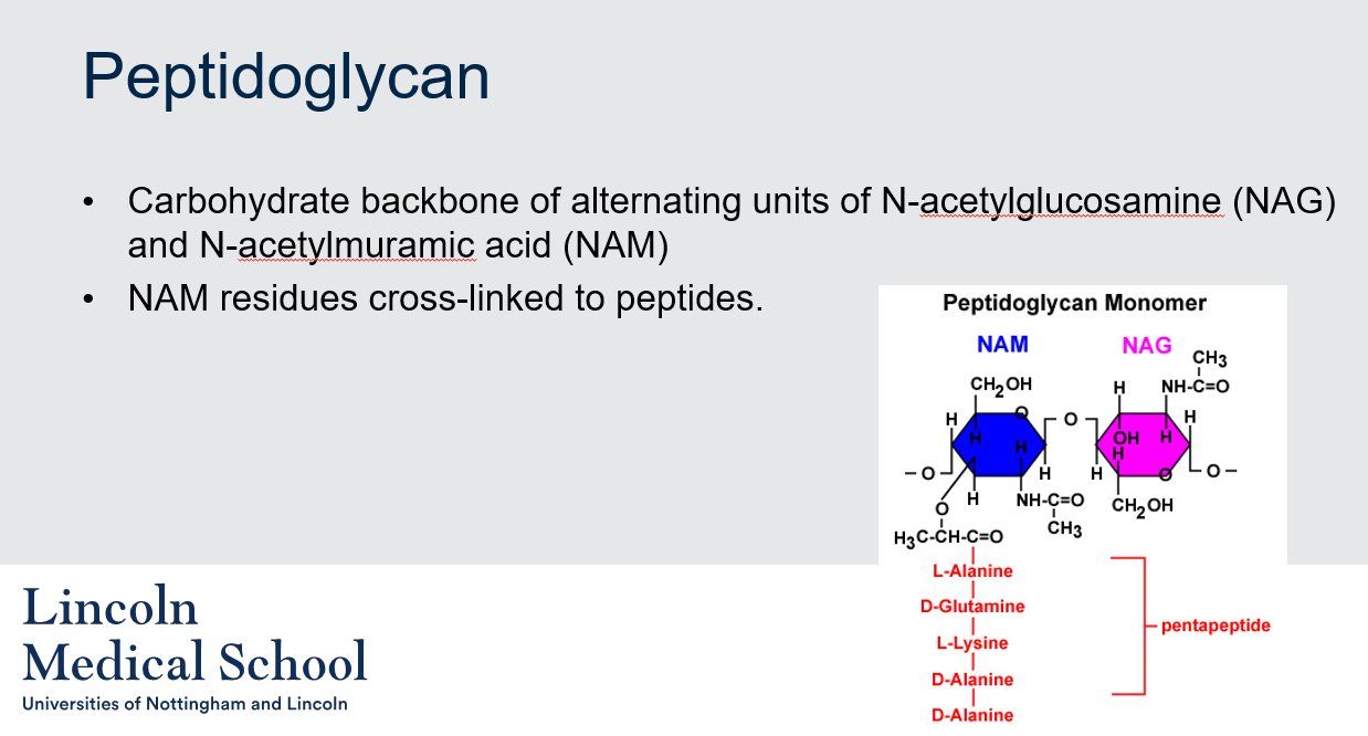
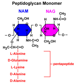
The basic structure of peptidoglycan in bacteria consists of a carbohydrate backbone made up of alternating units of N-acetylglucosamine (NAG) and N-acetylmuramic acid (NAM). The NAM residues are cross-linked to peptides, forming a strong, mesh-like layer that surrounds the bacterial cell membrane and provides structural support.
24
New cards
@@Peptidoglycan@@
Can you label, describe and explain what this diagram is/shows?
Can you label, describe and explain what this diagram is/shows?
25
New cards
Can you label, describe and explain what this diagram is/shows?
26
New cards
Can you label, describe and explain what this diagram is/shows?
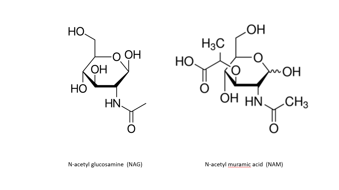
27
New cards
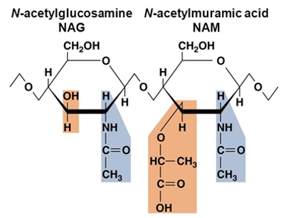
Can you label, describe and explain what this diagram is/shows?
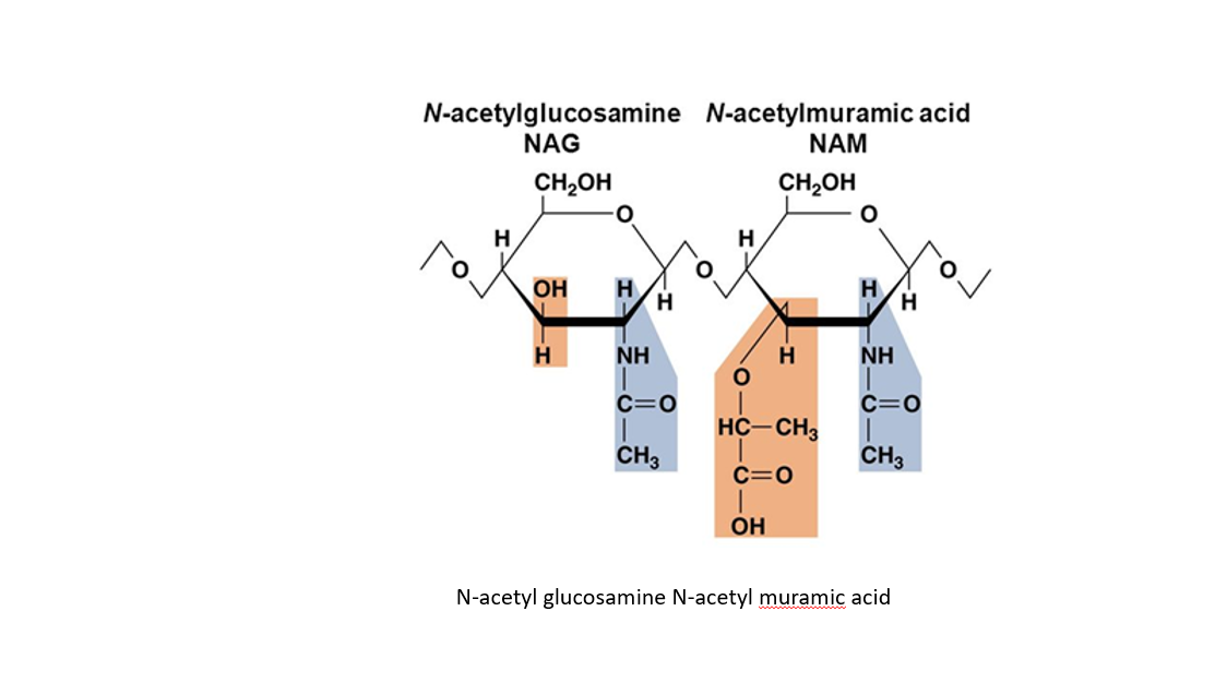
28
New cards
@@Penicillin@@
How does penicillin kill susceptible bacteria?
How does penicillin kill susceptible bacteria?
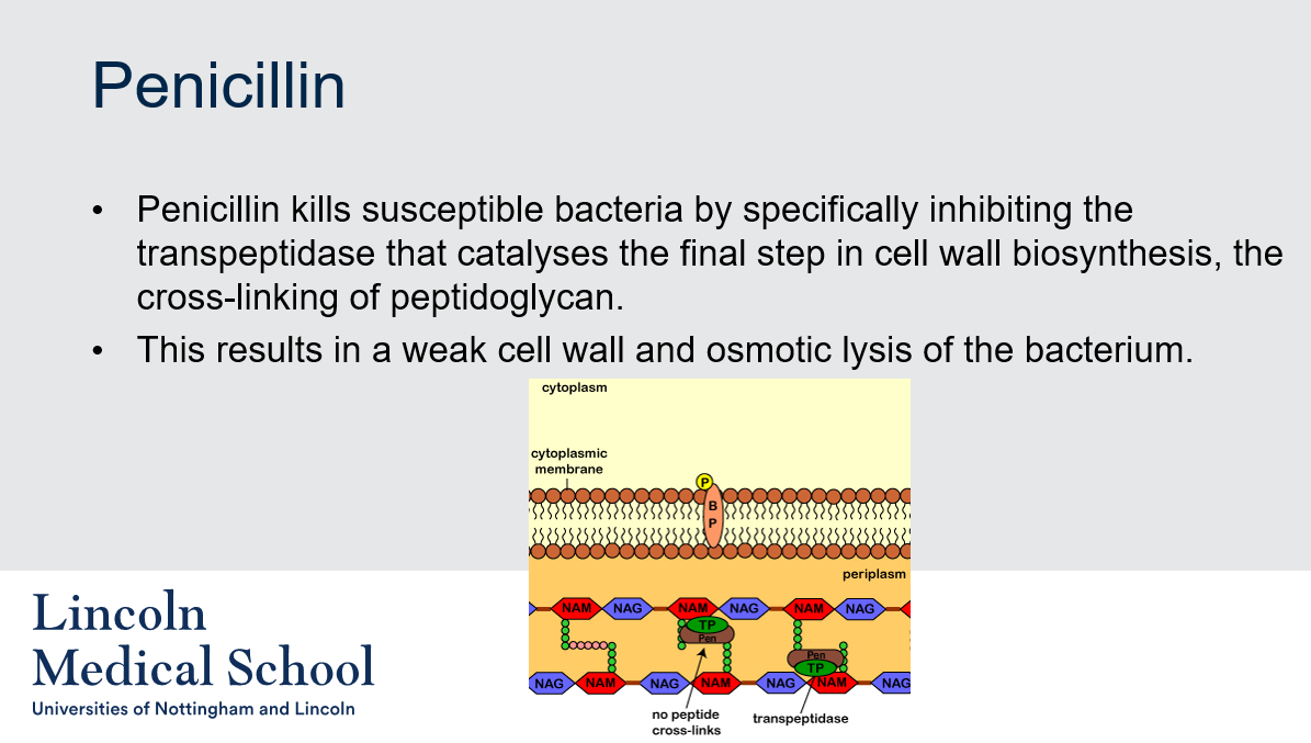
Penicillin kills susceptible bacteria by specifically inhibiting the transpeptidase that catalyses the final step in cell wall biosynthesis, the cross-linking of peptidoglycan. This results in a weak cell wall and osmotic lysis of the bacterium.
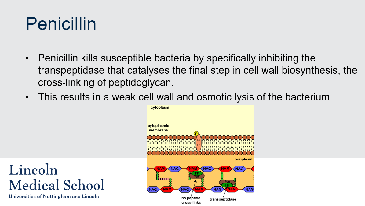
29
New cards
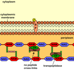
@@Penicillin@@
Can you label, describe and explain what this diagram is/shows?
Can you label, describe and explain what this diagram is/shows?
30
New cards
@@Pseudopeptidoglycan@@
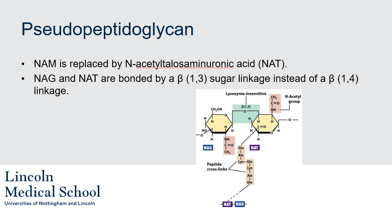
What is pseudopeptidoglycan and how is it different from regular peptidoglycan?
What is pseudopeptidoglycan and how is it different from regular peptidoglycan?
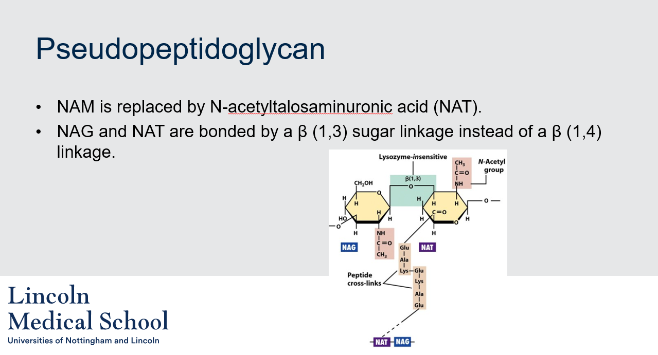
Pseudopeptidoglycan is a type of cell wall found in some archaea, which is similar in function to peptidoglycan in bacteria. However, it differs in its chemical composition, as it contains N-acetyltalosaminuronic acid (NAT) instead of N-acetylmuramic acid (NAM), and the sugar linkage between NAG and NAT is β (1,3) instead of β (1,4) in peptidoglycan. This difference in structure makes pseudopeptidoglycan resistant to the action of lysozyme, an enzyme that breaks down the β (1,4) linkage in peptidoglycan.
31
New cards
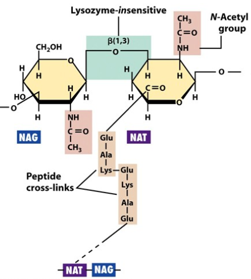
@@Pseudopeptidoglycan@@
Can you label, describe and explain what this diagram is/shows?
Can you label, describe and explain what this diagram is/shows?
32
New cards
@@Bacterial and Archaeal Cell Walls@@
1. What is the main component of bacterial cell walls?
2. Do archaea have peptidoglycan in their cell walls?
1. What is the main component of bacterial cell walls?
2. Do archaea have peptidoglycan in their cell walls?
1. The main component of bacterial cell walls is peptidoglycan.
2. No, archaea do not have peptidoglycan in their cell walls. They often have pseudopeptidoglycan instead.
33
New cards
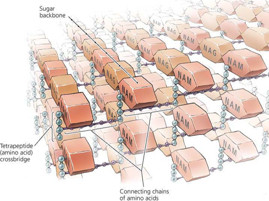
Can you label, describe and explain what this diagram is/shows?
34
New cards
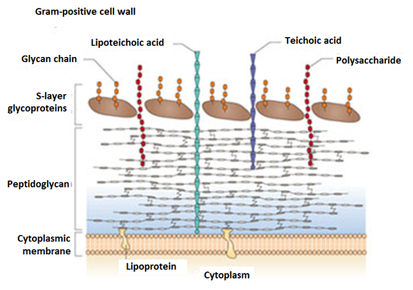
Can you label, describe and explain what this diagram is/shows?
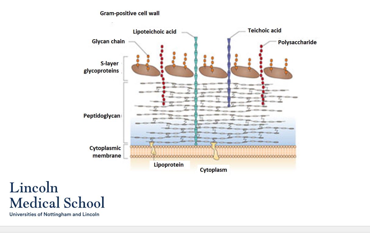
35
New cards
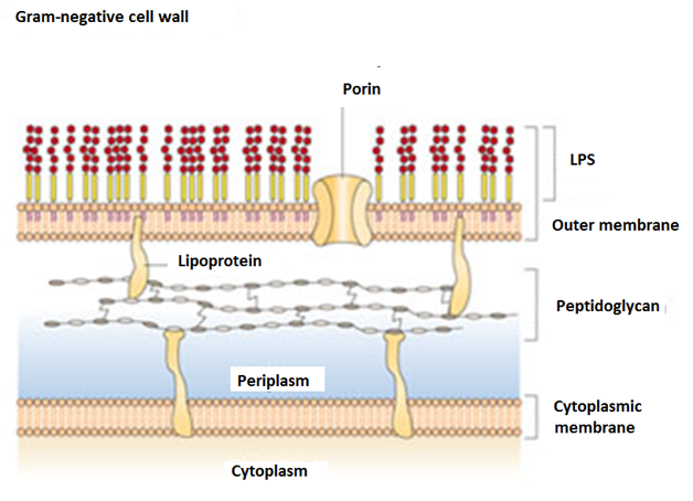
Can you label, describe and explain what this diagram is/shows?
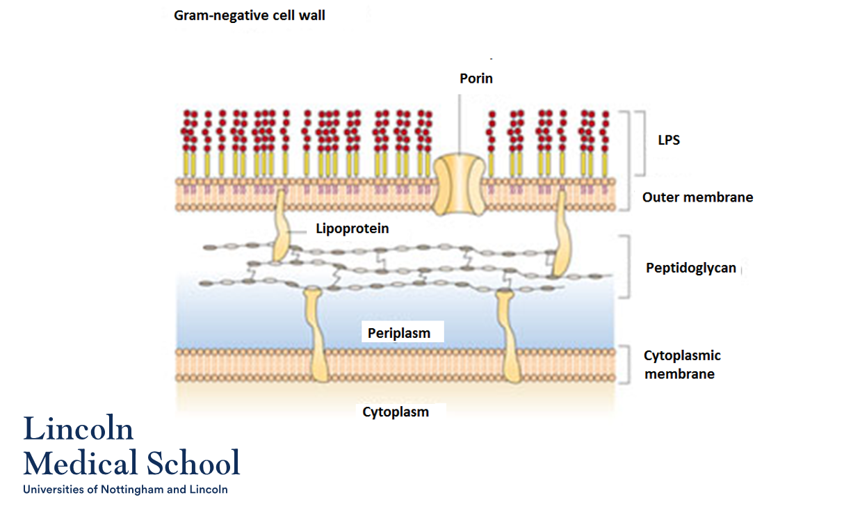
36
New cards
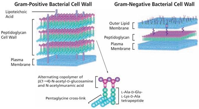
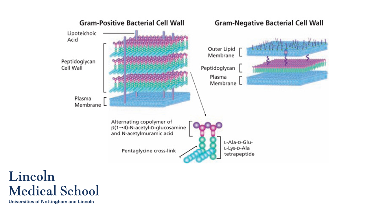
Can you label, describe and explain what this diagram is/shows?
37
New cards
@@Gram Staining@@
1. What is the Gram stain?
2. What is the purpose of the Gram stain?
1. What is the Gram stain?
2. What is the purpose of the Gram stain?
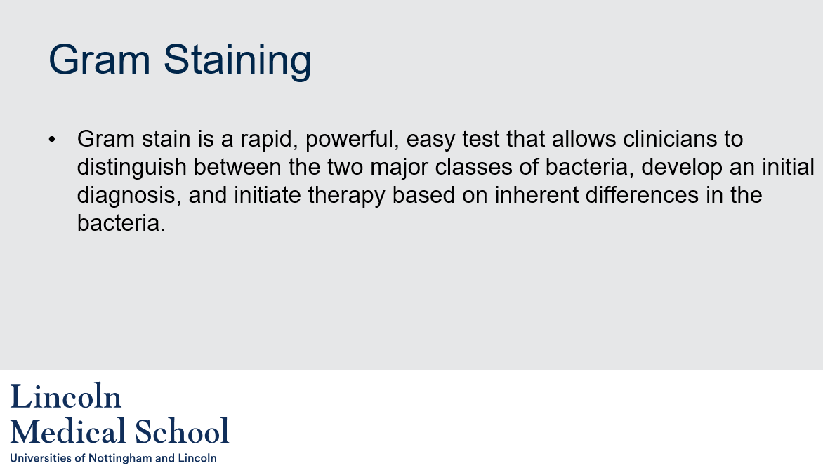
1. Gram stain is a rapid, powerful, easy test that allows clinicians to distinguish between the two major classes of bacteria based on their cell wall structure.
2. The purpose of the Gram stain is to help clinicians develop an initial diagnosis and initiate therapy based on inherent differences in bacteria.
38
New cards
@@Gram Staining@@
Can you label, describe and explain what this diagram is/shows?
Can you label, describe and explain what this diagram is/shows?
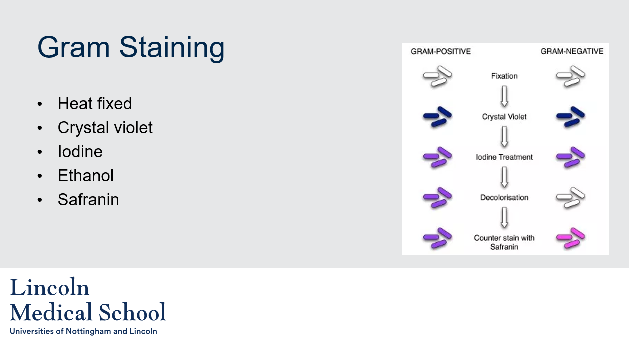
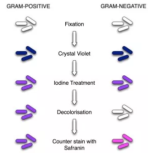
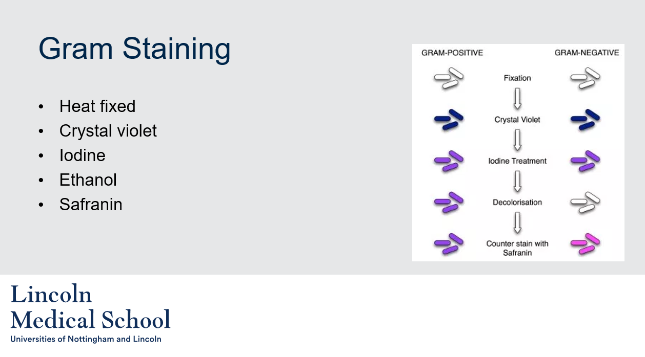
Gram staining involves the following steps:
1. Heat fixing: The bacterial sample is heat-fixed onto a glass slide to attach the cells to the slide and kill the bacteria.
2. Crystal violet: The slide is flooded with crystal violet, which stains all cells purple.
3. Iodine: Iodine is added to the slide, which acts as a mordant and fixes the crystal violet in the cells.
4. Ethanol: The slide is washed with ethanol, which acts as a decolorizing agent. In Gram-negative bacteria, the outer membrane is dissolved by the ethanol, allowing the crystal violet to be washed out. In Gram-positive bacteria, the thick peptidoglycan layer retains the crystal violet, and the bacteria remain purple.
5. Safranin: Finally, the slide is stained with safranin, which stains Gram-negative bacteria pink or red, while Gram-positive bacteria remain purple.
39
New cards
@@Gram Staining@@
Can you label, describe and explain what this diagram is/shows?
Can you label, describe and explain what this diagram is/shows?
40
New cards
@@Gram Staining@@
What happens to Gram positive and Gram negative bacteria during the Gram staining process?
What happens to Gram positive and Gram negative bacteria during the Gram staining process?
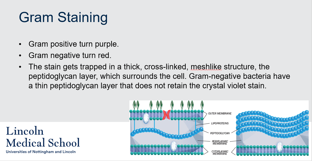
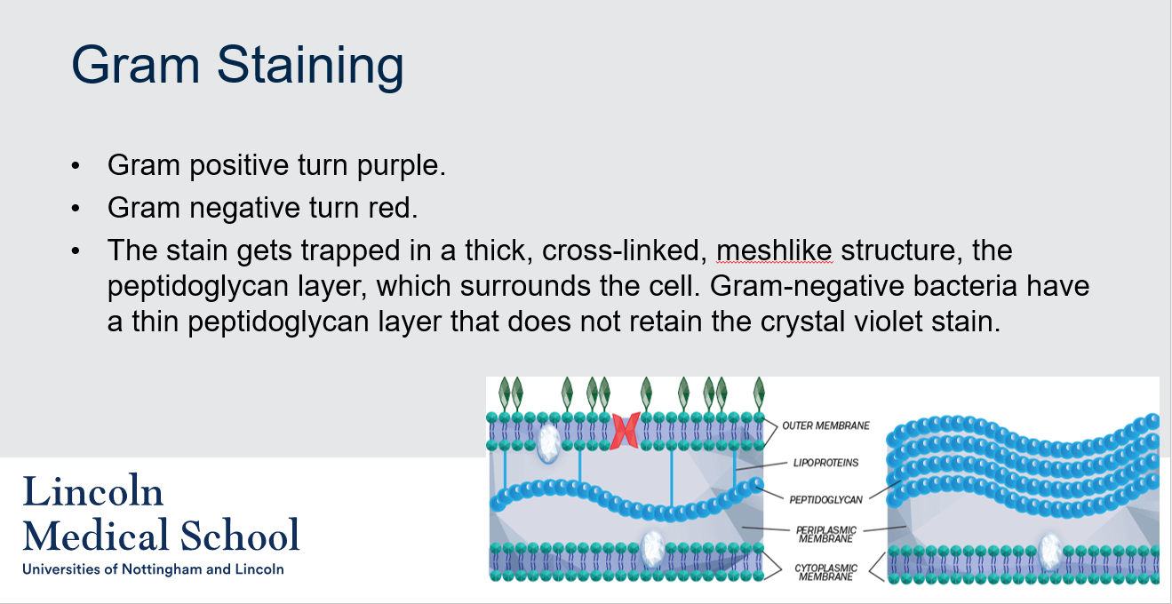
Gram positive bacteria retain the crystal violet stain and turn purple, while Gram negative bacteria do not retain the stain and turn red. This is because the crystal violet stain gets trapped in the thick, cross-linked, mesh-like structure of the peptidoglycan layer, which surrounds the cell in Gram-positive bacteria. In contrast, Gram-negative bacteria have a thin peptidoglycan layer that does not retain the crystal violet stain, making them appear red after the counterstain with safranin.
41
New cards

@@Gram Staining@@
Can you label, describe and explain what this diagram is/shows?
Can you label, describe and explain what this diagram is/shows?
42
New cards
@@Bacterial and Archaeal Cell Membranes@@
How do archaeal phospholipids differ from those found in Bacteria and Eukarya?
How do archaeal phospholipids differ from those found in Bacteria and Eukarya?
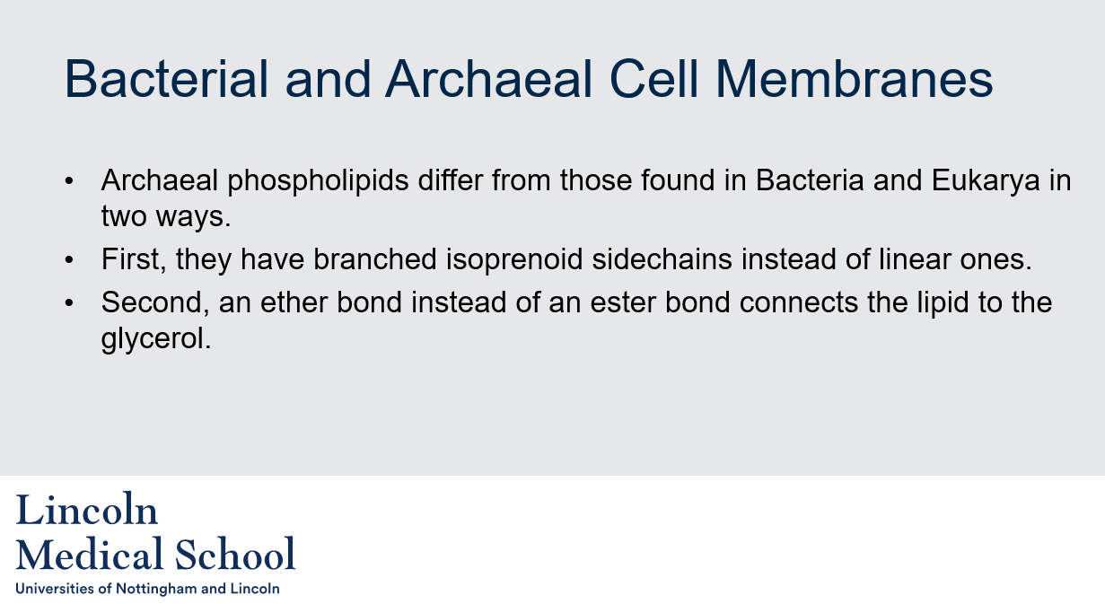
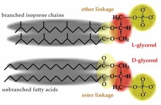
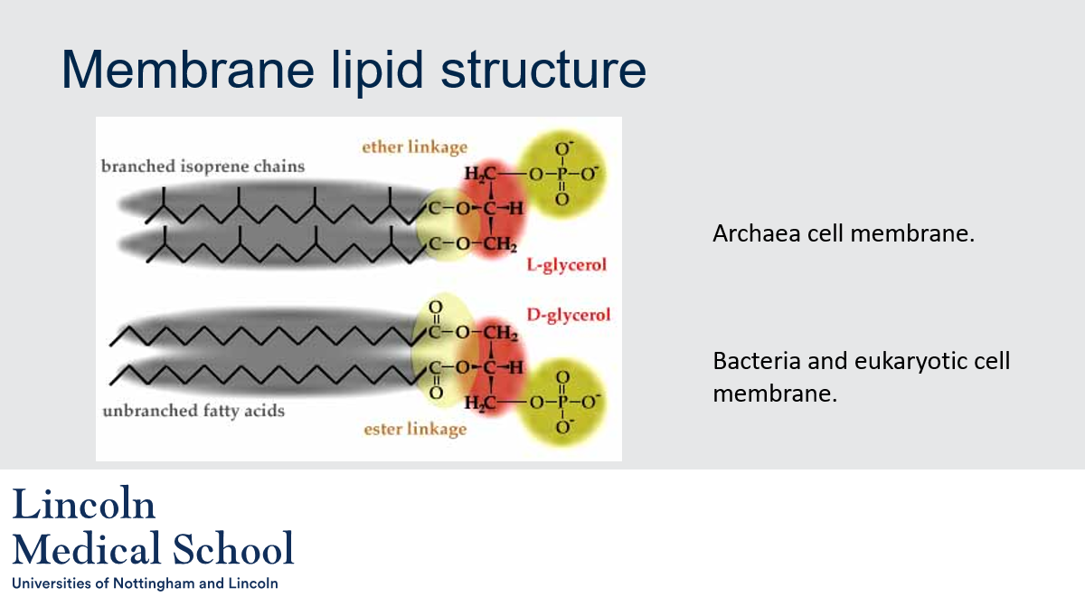
Archaeal phospholipids differ from those found in Bacteria and Eukarya in two ways. Firstly, they have branched isoprenoid sidechains instead of linear ones. Secondly, an ether bond instead of an ester bond connects the lipid to the glycerol.
43
New cards
@@Membrane lipid structure@@
Can you label, describe and explain what this diagram is/shows?
Can you label, describe and explain what this diagram is/shows?
44
New cards
@@Membrane lipid structure@@
What are the fundamental differences between the archaeal membrane and those of all other cells?
What are the fundamental differences between the archaeal membrane and those of all other cells?
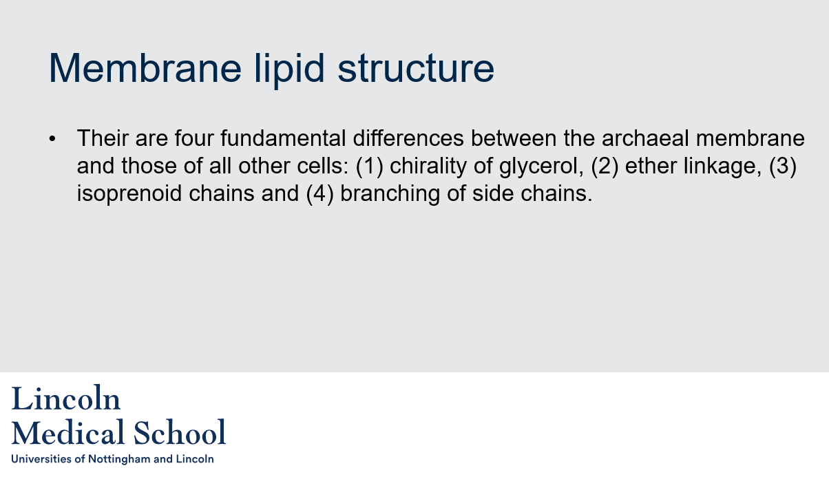
There are four fundamental differences:
* chirality of glycerol
* ether linkage
* isoprenoid chains
* branching of side chains
* chirality of glycerol
* ether linkage
* isoprenoid chains
* branching of side chains
45
New cards
@@Membrane lipid structure@@
What are the four fundamental differences between the archaeal membrane and those of other cells?
What are the four fundamental differences between the archaeal membrane and those of other cells?
The four fundamental differences between the archaeal membrane and those of other cells are:
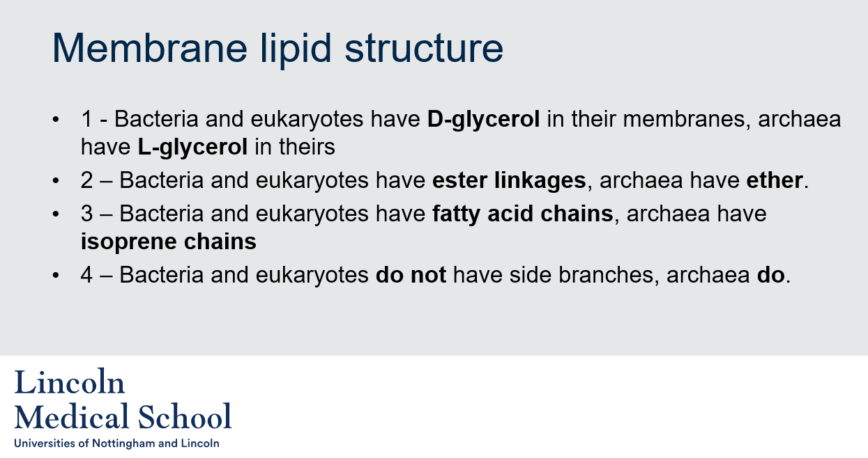
1. Bacteria and eukaryotes have D-glycerol in their membranes, archaea have L-glycerol in theirs
2. Bacteria and eukaryotes have ester linkages, archaea have ether linkages.
3. Bacteria and eukaryotes have fatty acid chains, archaea have isoprene chains.
4. Bacteria and eukaryotes do not have side branches, while archaea do.
