2.0 Central Nervous System
 Call Kai
 Learn
Learn Practice Test
 Spaced Repetition
 Match
Match Flashcards
 Knowt Play
1/46

There's no tags or description
Looks like no tags are added yet.
Name | Mastery | Learn | Test | Matching | Spaced | Call with Kai | Chat |
|---|
No analytics yet
Send a link to your students to track their progress
47 Terms
Syllabus point
Structure & function of the divisions of the nervous system can be observed & compared at different levels in detecting & responding to the changes in the internal & external environments including:
central-peripheral
afferent-efferent
autonomic-somatic
sympathetic-parasympathetic
The parts of the central nervous system, including the brain (cerebrum, cerebellum, medulla oblongata, hypothalamus, corpus callosum) & spinal cord, have specific roles in the co-ordination of body functions & are protected by the meninges & cerebrospinal
The nervous system
The nervous system is the body's control and communication center.
The nervous system is very important in helping to maintain homeostasis.
It works with the endocrine system to regulate and maintain a constant internal environment.
Functions of the Nervous System
In order to carry out its role, the nervous system has three functions:
1. Monitoring changes:
it uses sitside tine bosensory receptors to monitor changes occurring both inside
these changes are called stimuli, and the gathered information is called sensory input.
2. Interpretation of sensory input:
It processes and interprets the sensory input and decides what should be done at each moment, a process called integration.
3. Effects response:
it then effects a response by activating muscles or glands (effectors) via motor
Organisation of the nervous system
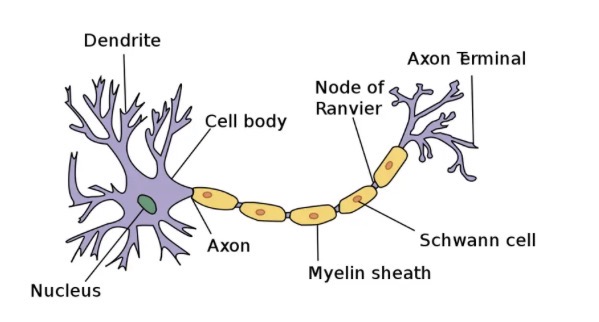
Neuron’s are the basic structural and functional unit of the nervous system.
The nervous system consists of the brain, spinal cord and all the neuron’s.
The nervous system is complex, and it is divided into divisions for ease of learning.
Divisions of the nervous system
Central nervous system (CNS)
Peripheral nervous system (PNS)
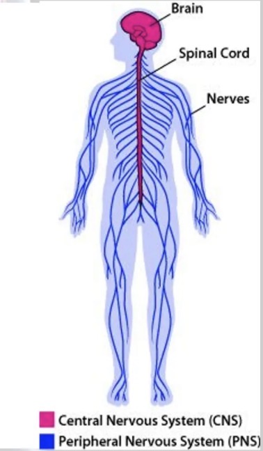
Central nervous system (CNS)
The CNS consists of the brain and spinal cord.
Acts as the integrating and command centres of the nervous system.
Peripheral nervous system (PNS)
The PNS is the part of the nervous system outside the CNS.
Consists mainly of the nerves that extend from the brain and spinal cord.
There are 12 cranial nerves and 31 pair of spinal nerves.
The PNS is further divided into divisions based on their functions.
Divisions of the nervous system
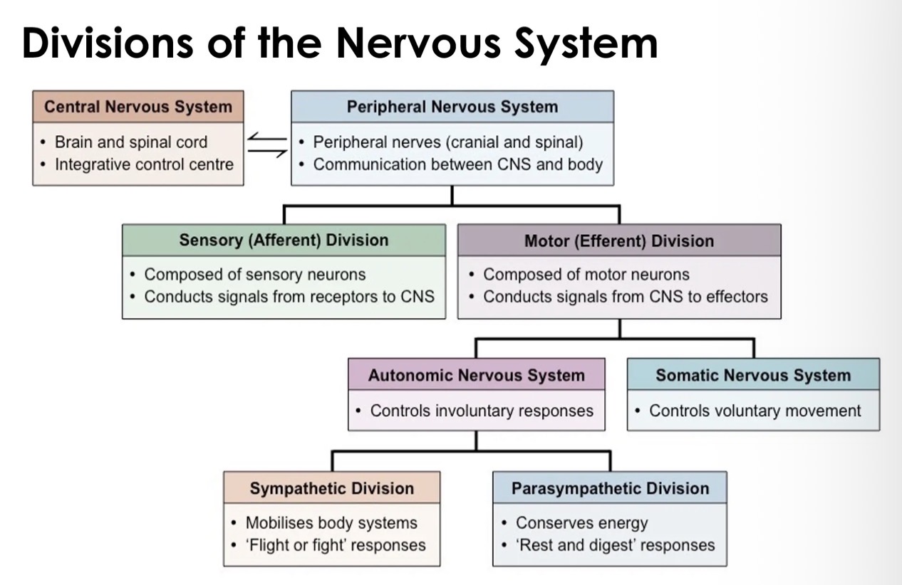
Overview of the workings of the nervous system
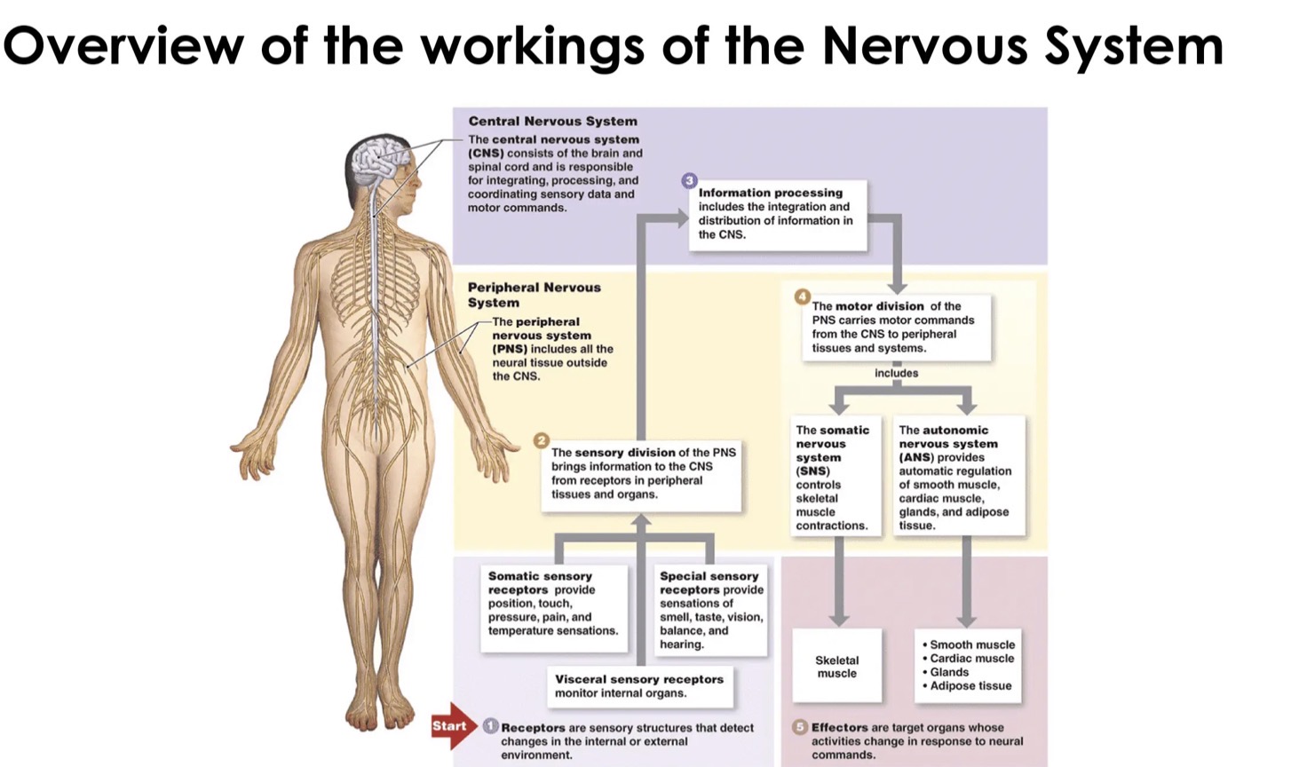
Central nervous system (CNS)
Consists of the brain and spinal cord.
Is the place where incoming messages are processed and outgoing messages are initiated.
The brain and spinal cord are very delicate so must be protected by the body.
Three structures help protect the brain and spinal cord:
bone
Membrane
dura mater
Arachnoid
Pia mater
cerebrospinal fluid (CSF)
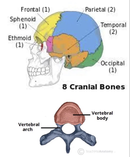
Bone
the brain is protected by the cranium
The spinal cord runs through an opening in the vertebrae called the vertebral canal
Membranes
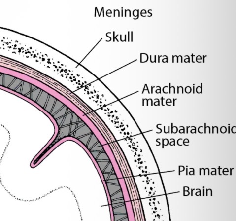
covers the surface of the brain and spinal cord.
Consists of three layers of connective tissue called the meninges.
Dura mater - outer layer is tough and fibrous. It sticks to the bones of the skull. Contains blood vessels.
Arachnoid - middle layer is a loose meshwork of connective fibres and contains the cerebrospinal fluid.
Pia Mater - inner layer is thin fibrous and contains most blood vessels. It sticks closely to the surface of the brain and spinal cord.
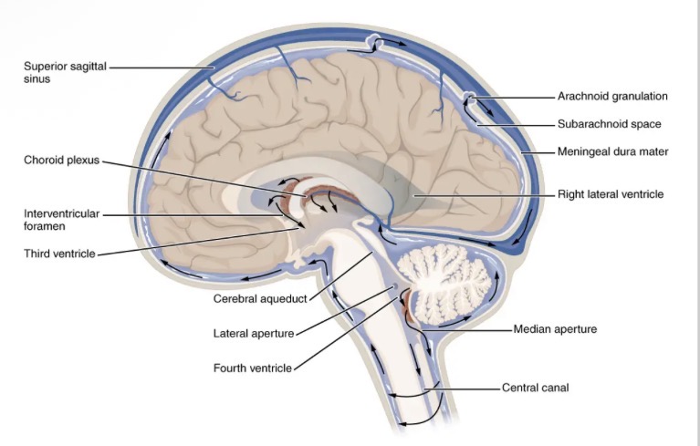
Cerebrospinal fluid (CSF)
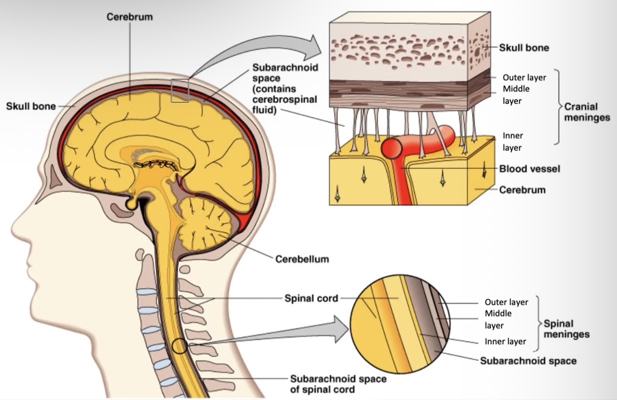
Is a clear, watery fluid containing glucose, protein, urea and salts.
Acts as a shock absorber, cushioning any blows to the CNS. - Found in the space between middle and inner layers of the meninges.
Circulates through a canal in the centre of the spinal cord.
Brain floats in the CSF.
CSF is formed from blood plasma in the ventricles. It circulates through the brain and spinal cord and is eventually reabsorbed back into the blood.
As it circulates it takes nutrients to cells of the brain and spinal cord and takes away their waste.
Brain
Although the brain is only 2% of the body's weight, it uses 20% of the oxygen supply and gets 20% of the blood flow.
The brain is about 73% water.
The brain contains about 86 billion brain cells.
The brain is not fully developed until around 25 years old.
3 functions of Cerebrospinal fluid
Protection - by acting as a shock absorber.
Support - brain is suspended in the cranium by the CSF.
Transport - takes nutrients to the cells of the brain and spinal cord.
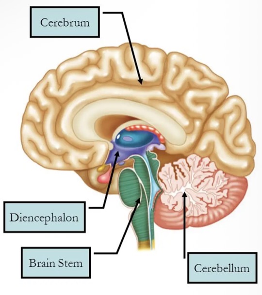
the brain is the largest and most complex mass of nervous tissue in the body, it is commonly discussed in terms of its four major regions:
Cerebrum
Diencephalon
thalamus
Hypothalamus
Brain stem
medulla
Pons
Midbrain
Cerebellum.
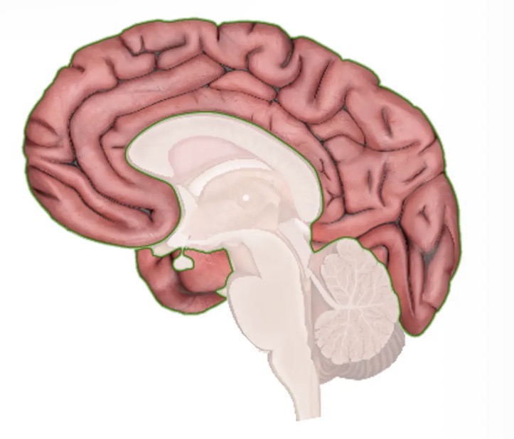
The cerebrum
The paired cerebral hemispheres are collectively called the cerebrum.
Largest part of the brain.
It performs higher functions like interpreting touch, vision and hearing, as well as speech, reasoning, emotions, learning, and control of voluntary movement.
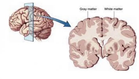
The cerebrum consists of
Cerebral cortex: the outer layer of grey matter
Cerebral white matter: below the grey matter.
Cerebral cortex consists of
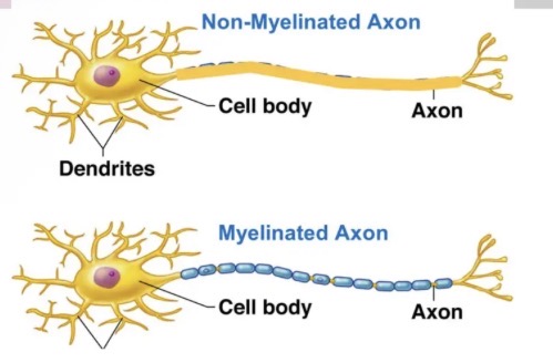
neuron cell bodies
Dendrites
Unmyelinated axons
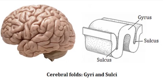
Gyri and sulci
Synapses
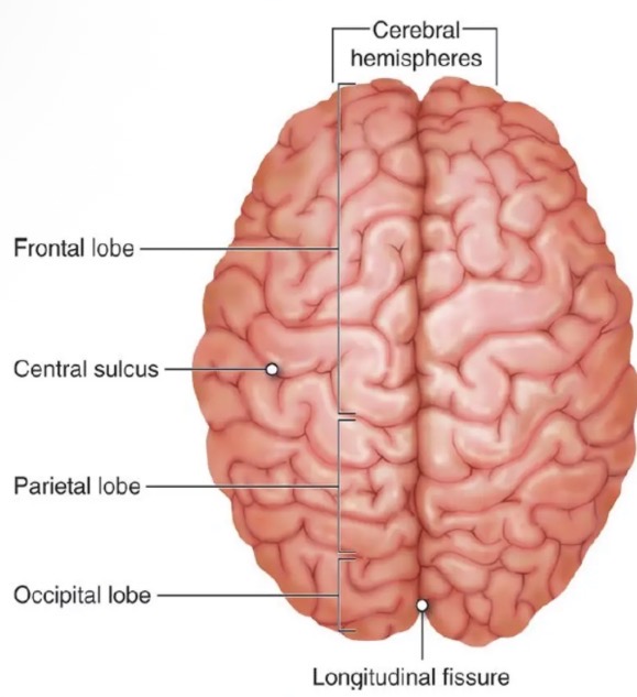
Fissures - longitudinal fissure
Corpus callosum
Left and right hemispheres
Gyri and sulci
The entire surface is folded so to increase surface area.
These folding produces elevated ridges of tissue called gyri, separated by shallow grooves called sulci.
Synapses are located here, and this is where sensory information is processed.
Fissures
Less numerous are the deeper grooves of tissue called fissures, which separate large regions of the brain. The cerebral hemispheres are separated into the right and left hemispheres by a single deep fissure, the longitudinal fissure.
Other fissures or sulci divide each hemisphere into several lobes, named for the cranial bones
that lie over them.
Longitudinal fissure - corpus callosum
At the base to the longitudinal fissure is an area of white matter called the corpus callosum. This connects the left and right hemispheres by nerve fibres and allows the two hemispheres to communicate with each other.
Lobes of the cerebrum
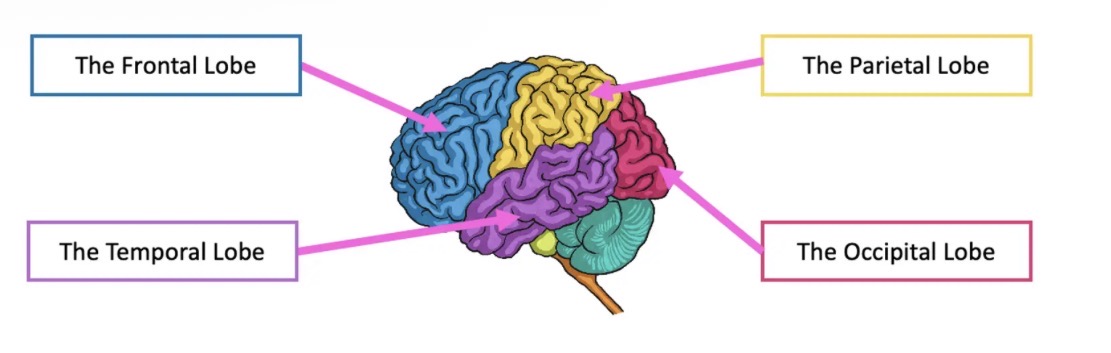
The cerebrum is split into different areas called 'lobes'
Each of the different lobes perform different functions and are linked to different senses.
Frontal
Temporal
Parietal
Occipital
The frontal lobe
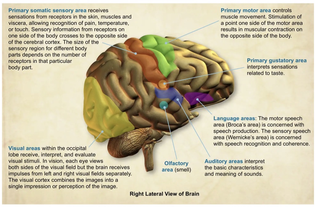
The frontal lobe houses the primary motor cortex, which helps coordinate voluntary movements, including walking and running.
Managing higher level executive functions. Executive functions refer to a collection of cognitive skills including the capacity to plan, organise, initiate, self-monitor and control one's responses in order to achieve a goal.
Considered our behaviour and emotional control centre and home to our personality.
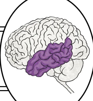
The temporal lobe
The temporal lobes sit behind the ears and are the second largest lobe.
They are most commonly associated with processing auditory information and with the encoding of memory.
Involved in understanding language
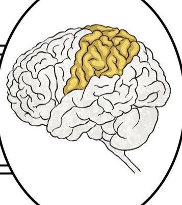
The parietal lobe
The parietal lobes are located near the back and top of the head.
They are important for processing and interpreting somatosensory input. For example, they inform us about objects in our external environment through touch and about the position and movement of our body parts (proprioception).
The parietal lobes are also responsible for integrating sensory input, such as pain and temperature.
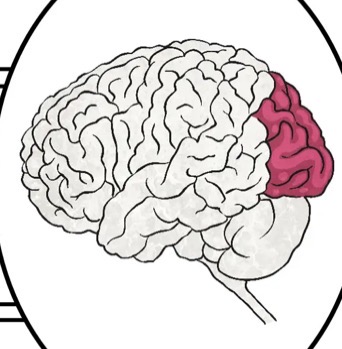
The occipital lobe
The occipital lobe (lubus occipitalis) is all about visual processing and it gets its information from the eyes.
The occipital lobes sit at the back of the head and are responsible for visual perception, including colour, form and motion.
Occipital' is Latin for 'back of the head'.
Functional areas of the cerebral cortex
Cerebral white matter
Located between the cerebral cortex(grey matter) and the basal ganglia (grey matter).
Composed of nerve fibres (axons) that are myelinated.
The axons of white matter transmit signals trom various grey matter areas (the locations of nerve cell bodies, of the cerebrum to each other.
Bundles of nerve fibres are called tracts.
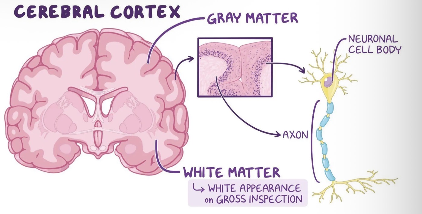
Three types of tracts occur in the cerebral white matter:
Tracts that connect areas within each hemisphere
Tracts that carry impulses between the left and right hemispheres
Tracts the connect the cortex to other parts of the brain or spinal cord.
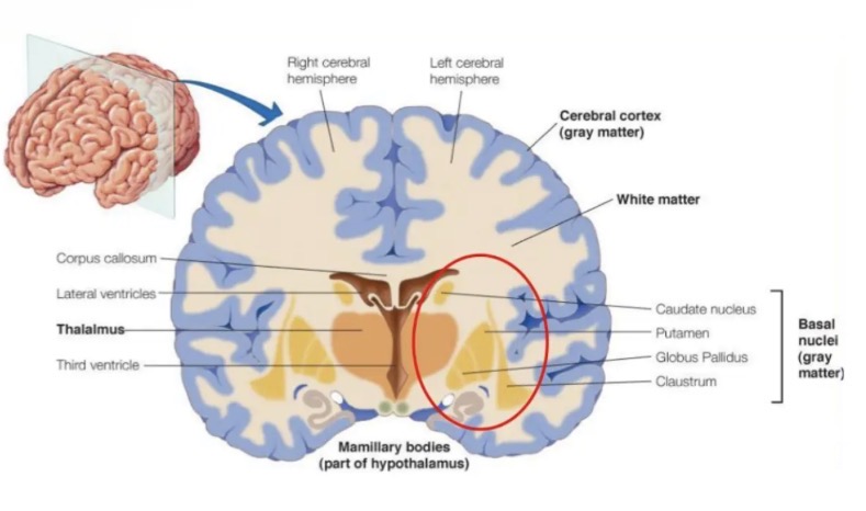
Basal ganglia
There are several islands of grey matter, called the basal nuclei, or basal ganglia, buried deep within the white matter of the cerebral hemispheres.
Consists of groups of nerve cell bodies.
Associated with the control of skeletal muscles. Help in the initiation of desired movements and inhibiting unwanted movements.
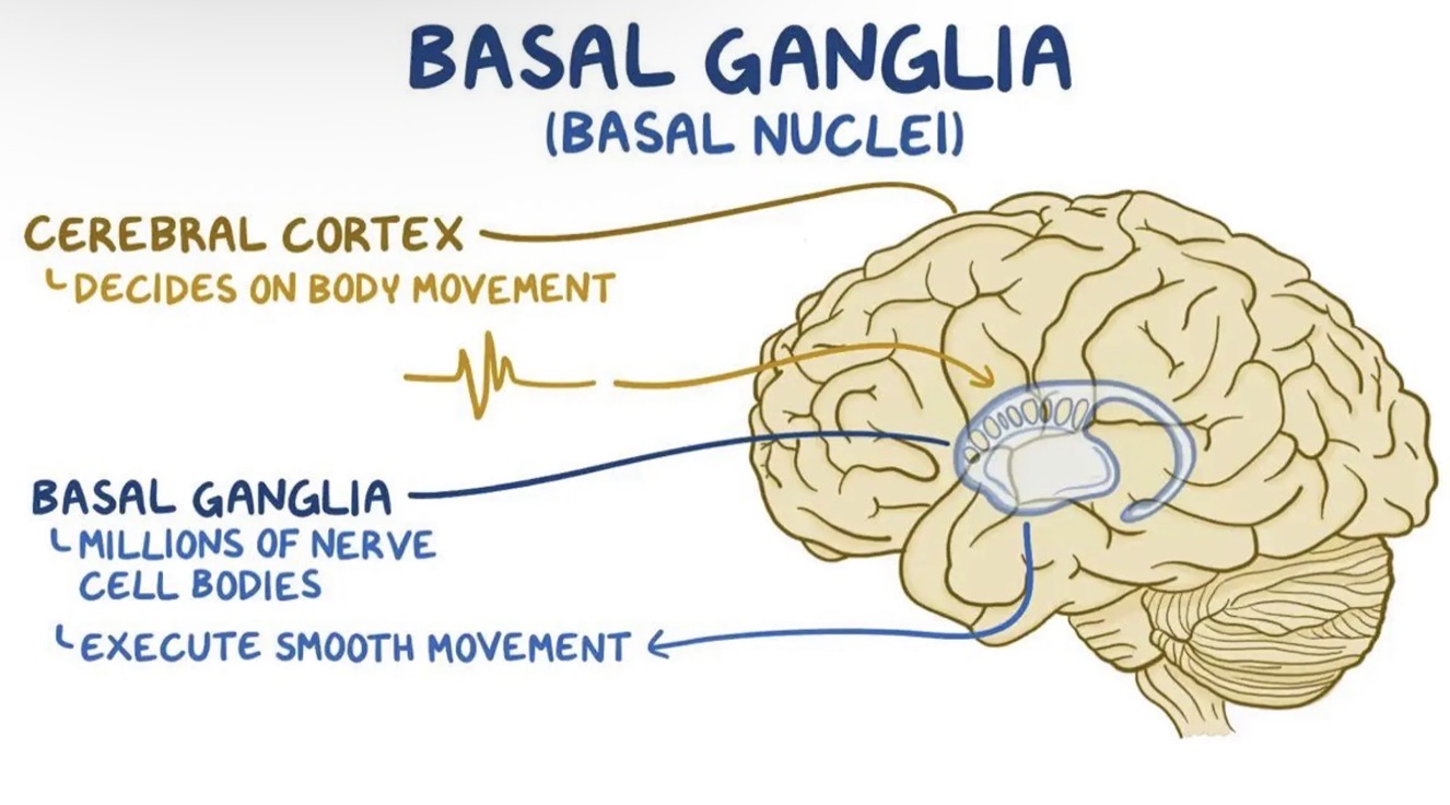
The diencephalon
the diencephalon or interbrain sits atop the brain stem and is enclosed by the cerebral hemispheres
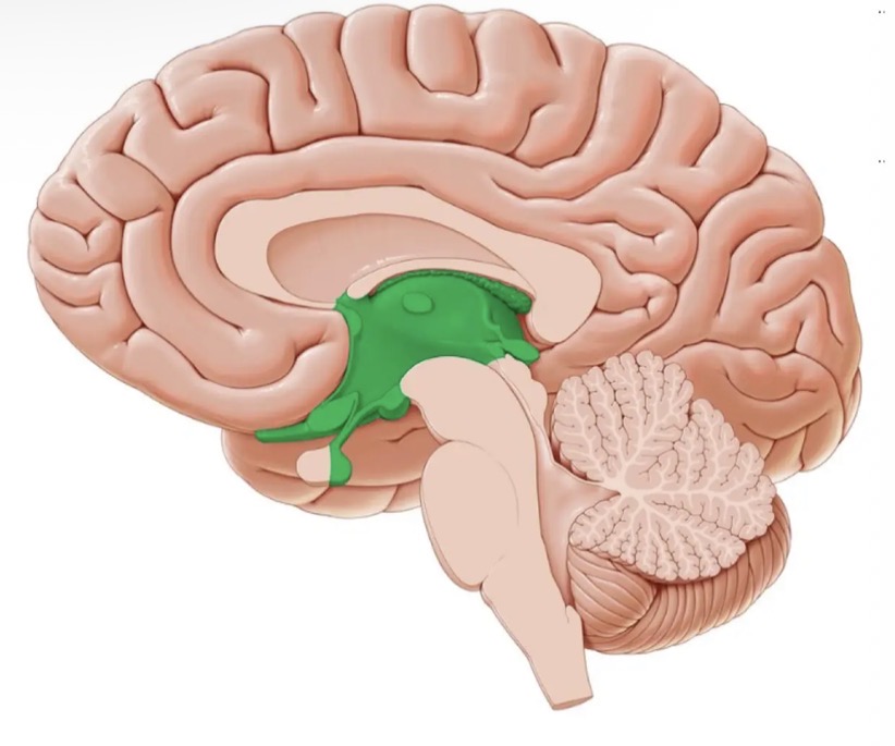
The diencephalon consists of
Thalamus
Hypothalamus
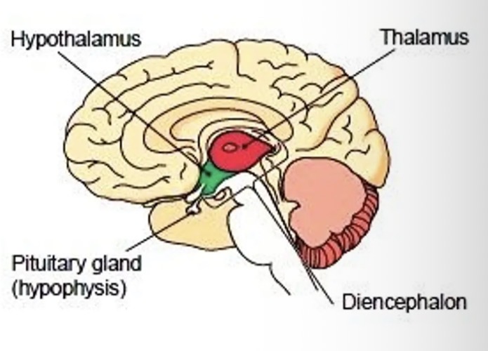
Thalamus
the main relay centre for all sensory messages that enter the brain before they are transmitted to the cerebrum.
Hypothalamus
Links the endocrine and nervous system
Regulates the pituitary gland and produces two hormones of its own.
Regulates body temperature
patterns of waking and sleeping
Regulates water balance and metabolism
Regulates emotional responses such as fear, anger, aggression and pleasure
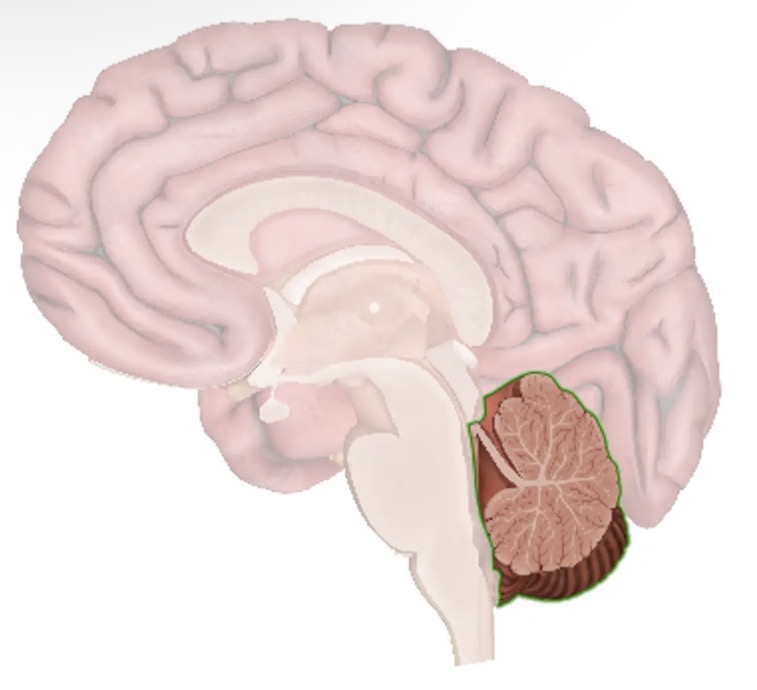
The cerebellum
Lies under the occipital lobe of the cerebrum.
Second-largest part of the brain.
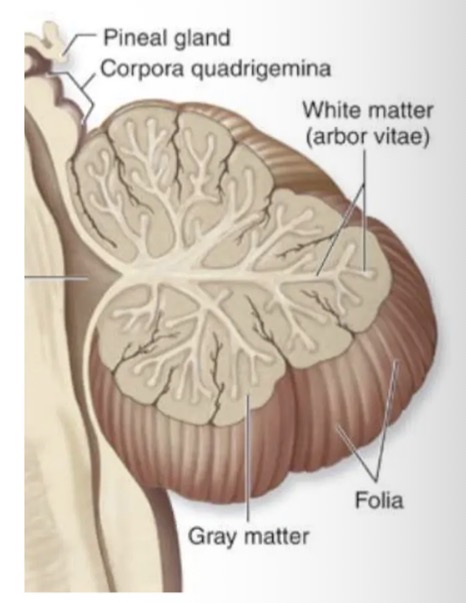
Like the cerebrum, the cerebellum has two hemispheres and a convoluted surface; it also has an outer cortex made up of grey matter and an inner region of white matter.
The cerebellum provides coordination of smooth motor movement and controls balance, posture and muscle tone.
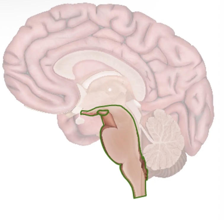
The brain stem
The brain stem is about the size of a thumb in diameter and consists of the:
Midbrain
Pons
Medulla oblongata
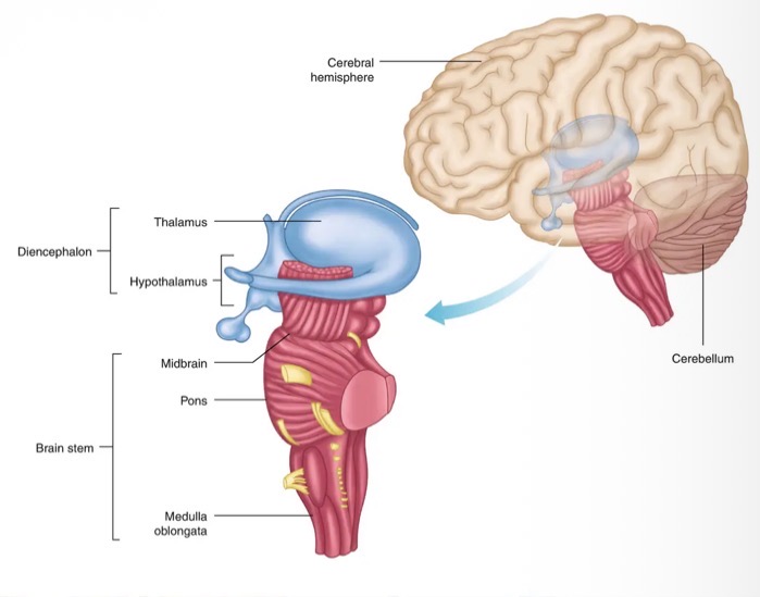
Medulla oblongata
Continuous with the spinal cord
Many nerve fibres pass through the medulla going to and from other parts of the brain
Regulates reflexes of swallowing, sneezing, coughing and vomiting
Medulla oblongata congains
Cardiac centre - regulates rate and force of heartbeat
Respiratory centre - control rate and depth of breathing
Vasomotor centre - regulates the diameter of blood vessels
Spinal cord
Cylindrical spinal cord is a continuation of the brain stem.
Extends from the foramen magnum (opening at the base of the skull) to the second lumbar vertebra.
The spinal cord provides a two-way conduction pathway to and from the brain, and it is a major reflex center.
The spinal cord is enclosed in the vertebral canal.
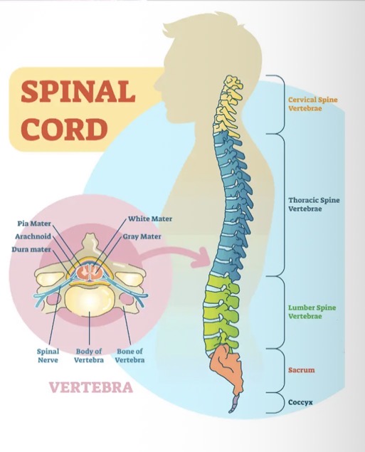
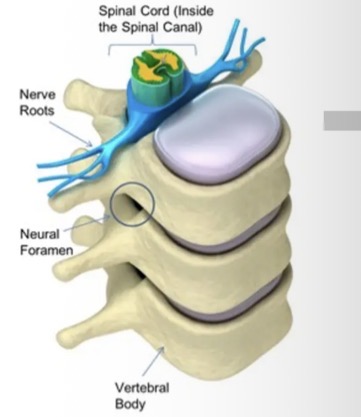
Spinal cord - 3 meningeal layers
The spinal cord is protected by the three meningeal layers.
In the spinal cord the outer layer of the meninges is not joined to the bone but contains a space containing fat, connective tissue and blood vessels.
This allows the spinal cord to bend and the fat aids in protecting the spinal cord.
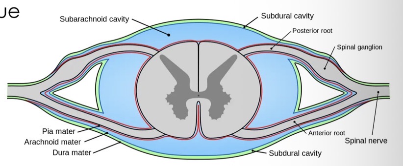
Spinal cord - central canal
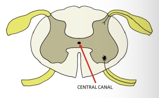
The central canal is the cerebrospinal fluid-filled space that runs through the spinal cord.
The central canal carries CSF that is rich in nutrients such as glucose.
These nutrients are delivered throughout the spinal cord via the central canal.
It also protects the spinal cord by cushioning the impact of a force on the spine.
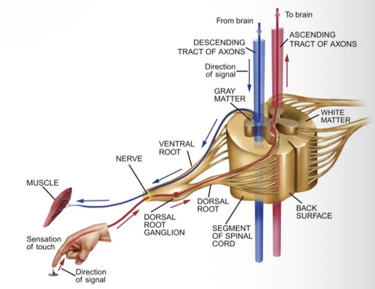
Spinal cord - roots
Spinal cord - grey matter
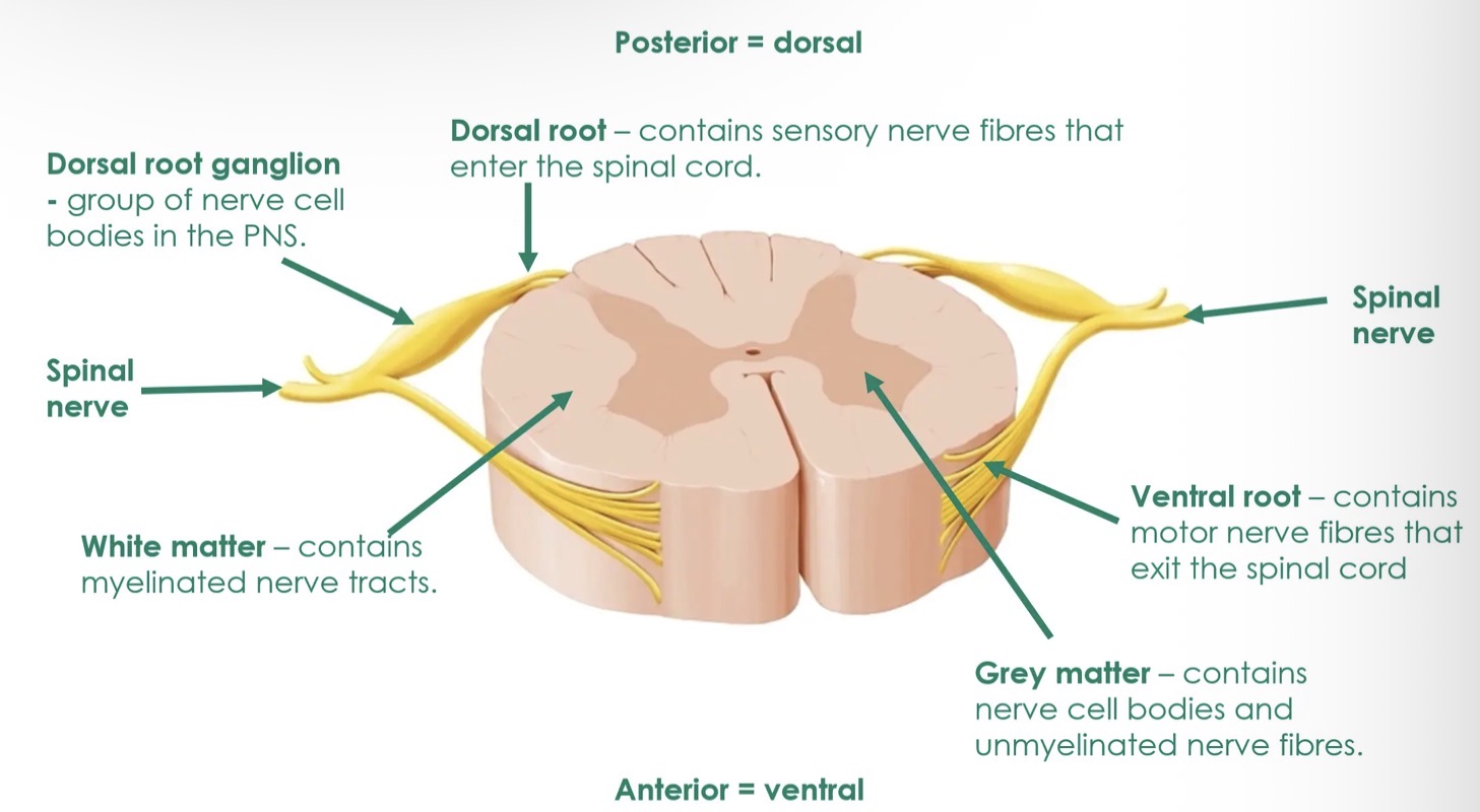
spinal cord consists of both grey and white matter
The grey matter is at the centre.
Composed of nerve cell bodies and unmyelinated nerves fibres.
The grey matter of the spinal cord looks like a butterfly or a letter H in cross section.
Some motor and sensory neurons synapse with interneurons neurons in the grey matter.
The grey matter surrounds the central canal of the cord, which contains CSF.
Spinal cord - white matter
Found outside the grey matter.
White matter of the spinal cord is composed of myelinated fibre tracts.
Nerve fibres are arranged in bundles and known as ascending and descending tracts.
Ascending tracts are bundles of axons that carry sensory information from the spinal cord to the brain.
Descending tracts are bundles of axons that carry motor commands from the brain down the spinal cord.
Spinal cord
the spinal nerves arise from the spinal cord at each segment by way of two
Dorsal roots carry input from the sensory receptors. Afferent sensory nerve fibres enter the spinal cord at the dorsal root.
Efferent motor nerve fibres exist the spinal cord at the ventral root.
Each spinal nerve is formed by the combination of nerve fibres from the dorsal and ventral roots of the spinal cord.