N230 - Exam #3 Study Guide
 Call Kai
 Learn
Learn Practice Test
 Spaced Repetition
 Match
Match Flashcards
 Knowt Play
1/25

There's no tags or description
Looks like no tags are added yet.
Name | Mastery | Learn | Test | Matching | Spaced | Call with Kai | Chat |
|---|
No analytics yet
Send a link to your students to track their progress
26 Terms

Postpartum Complications
Postpartum infection
Also called a puerperal infection
Any clinical infection of the genital tract that occurs within 28 days after miscarriage, induced abortion, or birth
Defined as the presence of a fever of 100.4 degrees F or 38 degrees C in the first 24 hours after birth
Endometritis (endomyometritis):
Infection of the lining of the uterus
Not endometriosis
Most common puerperal infection
Occurs in ~2% after vaginal birth, 10-15% after cesarean birth
Highest incidence with cesarean birth after prolonged labor and membrane rupture
Signs & Symptoms:
Fever (usually >38°C), increased pulse, chills
Anorexia, nausea, fatigue, lethargy
Pelvic pain, uterine tenderness
Foul-smelling lochia
Leukocytosis, elevated RBC sedimentation rate, possible anemia
Wound Infections:
Common after cesarean birth (3-5% rate)
Can develop in perineal lacerations or episiotomy sites
Signs & Symptoms:
Fever, erythema, edema, warmth
Tenderness, pain, seropurulent drainage
Wound separation
Treatment
Endometritis:
IV broad-spectrum antibiotics (clindamycin and gentamicin, cephalosporins, penicillins)
Supportive care: hydration, rest, pain relief
Antibiotics discontinued 24 hours after afebrile and asymptomatic
Comfort measures: cool compresses, warm blankets, perineal care, sitz baths
Wound Infections:
IV antibiotic therapy
Wound opened and drained if pus or significant effusion present
Irrigated with normal saline, redressed several times daily
Wound vacuum device may be used
Continued until wound base is clear without cellulitis signs
Nursing Care
Assessment:
Frequent vital signs and temperature monitoring
Lochia assessment
Wound assessment and care
Patient Teaching:
Hygienic care techniques: changing perineal pads, cleansing front to back, hand hygiene before/after perineal care
Signs of infection and when to contact provider
Side effects of therapy, prevention of infection spread
Wound care and dressing changes for home
Support:
Maintain mother-infant interactions and breastfeeding
Home visits for wound assessment and teaching reinforcement
Telephone follow-up and access to support resources
S/S → fundal tenderness, febrile
Causes → Prolonged rupture of membranes, manual introduction of bacteria
Other reasons for fever/infection symptoms:
PE, DVT, pneumonia, flu/virus
Mastitis (treated w/ dicloxacillin, cephalexin)
Misoprostol can cause a transient fever, not too high and no other symptoms

Postpartum bleeding and hemorrhage
Definition
Postpartum hemorrhage (PPH) is an obstetric emergency and a leading cause of maternal morbidity and mortality.
Current Definition (ACOG):
Cumulative blood loss ≥1000 mL, OR
Bleeding with signs/symptoms of hypovolemia within 24 hours of birth, regardless of delivery type
Classification:
Early/Primary PPH: Within 24 hours of birth
Late/Secondary PPH: More than 24 hours up to 12 weeks after birth
Causes
Most Common: Uterine Atony
Failure of uterine muscle to contract firmly
Relaxed uterus distends with blood and clots
Blood vessels at placental site not clamped off
Often results from retained placental fragments
Other Causes:
Vaginal or vulvar hematomas
Unrepaired lacerations of vagina or cervix
Lacerations of birth canal during placental separation
Incomplete placental separation
Undue fundal manipulation or excessive cord traction
Uterine prolapse
Late PPH: subinvolution, infection, retained placental fragments, coagulopathy
Assessment
Blood Loss Estimation:
Quantification by weighing (1 mL = 1 g) is the most accurate method
Visual estimation: scant, light, moderate, or heavy
Time factor is critical: saturating pad in 1 hour vs. hours indicates very different bleeding rates
First Step When Bleeding Appears Excessive:
Evaluate uterine contractility
If uterus is firmly contracted and bleeding continues, identify other sources
Assessment may include visual/manual inspection of perineum, vagina, uterus, cervix, rectum
Laboratory studies: hemoglobin, hematocrit, coagulation studies, platelet count
Prevention
Two Most Important Interventions:
Maintaining good uterine tone
Preventing bladder distention
Management
If uterus is hypotonic or boggy, management focuses on increasing contractility and minimizing blood loss
Treatment depends on bleeding source
Treated even if blood loss doesn’t meet parameters
Boggy fundus, tachycardia, hypotension, postural hypotension, sweating, anxiety, pallor, tachypnea, restlessness, lightheaded, altered mental status.
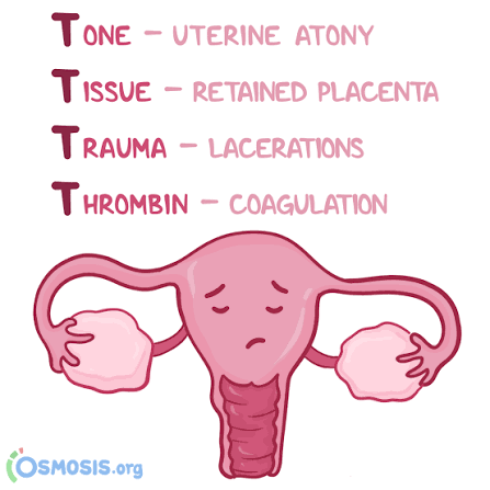
Early Postpartum Hemorrhage Causes: 4 T’s
Tone
Trauma
Tissue
Thrombin
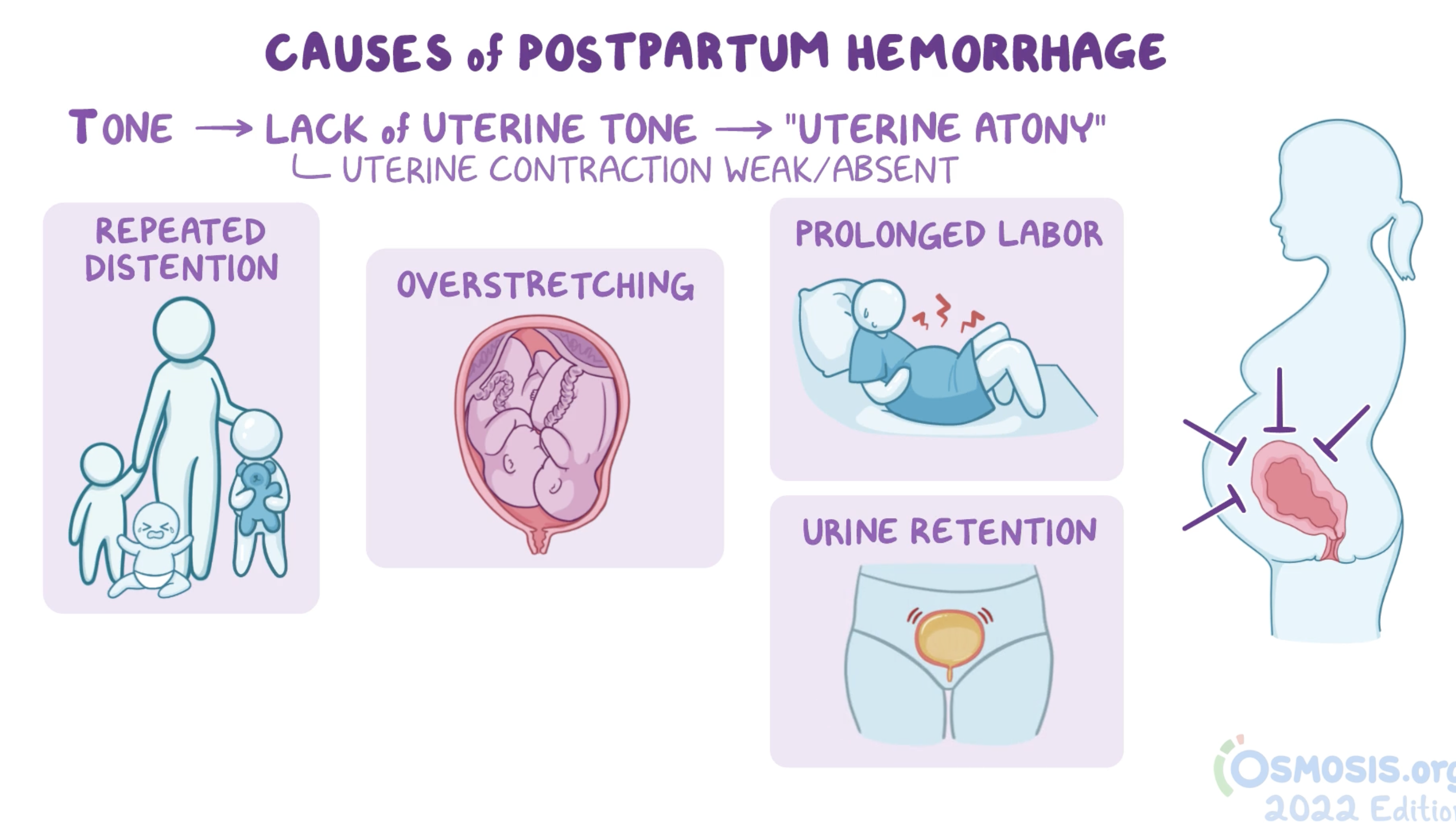
Tone
Uterine atony (Marked hypotonia of uterus)
Most common
Blood supply to the uterus to supply the placenta is cut off by myometrium contractions
Lattice muscle fibers surround large blood vessels
Contractions cause pressure on those vessels, which activate coagulation pathways
Initiated after delivery of placenta
Risk factors:
High parity
Polyhydramnios
Macrosomia
Multiple gestation
Uterine fatigue/long labor
Tocolytic drugs
The greatest risk for early PPH is during the first hour after birth. The body of the uterus is like a basket-weave of strong, interlacing smooth muscle bundles through which many large maternal blood vessels pass. After the expulsion or removal of the placenta, bleeding is controlled when these smooth muscle fibers in the uterus are well contracted. If inadequate uterine contraction occurs, the uterus remains flaccid, and rapid blood loss can follow. This hypotonia of the uterus is called uterine atony.
Uterine atony is the leading cause of early PPH. It is associated with high parity, polyhydramnios, fetal macrosomia, obesity, and multiple gestation. In such conditions the uterus is more distended and therefore may not contract as well after birth.
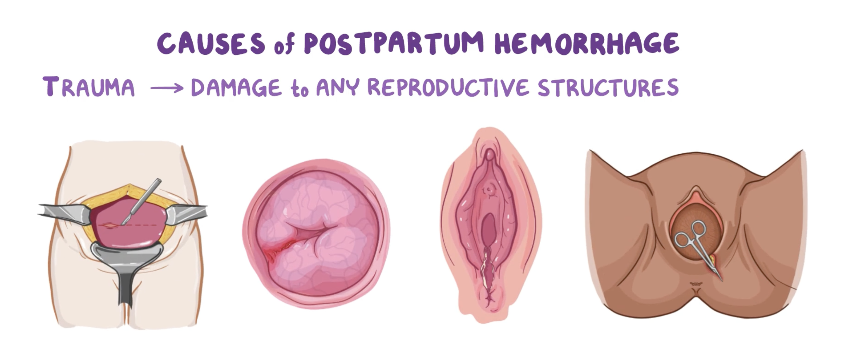
Trauma
Lacerations of the genital tract
Uterus, cervix, vagina, perineum
Hematomas
Lacerations of the cervix, vagina, and perineum are also causes of PPH. Hemorrhage related to lacerations should be suspected if bleeding continues despite a firm, contracted uterine fundus. This bleeding can be a slow trickle, an oozing, or frank hemorrhage. Factors that influence the causes and incidence of obstetric lacerations of the lower genital tract include operative birth, precipitous or rapid birth, congenital abnormalities of the maternal soft tissue, and contracted pelvis. Other possible causes of lacerations are increased size, abnormal presentation, and position of the fetus; relative size of the presenting part and the birth canal; previous scarring from infection, injury, or surgery; and vulvar, perineal, and vaginal varicosities.
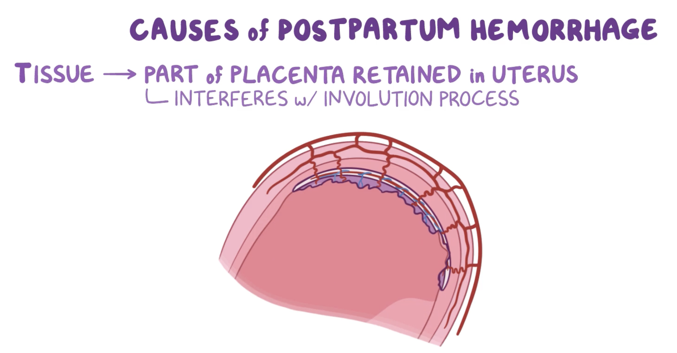
Tissue
Retained products of the placenta
Placental anomaly → sometimes connects further into the abdominal wall
When the placenta has not been expelled within 30 minutes after birth despite an upright posture of the woman, gentle traction on the umbilical cord, and uterine massage, it is described as a retained placenta. Initial management of a retained placenta consists of manual separation and removal by the midwife or physician. This involves the provider reaching into the uterus and gently separating the placenta from the uterine wall and removing it manually. When the client has regional anesthesia for labor, supplementary anesthesia is usually not needed. For other women, administration of light nitrous oxide and oxygen inhalation anesthesia or intravenous (IV) pain medications should be considered. After removal of a retained placenta, the woman has an increased risk for PPH and infection.
Fragments of the placenta and/or amniotic membranes can remain in the uterus after spontaneous separation of the placenta during the third stage of labor. In this case the client may have excessive bleeding and the uterus feels boggy (soft) due to uterine atony. The midwife or physician may attempt manual exploration to remove the fragments; uterine curettage (removal of uterine contents using a curette or vacuum suction) may be necessary
PPH can also be due to an abnormally implanted, invasive, or adherent placenta; this is known as placenta accreta syndrome. Abnormal placental adherence can be total, partial, or focal, depending on how much placental tissue is involved. The following degrees of abnormal placental attachment are recognized:
• Placenta accreta—slight penetration of myometrium
• Placenta increta—deep penetration of myometrium
• Placenta percreta—perforation of myometrium and uterine serosa, possibly involving adjacent organs
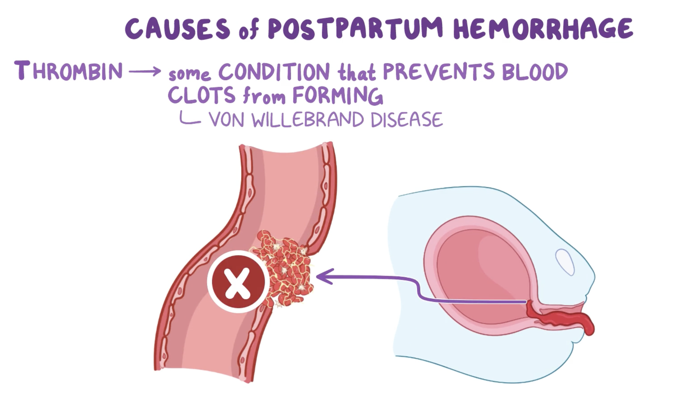
Thrombin (Coagulation)
When no identifiable cause, think coagulopathy
TXA first-line therapy
Von Willebrand
Most common inherited bleeding disorder. Missing clotting factor
Idiopathic or immune thrombocytopenic purpura (ITP)
Taking anticoagulants
Coumadin is generally contraindicated; should be avoided in the first trimester due to tetragenicity
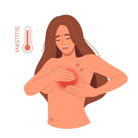
Mastitis
Definition
Mastitis is an infectious process of the breast characterized by inflammation that may be accompanied by infection and pain. While most commonly associated with lactating and breastfeeding women, it can occur in non-lactating women as well.
Signs & Symptoms
Sudden onset of influenza-like symptoms: fever, chills, malaise, body aches, headache, nausea, vomiting
Localized breast pain and tenderness
Hot, reddened area on the breast
Most commonly occurs in the upper outer quadrant of the breast
One or both breasts can be affected
Timing
Most cases occur during the first 2 to 4 weeks postpartum, although mastitis can occur at any time.
Risk Factors
Breastfeeding-Related:
Inadequate emptying of breasts (engorgement, plugged ducts)
Sudden decrease in number of feedings or abrupt weaning
Wearing underwire bras
Sore, cracked nipples (provide portal of entry for bacteria)
Other Factors:
Stress, fatigue, maternal illness, ill family members
Breast trauma, poor maternal nutrition
Smoking and nipple piercings (bacteria can enter through milk duct)
Common Causative Organisms: Staphylococcus, Streptococcus, and Escherichia coli
Treatment
Bedrest
Antibiotics: dicloxacillin, cephalexin, or clindamycin
Pain and swelling reduction: anti-inflammatory medications and cold compresses
Continued lactation with complete emptying of breasts through breastfeeding, hand expression, or pumping
Adequate fluid intake and balanced diet
Important: The infection cannot be transmitted to the infant
Complications
Breast abscess
Chronic mastitis
Fungal infections of the breast
Most complications can be prevented by early recognition and treatment.
Patient Education
Breastfeeding mothers should be taught the signs of mastitis before discharge from the birth facility and instructed to call the healthcare provider promptly if symptoms occur.
Important Note: Mastitis does not increase the risk for developing breast cancer in the future

Newborn Nutrition/Breastfeeding
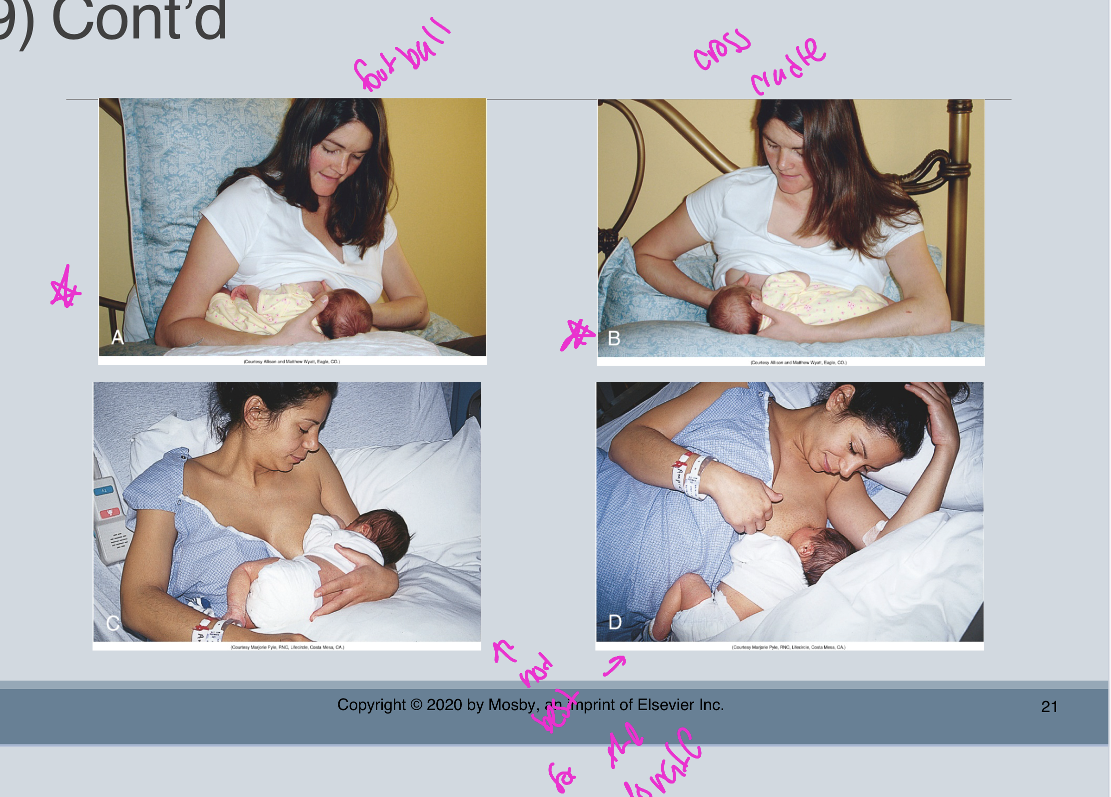
Breastfeeding positions
Initial Feeding Position
Semireclining Position (Laid-Back Breastfeeding):
Recommended for initial feedings
Mother in semireclining position with newborn prone, skin-to-skin on her bare chest
Mother's body supports the baby
Benefits: reduces/eliminates nipple pain, mother more relaxed with freedom of movement, baby uses inborn reflexes to latch and feed effectively
Some mothers prefer this position even after early days
Four Traditional Positions
Football or Clutch Hold (Under the Arm):
Often recommended for early feedings
Mother can easily see baby's mouth as she guides infant onto nipple
Preferred by mothers after cesarean birth
Works well for mothers with large breasts or small babies
Modified Cradle or Across-the-Lap (Cross-Cradle):
Works well for early feedings, especially with smaller babies
Mother has more control over baby's head positioning
Cradle Hold:
Most common position for infants who have learned to latch easily and feed effectively
Used once breastfeeding is well established
Side-Lying Position:
Allows mother to rest while breastfeeding
Preferred by women with perineal pain and swelling
Good for nighttime feedings
General Positioning Guidelines
Key Principles:
Mother should use position that most easily facilitates latch while allowing maximal comfort
Infant held securely at level of breast, supported by firm pillows or folded blankets
Baby facing toward mother
Baby's mouth directly in front of nipple
Mother supports baby's neck and shoulders with her hand (not pushing on occiput)
Baby's body held in alignment: ears, shoulders, and hips in straight line
Comfort Measures:
Mother should be as comfortable as possible
Empty bladder before feeding
Nurse/lactation consultant at mother's eye level when assisting
Before Discharge:
Help mother try all positions so she's confident using them at home
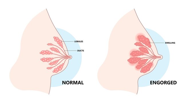
Engorgement
Definition
Engorgement is a common response of the breasts to sudden hormonal changes and the onset of significantly increased milk volume during lactogenesis stage II. It usually occurs 3 to 5 days after birth as milk transitions from colostrum to mature milk.
Pathophysiology
Increased blood flow to the breasts
Increased uptake of glucose and oxygen by the breasts
Milk production rapidly increases, volume can exceed storage capacity of the alveoli
If milk is not removed, alveoli become distended, causing impaired capillary blood flow
Blood vessels become congested, fluid leaks into surrounding tissue causing edema
Milk ducts compressed by tissue edema, preventing easy milk flow
Signs & Symptoms
Breasts become firm, tender, and hot
Appear shiny and taut
Areolae become edematous
Nipples can flatten, making it difficult for infant to latch
May feel hard and warm, enlarged and painful
Timing
Not unusual in first 24 to 48 hours after breasts fill with milk
Later development may signal onset of mastitis
Consequences
Back pressure on full milk glands inhibits milk production - if milk is not removed from breasts, milk supply can diminish.
Note: Engorgement does not occur in all breastfeeding mothers.
Management
Key Intervention: Remove milk from the breasts through frequent breastfeeding and/or milk expression
Assessment:
Determine if breasts are adequately supported with a properly fitting bra
Palpate breasts to determine degree of softness
Distinguish between full breasts (firm, dense, slightly enlarged) and engorged breasts (hard, warm, enlarged, shiny, painful)
Prevention:
Frequent, effective breastfeeding
Proper breast support without overly tight bras

Neonates
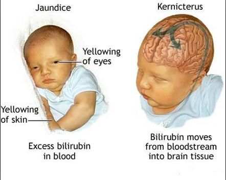
Jaundice
Jaundice (icterus) occurs when bilirubin levels exceed 5 mg/dL and becomes visible in the skin 2. It's observed in approximately 60% of term infants and 80% of preterm infants during the first week after birth.
Physiologic Jaundice (Normal)
Timing & Pattern:
Appears after 24 hours of age
Peaks between days 2-4 at 5-6 mg/dL
Gradually decreases between days 5-71
Resolves by 2 weeks of age in term infants
Usually mild (total bilirubin <15 mg/dL) and self-limiting
Causes:
Immature liver uptake and conjugation of bilirubin
Higher hemoglobin levels and shorter RBC lifespan in newborns
Not associated with any pathologic process
Breastfeeding-Related Jaundice
Early-Onset (Breastfeeding-Associated):
Begins at 2-4 days of age
Related to poor latch or inadequate milk supply
Caused by decreased caloric/fluid intake and increased enterohepatic circulation
Late-Onset (Breast Milk Jaundice):
Occurs in up to 30% of breastfed infants
Peaks between days 5-15
May persist beyond 1 month, lasting up to 3 months
Safe—do not discontinue breastfeeding
Pathologic Jaundice (Abnormal)
Warning Signs:
Jaundice appearing within first 24 hours
Total bilirubin >15-17 mg/dL
Bilirubin increasing >5 mg/dL per day
Jaundice persisting beyond 2 weeks in term infants
Risk Factors:
Prematurity
Blood group incompatibility (ABO, Rh)
G6PD deficiency
Cephalhematoma or significant bruising
Asian or Asian American race
Exclusive breastfeeding with poor intake
Family history of hyperbilirubinemia
Maternal diabetes
Evaluation
Visual Assessment: Jaundice progresses head to toe, but visual determination can be erroneous. Diagnosis requires serum bilirubin measurement or transcutaneous monitoring.
Important: Evaluation considers timing, gestational age, feeding method, family history, and serial bilirubin trends—not just a single value
Yellow skin
Immature liver (Not usually the liver’s fault); liver can’t keep up (physiologic)
Usually isn’t a problem if it occurs 24 hours after birth; still needs treatment → UVB light therapy
As it gets worse, it heads down the body, starts at face, goes down the body
If jaundice occurs in the first 24 hours, it can be a pathologic problem! Like a blood incompatibility with mom
Newborns produce bilirubin 2-2.5 times that of an adult

Fetal loss communication
Core Principles of Communication After Fetal Loss
Validate the Experience: One of the most important goals is to validate the experience and feelings of the parents by encouraging them to tell their stories and listening with care. Help parents talk about their loss, the meaning it has for their lives, and share their emotional pain.
Simple Acknowledgment: At the very least, acknowledge the loss with a simple but sincere, "I am sorry".
Listen Patiently: Listen patiently during the story of loss or grief, even though it can be difficult and sometimes painful. Use therapeutic communication techniques like:
Leaning forward
Nodding the head
Using words such as "Tell me more" to encourage the bereaved person to tell their story
Sitting through the silence—this gives the bereaved person an opportunity to collect thoughts and process what they are sharing
Include Both Parents
Don't forget the partner: Because nurses tend to focus on the physical and emotional needs of the mother, it's especially important to ask the father or partner directly about their views of what happened and the associated feelings of loss.
What to Avoid
Resist unhelpful responses:
Don't give advice or use clichés
Don't try to reduce their pain with quick fixes—although the impulse to say or do something to reduce helplessness is natural, such responses can stifle the further expression of emotion
Be cautious with touch: Careful assessment is important before using touch as a therapeutic technique. For some, touch is meaningful; for others, it's an invasion of privacy or contrary to their cultural or religious beliefs.
Create Adequate Time
Don't rush: The nurse must create enough time to engage with bereaved families without being rushed. Nurses caring for families whose baby has died may need assignments that allow time to give adequate attention to newly bereaved individuals.
Professional Self-Awareness
Nurses need to be comfortable with their own feelings of loss and grief to support and care for bereaved individuals effectively. Have a presence of self and willingness to be alongside, quietly supporting the bereaved person in whatever expressions of feelings or emotions are appropriate for them.
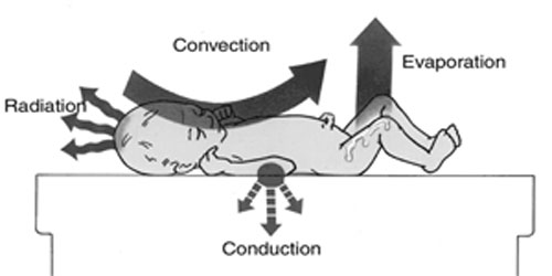
Neonatal heat loss
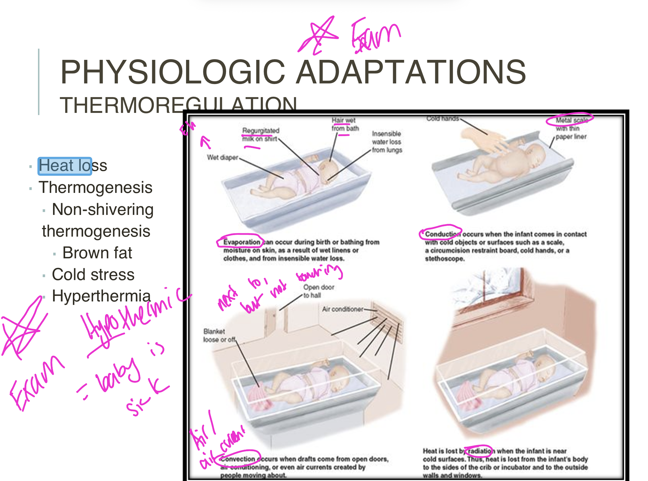
Four Modes of Heat Loss
Newborns lose heat through four distinct mechanisms:
Convection - Heat loss to cooler air currents flowing over the infant's skin
Conduction - Heat transfer to cooler surfaces in direct contact with the infant
Radiation - Heat loss to cooler solid objects not in direct contact (e.g., cold walls, windows)
Evaporation - Heat loss through moisture on the skin converting to vapor
Why Newborns Are Vulnerable
Anatomic & Physiologic Factors:
Large surface area relative to weight facilitates environmental heat loss
Thin subcutaneous fat layer provides minimal insulation
Temperature gradient between core (higher) and surface (lower) body temperature drives heat transfer
Cannot shiver - instead produce heat through nonshivering thermogenesis (NST)
High-Risk Infants Face Additional Challenges:
Smaller muscle mass
Fewer brown fat deposits
Lack of insulating subcutaneous fat
Poor reflex control of skin capillaries
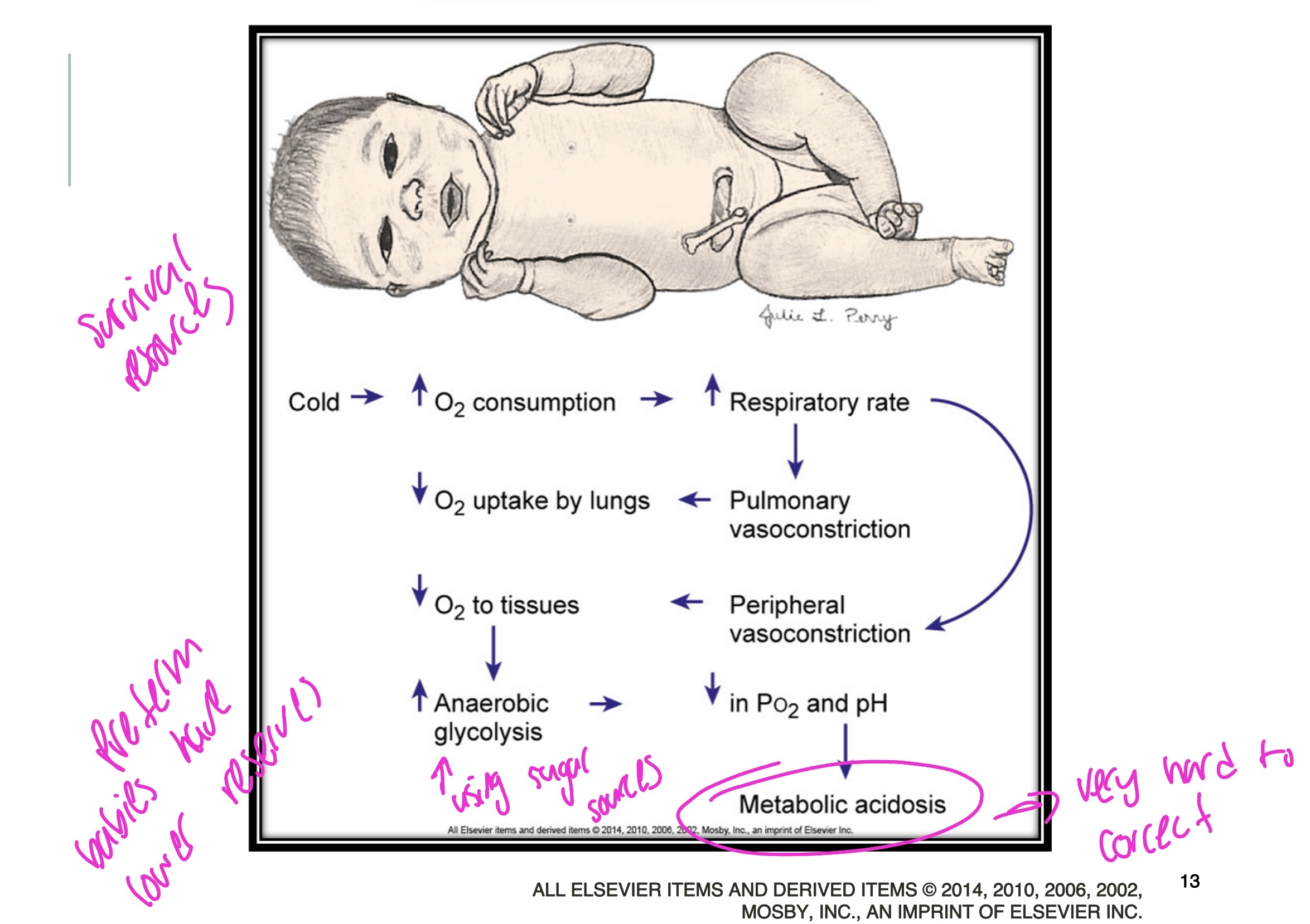
Consequences of Cold Stress
Cold stress produces serious complications:
Hypoxia
Pulmonary vasoconstriction
Metabolic acidosis
Hypoglycemia
Critical statistic: For every 1°C temperature decrease from normal, infant mortality risk increases by 28%.
Prevention Strategies
Immediate Interventions:
Dry the infant quickly after birth
Skin-to-skin contact with mother reduces conductive and radiant heat loss
Cover with warm blanket
Place cap on head to conserve heat
For High-Risk Infants:
Plastic wrap or heated humidified gas in delivery room
Prewarmed incubators
Heated gel mattress with radiant heat
Head coverings (fabric-insulated or wool caps)
Double-walled incubators
Goal: Neutral Thermal Environment
A neutral thermal environment (NTE) allows the newborn to maintain normal core temperature with minimum oxygen consumption and calorie expenditure.
Target temperatures:
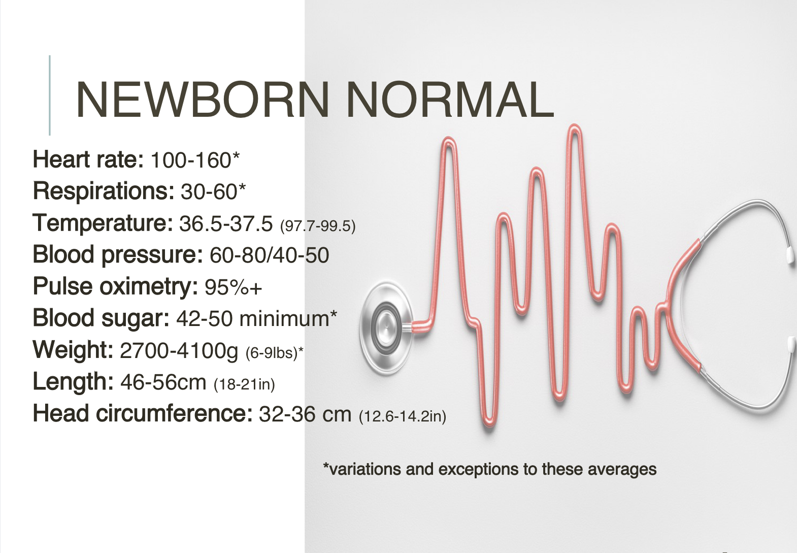
Term infants: 36.5°C to 37.5°C (97.7°F–99.5°F) axillary
Preterm infants: 36.5°C to 37.2°C (97.7°F–98.9°F)
Apgar score
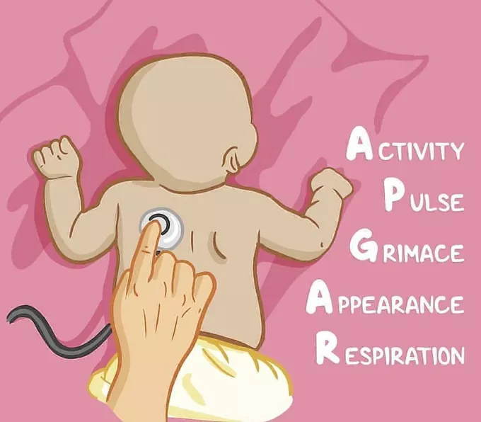
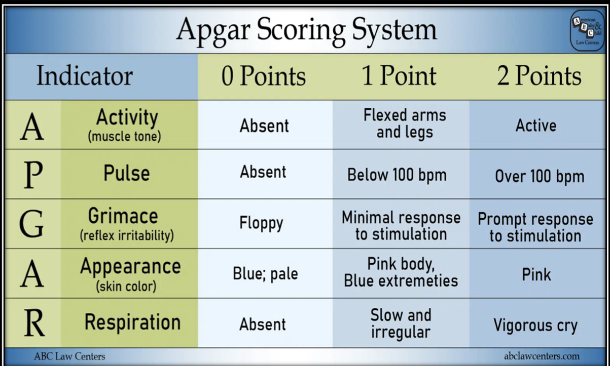
The Apgar score is a rapid assessment of the newborn's overall status and immediate adjustment to extrauterine life. It evaluates five physiologic signs, each scored 0, 1, or 2 1:
Heart rate - Auscultation with stethoscope or palpation of umbilical cord
Respiratory effort - Observed chest wall movement
Muscle tone - Degree of flexion and movement of extremities
Reflex irritability - Grimace, crying, or active withdrawal
Generalized skin color - Pallid, cyanotic, or pink
Timing & Interpretation
When assessed:
At 1 and 5 minutes after birth
Repeated every 5 minutes for up to 20 minutes if score <7 at 5 minutes
Score interpretation:
0-3 = Severe distress
4-6 = Moderate difficulty
7-10 = Minimal or no difficulty adjusting to extrauterine life
Important Limitations
The Apgar score does NOT:
Predict future neurologic outcome or physical status
Stand alone to interpret past events
Determine need for resuscitation (resuscitation should begin before the 1-minute score if needed)
Factors affecting accuracy:
Physiologic immaturity
Infection or congenital anomalies
Maternal sedation/analgesia
Hypovolemia
Neuromuscular disorders
Assessment of skin color in darker skin tones may lead to under-recognition of cyanosis
Clinical Use
The Apgar score is useful for describing the newborn's transition to extrauterine environment and response to resuscitative efforts. It represents an assessment of physiologic condition at one specific timepoint.
Remember: A score of 8-10 indicates the baby is adapting well, but the score should be interpreted alongside other clinical findings and the infant's overall condition.
Head-to-toe assessment findings
General Appearance & Posture
Term newborn position: Complete flexion with head flexed, chin on chest, arms flexed with clenched hands, legs flexed at knees and hips with thighs on abdomen, and feet dorsiflexed. The vertebral column is also flexed. Any deviation from this characteristic fetal position should be noted.
Behavior assessment: Observe degree of alertness, drowsiness, and irritability, which may reflect neurologic problems.
Vital Signs & Measurements
Gestational age indicators:
Full-term (40 weeks): Many sole creases, 4-mm breast nodule, ear cartilage present, descended testes with scrotal rugae
Increasing muscle tone with predominantly flexed extremities indicates maturity
Skin
Nail beds: Should be pink, though slight blueness in acrocyanosis is normal. Persistent cyanosis may indicate hypoxia; yellowing suggests intrauterine distress, postterm birth, or hemolytic disease.
Nail length: Short/absent in preterm; long nails extending over fingertips in postterm infants.
Extremities
Digits: Count fingers and toes; note polydactyly (extra digits) or syndactyly (fused digits). Partial syndactyly between 2nd and 3rd toes is a common normal variation.
Palmar creases: Should have usual creases; transverse palmar crease (simian crease) suggests Down syndrome but may be normal.
Sole creases:
Full-term: Creases cover entire sole
Postterm: Deep creases covering sole
Preterm: Partial or absent creases
Neurologic Assessment
Reflexes to elicit:
Grasp reflex: Touching palms or soles near digit base causes flexion
Babinski reflex: Stroking outer sole upward causes big toe dorsiflex and other toes hyperextend
Important: Test mass (total body) reflexes at examination end as they may disturb the infant. Record and report absence, asymmetry, persistence, or weakness of reflexes.
Key Principle
Physical assessment should establish baseline data and identify normal characteristics versus abnormalities requiring intervention. Ongoing assessments are critical nursing priorities
Neonatal hypoglycemia
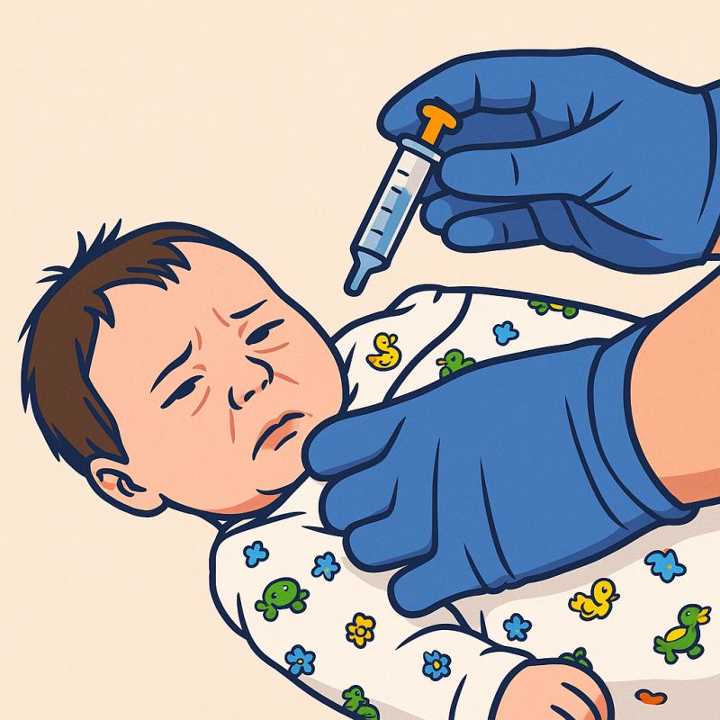
Definition & Thresholds
Hypoglycemia is defined as blood glucose <40 mg/dL in the first 4 hours of life and <45 mg/dL between 4-24 hours. For asymptomatic infants, treatment is recommended when glucose is <25 mg/dL if <4 hours old and <35 mg/dL between 4-24 hours. When symptoms are present, begin treatment at <40 mg/dL with a goal to maintain prefeeding glucose of 45 mg/dL.
Some sources cite the lower limit for normal plasma glucose during the first 72 hours as 40-45 mg/dL.
At-Risk Populations
Infants of Diabetic Mothers (IDM):
Most common in macrosomic or large-for-gestational-age infants
Caused by fetal pancreatic hypertrophy and hyperplasia from chronic maternal hyperglycemia
High maternal glucose stimulates fetal insulin production; at birth, abrupt loss of maternal glucose supply causes hyperinsulinism and hypoglycemia within 0.5-4 hours
Growth-Restricted & Preterm Infants:
Decreased glycogen stores
Inadequate gluconeogenesis
Increased metabolic demands from illness
Clinical Presentation
Symptoms include:
Jitteriness
Apnea and tachypnea
Hypotonia and decreased activity
Cyanosis
Poor feeding and hypothermia
Weak cry, lethargy, floppy posture
Seizures with profound hypoglycemia
Critical concern: Many infants remain asymptomatic. Asymptomatic hypoglycemia is associated with dose-dependent increased risk of poor executive function and visual motor function.
Management
Monitoring:
Begin screening soon after birth in all high-risk infants
Use point-of-care testing (glucometers) confirmed by laboratory
Frequent determinations for first 2-4 days
Treatment:
Initiate feeding within first hour if cardiorespiratory status stable (breast milk or formula preferred)
Avoid oral glucose - triggers massive insulin release causing rebound hypoglycemia
IV dextrose (10%) for poorly controlled IDM or symptomatic infants
Goal: maintain glucose >45 mg/dL
Physiologic correlation: Direct relationship exists between temperature regulation, glucose homeostasis, and respiratory distress

Skin-to-skin care
Skin-to-skin contact (also called kangaroo care) involves placing the undressed infant (wearing only a diaper) in a vertical position on the parent's bare chest, which permits direct eye contact, skin-to-skin contact, and close proximity. The parent is bare-chested or wears a loose-fitting, open-front top with a modified marsupial-like pocket carrier.
Benefits for Preterm & High-Risk Infants
Physiologic stability:
Maintains thermal stability and oxygen saturation
Promotes stability of heart and respiratory function
Reduces risk of mortality and nosocomial infections
Decreases length of hospital stay
Developmental benefits:
Fosters appropriate neurobehavioral development
Minimizes purposeless movements
Improves infant's behavioral state
Permits self-regulating behaviors
Maintains organized state
Feeding & growth:
Increased feeding vigor and enhanced breastfeeding
Improved growth outcomes
Mothers more likely to continue breastfeeding
Pain management:
Strong analgesic effect during procedures like heel lance in full-term newborns
Decreased pain perception during painful heelsticks in preterm infants
Benefits for Parents
Maternal healing:
Positive healing effect for mothers who had high-risk pregnancies
Psychologic healing related to preterm delivery
Helps mothers regain the mothering role through early contact
Permits early contact even with mechanically ventilated infants
Safety & Appropriateness
Skin-to-skin contact is appropriate for stable preterm infants because of the maintenance of appropriate body temperature. This practice has minimal untoward effects of being held.
Temperature Regulation
Skin-to-skin contact is an effective method for conserving newborn body heat. At birth, rapidly drying the skin and hair with a warmed towel and placing the infant in skin-to-skin contact with mother, covered by a blanket, minimizes heat loss through evaporation
Neonatal infection
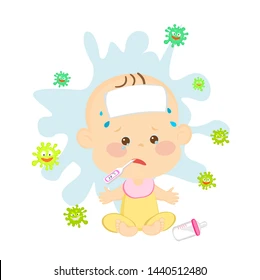
Definition & Susceptibility
Sepsis (septicemia) refers to a generalized bacterial infection in the bloodstream. Neonates are highly susceptible due to diminished immune function:
Impaired phagocytosis
Delayed chemotactic response
Minimal or absent IgA and IgM
Decreased complement levels
Critical challenge: Infants show no local inflammatory reaction at the portal of entry, and symptoms are vague and nonspecific, delaying diagnosis and treatment. Maintain a high suspicion for infection.
Early-Onset Sepsis (EOS)
Timing: Less than 3 days after birth
Acquisition routes:
Direct contact with maternal GI/GU tract organisms
Transplacental transfer from maternal bloodstream
Aspiration of infected amniotic fluid during labor
Contact with mother with active infection during delivery
Common pathogens:
Term infants: Group B streptococcus (GBS) - most common
Preterm infants: Escherichia coli - most common
Others: Viridans streptococci, Listeria monocytogenes, enteroviruses, coagulase-negative Staphylococcus
Vaginal pathogens: Syphilis, gonococci, C. albicans, HSV type II, Chlamydia
Risk factors:
Prolonged rupture of membranes
Preterm labor (<37 weeks)
Maternal intrauterine infection
Maternal GBS colonization
Current rates: Despite maternal screening and prophylaxis, early-onset GBS infection remains at approximately 0.25 per 1000 live births.
Treatment
Empiric antibiotic therapy:
Early-onset sepsis: Ampicillin + aminoglycoside
Late-onset sepsis: Vancomycin + aminoglycoside
Initiated before laboratory confirmation
Narrow therapy to confirmed pathogen ASAP due to antibiotic-resistance concerns
Supportive care:
Circulatory and respiratory support
Oxygen administration if needed
Fluid regulation and electrolyte correction
Temporary discontinuation of oral feedings
Blood product transfusions as needed
Nursing Priorities
Recognition is paramount - nurses typically identify that "something is wrong.” Obtain a minimum 1 mL blood for culture to detect low-level bacteremia 2. Monitor for superinfection from prolonged antibiotic therapy (C. albicans) 2, though prophylactic antifungal therapy is not standard
The newborn infant is susceptible to infection because of the immature immune system
Sepsis
One of the most significant causes of neonatal morbidity and
mortality: Systemic Inflammatory Response Syndrome (SIRS)
2 Types of Neonatal Sepsis
Early-onset sepsis
Late-onset sepsis
S/S: Respiratory distress, apnea, decreased tone, difficulty with thermoregulation
Viral infections:
Birth of baby exposed to TORCH infections
Hep B
HIV
Herpes simplex
Bacterial infections:
GBS
E. coli
Staphylococcus aureus
Chlamydia
Gonorrhea
Syphilis
Listeriosis
Protozoal infections → Toxoplasmosis
Fungal infection → Candidiasis
Neonatal skin findings
Normal Skin Characteristics
Texture & appearance: Velvety smooth and puffy, especially around eyes, legs, dorsal hands/feet, and scrotum or labia. By the second or third day, skin becomes drier and flakier.
Color variations:
Depends on racial and familial background
White infants have light pigment at birth
Black, Indigenous, Hispanic, Latino, Asian, and other children of color may have partial pigmentation that may change with development
Hyperpigmentation may appear shortly after birth in lips, axillae, genitalia, umbilicus, and fingertips in darker-skinned infants
Important: In darker skin tones, visual inspection alone may not reliably identify jaundice - rely on laboratory bilirubin testing.
Preterm Infant Skin
Characteristics:
Bright pink (often translucent depending on immaturity)
Smooth and shiny
Small blood vessels clearly visible under thin epidermis
Fine lanugo hair abundant over body (sparse on head)
Soft, pliable ear cartilage
Minimal sole/palm creases
Fragility concerns:
Thinner than full-term skin, lacks rete pegs (anchoring appendages)
Less cohesion between skin layers
Easily excoriated and denuded
Adhesive tape may separate epidermis from dermis
Use pectin barriers and hydrocolloid adhesives instead
Care precautions:
Avoid alkaline-based soaps
Use alcohol, chlorhexidine, and povidone-iodine with caution
Rinse skin with water after product use to prevent severe irritation and chemical burns in VLBW/ELBW infants
Increased permeability facilitates absorption of topical products
Benign Skin Findings
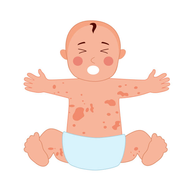
Erythema toxicum neonatorum:
Benign accumulation of macules, papules, or pustules
Appears 24-48 hours after birth (may develop until day 10)
Discrete, blotchy, macular erythematous rash
Light yellow/white papules/pustules (1-3 mm)
Affects any body surface except palms and soles
Self-limiting, resolves within weeks
No treatment required
Periauricular papillomas (skin tags): Fairly frequent, usually familial trait, no consequence
Concerning Findings
Report immediately:
Pallor, plethora (deep purplish color), central cyanosis, jaundice
Petechiae scattered over body (may indicate low platelets or infection)
Birth injuries: forceps marks, fetal monitoring lesions
Bruising (increases hyperbilirubinemia risk)
Neonatal vital signs
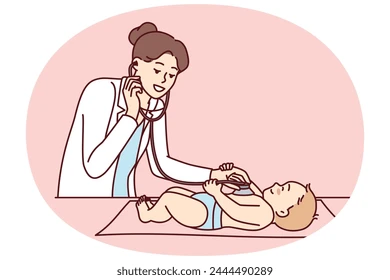
Heart Rate
Normal range: 120-160 beats/min
Variations:
During deep sleep: 80-100 beats/min
When crying: Up to 180 beats/min or higher
Shortly after birth: May range 100-180 beats/min
After stabilization: 120-140 beats/min
Reevaluation needed: Heart rate >160 beats/min or <100 beats/min should be reassessed within 30 minutes to 1 hour or when infant's activity changes.
Location: Point of maximal impulse (PMI) at fourth to fifth intercostal space, medial to left midclavicular line. Often visible and easily palpable due to thin chest wall.
Respiratory Rate
Normal range: 40-60 breaths/min
Variations:
Rate up to 80 breaths/min may be noted
During first reactive period: May reach 80 breaths/min
Infants delivered by cesarean section may have more rapid rate than vaginal births
Assessment: Count for 1 full minute
Blood Pressure
Normal ranges:
Systolic: 60-96 mm Hg
Diastolic: 30-62 mm Hg
Measurement: Often determined using electronic sphygmomanometer with Doppler or oscillometric technique
Clinical significance: Sustained increase in blood pressure is almost always significant. Hypertension may indicate thrombosis (from umbilical artery catheter), coarctation of aorta, renal disorders, congenital adrenal hyperplasia, or CNS disease.
Temperature
Maintaining appropriate temperature is best accomplished through skin-to-skin care with mother. Temperature may decrease during the first reactive period.
Key Assessment Points
Pulse variability: Greatest around time of birth and more marked in premature infants. Sustained tachycardia may indicate infection or cardiac rhythm disturbance (e.g., paroxysmal atrial contractions).
Irregular heart rate (sinus dysrhythmia): Common in first few hours but may need evaluation thereafter