Cell Division & Replication
1/23
There's no tags or description
Looks like no tags are added yet.
Name | Mastery | Learn | Test | Matching | Spaced |
|---|
No study sessions yet.
24 Terms
What is the cell cycle?
Produces two genetically identical daughter cells.
The cell cycle is the regulated sequence of events that occurs between one cell division and the next
The cell cycle has three phases:
interphase
nuclear division (mitosis)
cell division (cytokinesis)
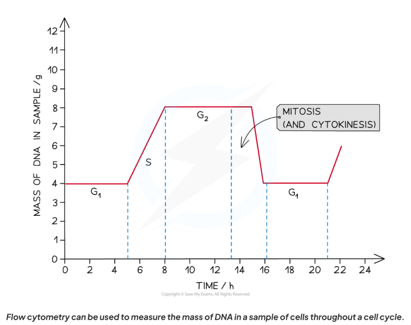
Stages of interphase
G₁ phase (Gap 1):
Proteins for organelle synthesis made (the RNA, enzymes and other proteins)
Organelles replicate.
Synthesis of cytoplasm
Cell increases in size.
S phase (Synthesis):
DNA replicated in nucleus.
G₂ phase (Gap 2):
Cell continues to grow.
Cell organelles grow & divide
Energy stores increase.
Duplicated DNA checked for errors.
Production of tubulin protein, which is used to make microtubules for the mitotic spindle)
Interphase = G1 + S + G2
The movement from one phase to another is triggered by…
…chemical signals called cyclins
Mitotic Phase
Mitosis = nucleus divides.
Cell growth stops during the M phase
Cytokinesis = cytoplasm divides, producing 2 genetically identical daughter cells.
constriction of the cytoplasm between the two nuclei
in plant cells, a new cell wall is formed
G₀ Phase (resting state)
Cell leaves cycle, temporarily or permanently.
Reasons:
Differentiation → cell specialised, cannot divide again.
DNA damage → cell no longer viable → permanent arrest.
Senescence → normal cells can only divide a limited number of times.
Aging → more senescent cells (linked to cancer, arthritis).
Some G₀ cells can be stimulated back into cycle (e.g. lymphocytes during immune response).
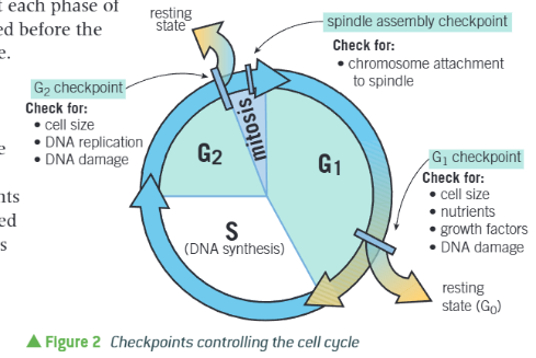
Regulation of the Cell Cycle
Vital for correct size, error-free DNA replication, and correct chromosome alignment.
Ensures fidelity of cell division → daughter cells genetically identical.
Protein complexes determine when the cell enters each stage of the cell cycle.
Specific proof-reading enzymes and repair enzymes are involved in this checking process
When possible enzymes will repair the error but in some cases the cell may destroy itself to prevent passing on harmful mutations
G₁ checkpoint:
End of G₁ phase, before S.
Checks: cell size, nutrients, growth factors, DNA damage.
If passed → begin DNA replication.
If failed → enters resting state (G₀).
chromosomes are checked for damage
If damage is detected then the cell does not advance into the S phase until repairs have been made
During S phase to check…
Chromosomes are checked to ensure they have been replicated.
If all the chromosomes haven't been successfully replicated then the cell cycle stops
G₂ checkpoint:
End of G₂, before mitosis.
Checks: DNA replication without error, cell size, DNA damage.
The cell cycle will be delayed until any necessary repairs are made
During metaphase to check…
Spindle assembly checkpoint (metaphase checkpoint):
During mitosis.
Checks: aligned at metaphase plate, whether the chromosomes are correctly attached to the spindle fibres prior to anaphase
Mitosis cannot continue until passed.
Mitotic Index
The mitotic index is the proportion of cells actively undergoing mitosis
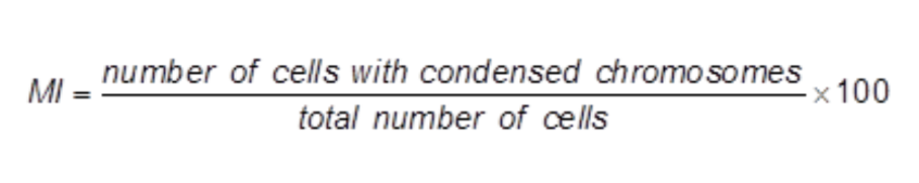
Table demonstrating what checkpoint actually check.
G1 Checkpoint | C2 Checkpoint | Spindle Assembly Checkpoint |
Cell Size | Cell Size | Chromosome attachment to spindle. |
Nutrients | DNA Replication | n/a |
Growth Factors | DNA Damage | n/a |
DNA Damage | n/a | n/a |
Cell regulation are controlled by cyclins and CDKs (cyclin-dependent kinases). What are they?
Cyclins: regulatory activating proteins that fluctuate in concentration.
CDKs: enzymes that phosphorylate proteins → activates them.
Process of cell regulation.
Cyclin binds to inactive CDK → forms complex, which determines the checkpoints that are passed.
Complex phosphorylates (adds phosphate groups to other proteins) target protein → changes tertiary structure → activates protein.
Activated proteins ensure cycle progresses and checkpoint passed.
After use, cyclins degraded by enzymes.
Cancer
Caused by uncontrolled cell division.
Abnormal mass = tumour:
Benign: does not spread, not usually harmful.
Malignant: invades other tissues, spreads (metastasis).
Tumours often result from mutation in checkpoint gene/protein → regulation lost.
Overexpression of cyclin gene → too much cyclin → uncontrolled division → tumour formation.
Importance of Mitosis
Involves nuclear division (mitosis) + cytoplasmic division (cytokinesis).
Functions:
Growth, repair, tissue replacement in multicellular organisms
Asexual reproduction → genetically identical offspring (unicellular eukaryotes, fungi, some animals, some plants) → clones
Allows unicellular zygote to grow into multicellular organism.
Ensures daughter cells are genetically identical with the same DNA and chromosome number as the parent cell.
Only occurs in eukaryotic cells (prokaryotic cells undergo binary fission)
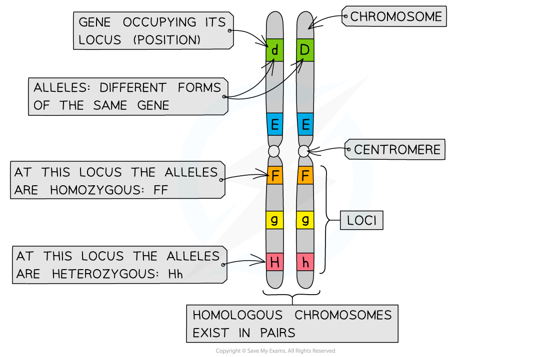
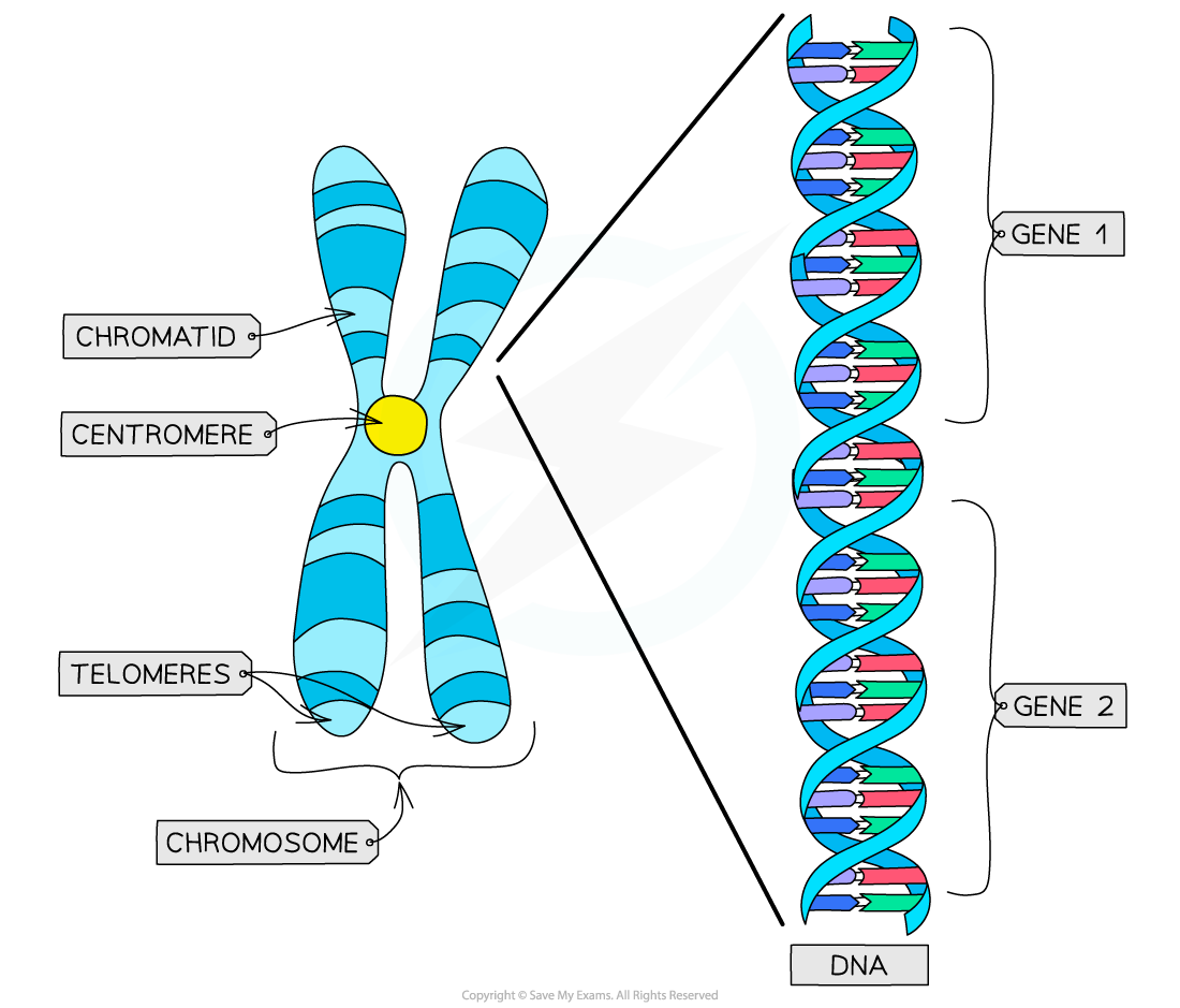
Chromosomes Structure
Each DNA molecule → two identical DNA molecules (sister chromatids).
Joined at centromere (ensures correct segregation).
Each chromatid separated into daughter cells during mitosis.
Locus → fixed position of a gene on a chromosome
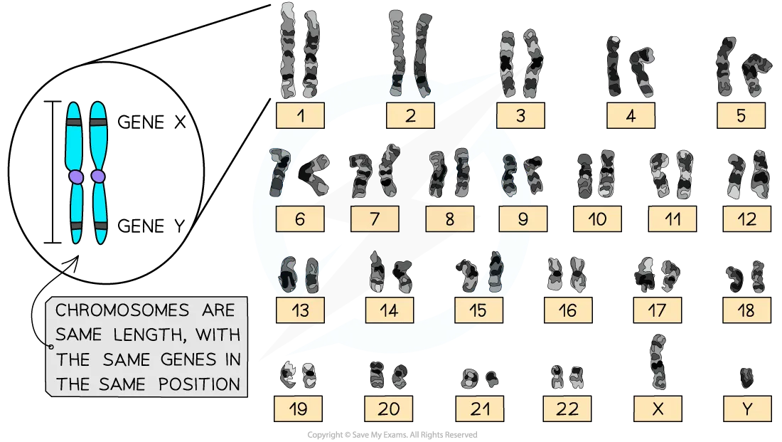
Karyotypes:
Karyogram is an image that shows all of the chromosomes in a cell, arranged by size, shape, and banding pattern, and placed with their homologous pairs
A karyogram shows the karyotype of an individual,
The appearance of a complete set of an individual's chromosomes, including their number, size, shape, and banding
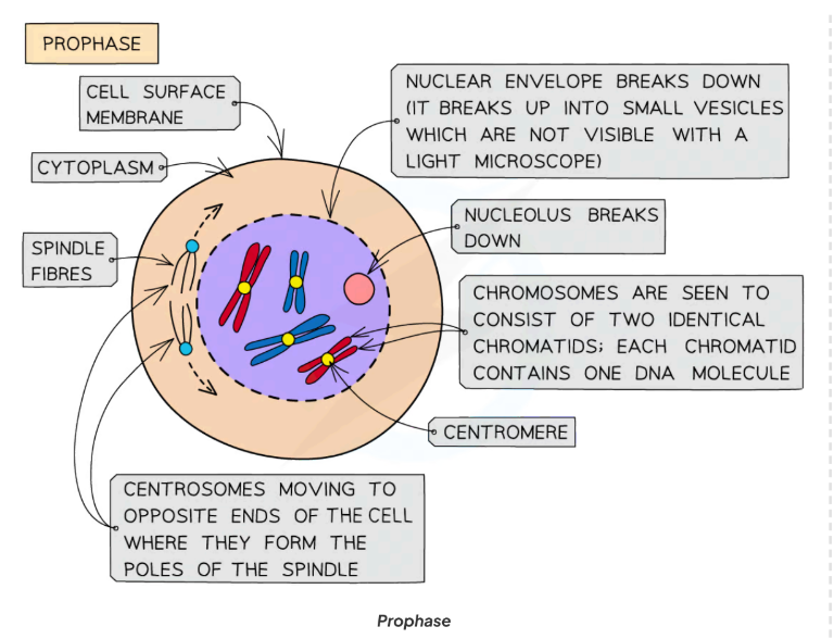
🔴 Prophase
Chromatin fibres condense into visible chromosomes.
Sister chromatids (each containing one DNA molecule)that are joined together at the centromere
The two centrosomes (replicated in the G phase just before prophase) move towards opposite poles
Nuclear envelope breaks down into small vesicles.
Nucleolus disappears; nuclear envelope begins to break down.
Spindle fibres (protein microtubules) form from centrioles (animal cells).
Centrioles migrate to poles.
In plant cells, spindle formed without centrioles.
Spindle fibres attach to centromeres.
By late prophase, nuclear envelope fully disintegrated.
🟠 Metaphase
Centrosomes reach opposite poles
Spindle fibres (protein microtubules) continue to extend from centrosomes
Chromosomes line up at the equator of the spindle (also known as the metaphase plate) so they are equidistant to the two centrosome poles
Spindle fibres (protein microtubules) reach the chromosomes and attach to the centromeres
This attachment involves specific proteins called kinetochores
Held in place by tension at centromeres.
Each sister chromatid is attached to a spindle fibre originating from opposite poles

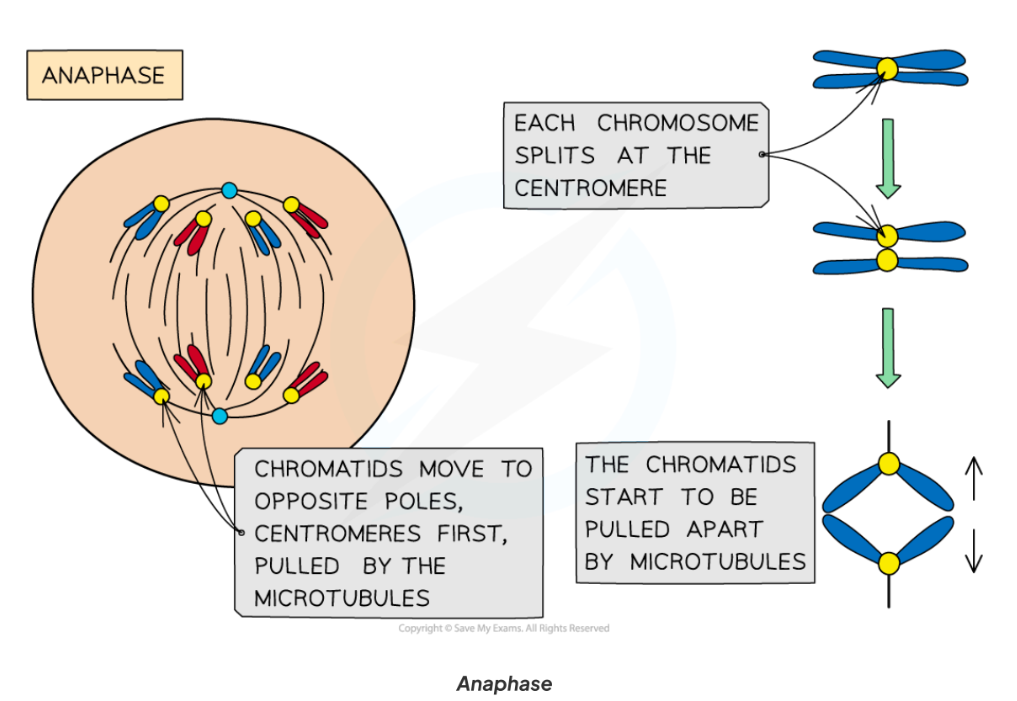
🟡 Anaphase
Centromeres divide (not split!).
The sister chromatids separate at the centromere (the centromere divides in two)
Spindle fibres (protein microtubules) begin to shorten
The separated sister chromatids (now called chromosomes) are pulled to opposite poles by the spindle fibres (protein microtubules)
Characteristic ‘V’ shape (chromatids dragged by centromeres).
🟢 Telophase
Chromosomes arrive at opposite poles and begin to decondense→ now called chromosomes again.
Chromosomes uncoil and become less visible.
Nuclear envelopes (nuclear membranes) begin to reform around each set of chromosomes
The spindle fibres break
New nucleoli form within each nucleus
Nucleolus reforms.
Cytokinesis begins.

Cytokinesis (Cytoplasmic Division)
Animal cells:
Cleavage furrow forms at centre.
Cytoskeleton pulls membrane inwards until fusion → two separate cells.
Figure 12–13 show cytokinesis in animal cells.
Plant cells:
Cell wall prevents cleavage furrow.
Vesicles from Golgi align at metaphase plate.
Vesicles fuse with each other + plasma membrane → cell plate forms.
New sections of cell wall form.
Identifying Mitosis in Plant Cells
Growth in plants occurs in specific regions called meristems
The root tip meristem can be used to study mitosis ( found just behind the protective root cap )
Pre-prepared slides of root tips can be studied or temporary slides can be prepared using the squash technique (root tips are stained and then gently squashed, spreading the cells out into a thin sheet
and allowing individual cells undergoing mitosis to be clearly seen)