A&P II Lab Practical 3
 Call Kai
 Learn
Learn Practice Test
 Spaced Repetition
 Match
Match Flashcards
 Knowt Play
1/61

There's no tags or description
Looks like no tags are added yet.
Name | Mastery | Learn | Test | Matching | Spaced | Call with Kai | Chat |
|---|
No analytics yet
Send a link to your students to track their progress
62 Terms
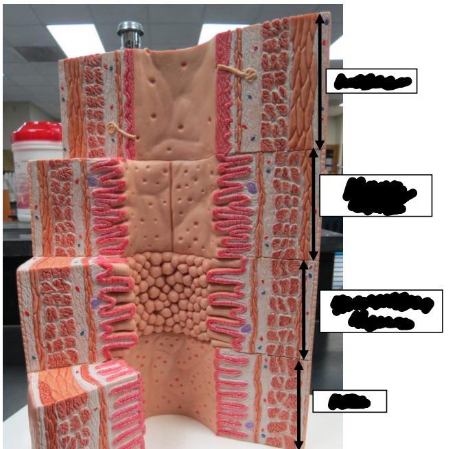
Label this image from top to bottom.
Esophagus, stomach fundus, small intestine jejunum, colon.
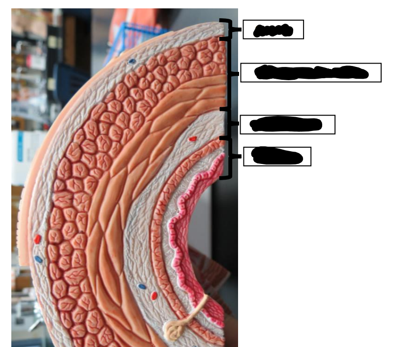
Label this image from top to bottom.
Serosa, muscularis externa, submucosa, mucosa.
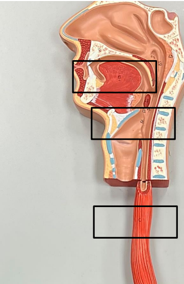
Label this image from top to bottom.
Tongue, larynx, esophagus.
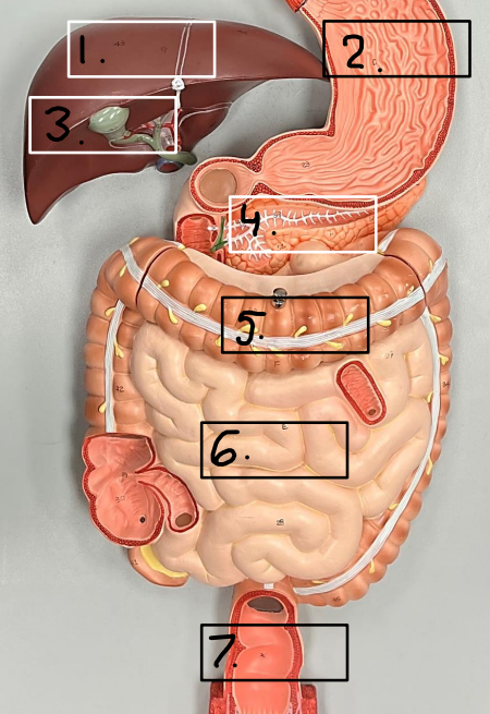
Label this image.
1. Liver, 2. stomach, 3. gallbladder, 4. pancreas, 5. large intestine, 6. small intestine, 7. rectum
What are the two parts of the rectum?
Internal (involuntary) and external (voluntary) sphincters.
What is an accessory structure?
A structure that food does not pass through.
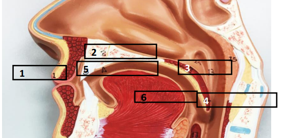
Label this image.
1. Lips, 2. hard palate, 3. soft palate, 4. uvula, 5. teeth, 6. tongue
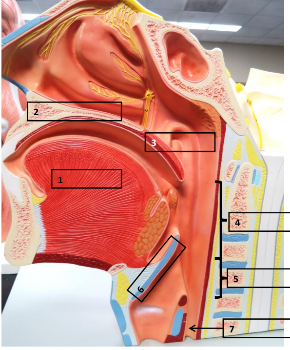
Label this image.
1 . Tongue, 2. hard palate, 3. soft palate, 4. oropharynx, 5. laryngopharynx, 6. epiglottis, 7. esophagus
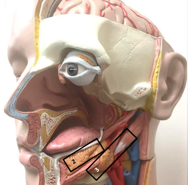
Label this image.
2. Sublingual salivary gland, 3. submandibular salivary gland
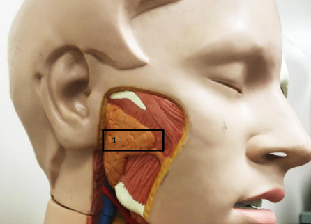
What is this?
Parotid salivary gland
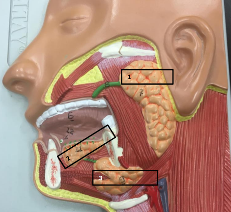
Label this image.
1. Parotid, 2. sublingual, 3. submandibular
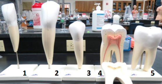
Label this image.
1 . Incisor, 2. canine (cuspid), 3. premolar (bicuspid), 4. upper molar, 5. lower molar
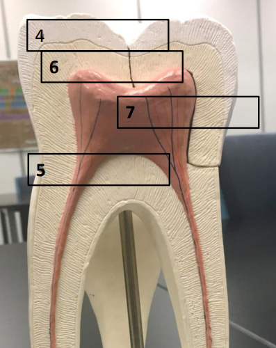
Label this image.
4. Enamel, 5. cementum, 6. dentin, 7. tooth pulp
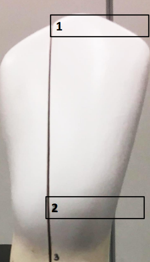
Label this image.
1. Crown, 2. neck
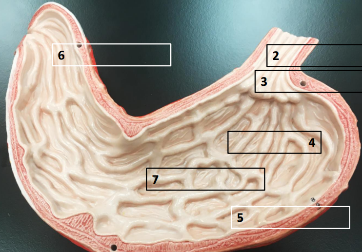
Label this image.
2 . Esophagus, 3. cardiac (esophageal) sphincter, 4. fundus of stomach, 5. body, 6. pylorus of stomach, 7. rugae of stomach
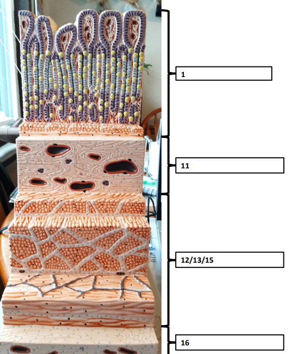
Label this image.
1. Mucosa, 11. submucosa, 12/13/15. muscularis externa, 16. serosa
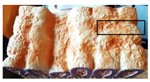
What is this?
The gastric pit which is a pitting of the mucosal layer where the gastric secretions are released from.
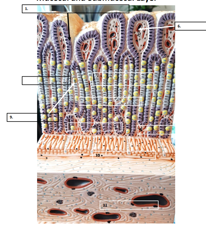
Label this image, 1-5.
1 . Mucosal epithelium, 2. mucous membrane, 3. gastric gland neck region, 4. gastric gland basal region, 5. lamina proper
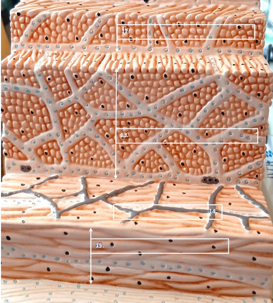
Label this image.
12 . Muscularis externa oblique, 13. muscularis externa longitudinal, 14. lymphatic vessels (gray lines across layer), 15. muscularis externa circular, 16. serosa
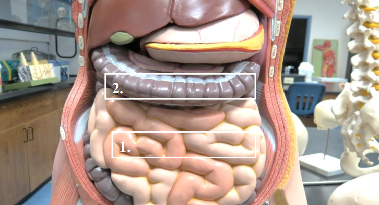
Label this image.
1 . small intestine, 2. large intestine
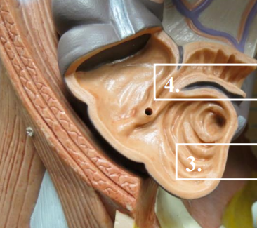
Label this image.
3. Cecum, 4. ileocecal valve
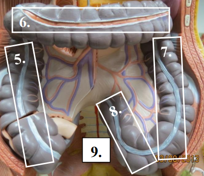
Label this image.
5 . Ascending colon, 6. transverse colon, 7. descending colon, 8. sigmoid colon, 9. rectum
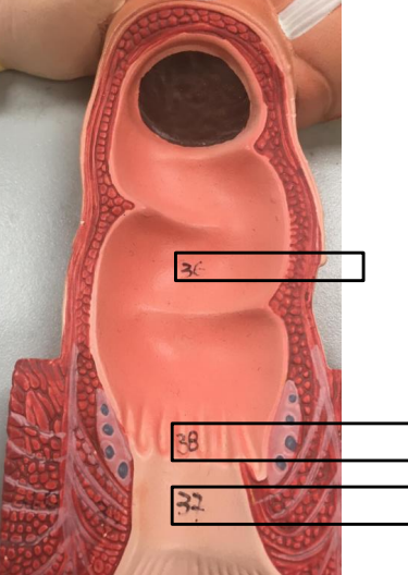
Label this image.
36 . rectum, 37. external anal sphincter (voluntary), 38. internal anal sphincter (involuntary)
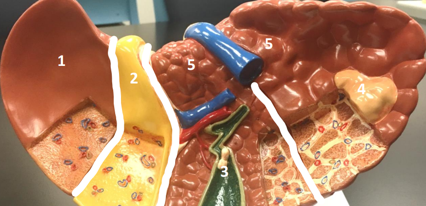
Label this image.
1 . liver in normal health, 2. biliary obstruction liver, 3. gallstone liver, 4. tumor liver, 5. nodular cirrhosis liver
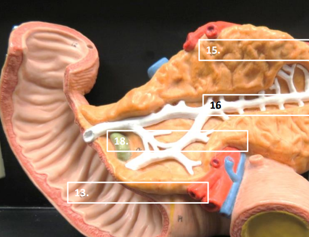
Label this image.
13 . Duodenum, 15. pancreas, 16. pancreatic duct, 18. bile duct
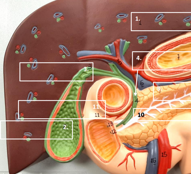
Label this image.
1 . Liver, 2. gallbladder, 3. cystic duct, 4. common bile duct, 9. pancreas, 10. pancreatic duct, 11. duodenum
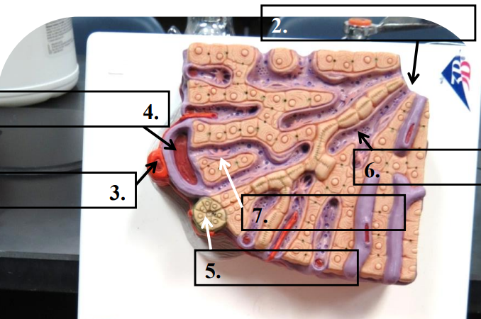
Label this image.
2 . Central vein, 3. interlobular artery, 4. hepatic portal vein branch, 5. interlobular bile duct, 6. liver sinusoids, 7. kupffer cell
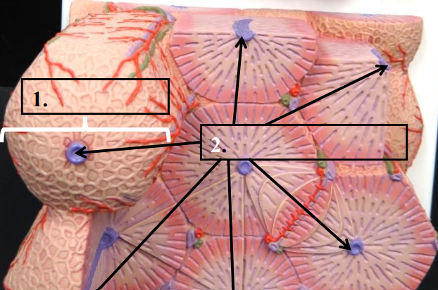
Label this image.
1 . Liver lobule, 2. central vein
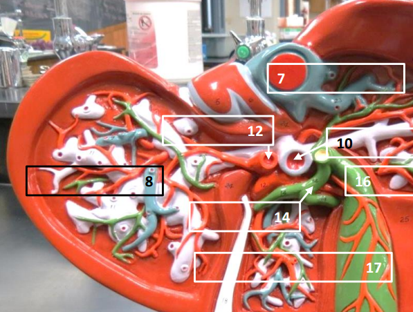
Label this image.
7 . inferior vena cava, 8. hepatic veins, 10. portal vein, 12. hepatic artery, 14. hepatic duct, 16. cystic duct, 17. gallbladder
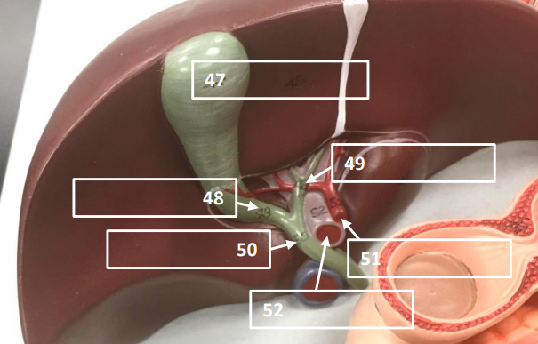
Label this image.
47. Gallbladder, 48. cystic duct, 49. hepatic duct, 50. common bile duct, 51. hepatic artery, 52. portal vein
What do the mucosa layer folds form?
Gastric pits
What is the brush border microvilli, and where are they found?
They help with absorption and are found in the small intestine.
What are folds in the colon for?
Water reabsorption and hosting of the microbial flora.
What is the lumen?
The tube.
What are the layers of the muscularis externa?
Longitudinal (outer layer), circular (inner layer).
What is saliva made up of?
Water, IgA antibodies, lysozymes, mucin, and amylase.
What is unique about the stomach?
It has an extra layer called the oblique smooth muscle, for peristalsis.
What are the parts of the small intestine?
Ileum, duodenum, cecum.
What is the exocrine portion of the pancreas for?
Secreting enzymes that travel through the pancreatic duct and combine with bile from the liver; eventually mixes with chyme in the duodenum.
What is the hepatic triad?
The interlobular artery, hepatic portal vein branch, interlobular bile duct
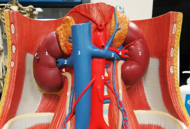
Label this image.
1 . Kidney (R/L), 2. renal vein (R/L), 3. inferior vena cava, 4. renal artery (R/L), 5. abdominal aorta, 6. ureter (R/L)
What is the curved part of the kidney called?
The hilum.
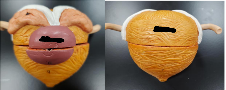
Label this image.
Left: Prostate, Right: Bladder
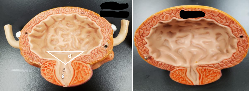
Label this image.
D: Trigone, 8: Urethra, Right: Bladder
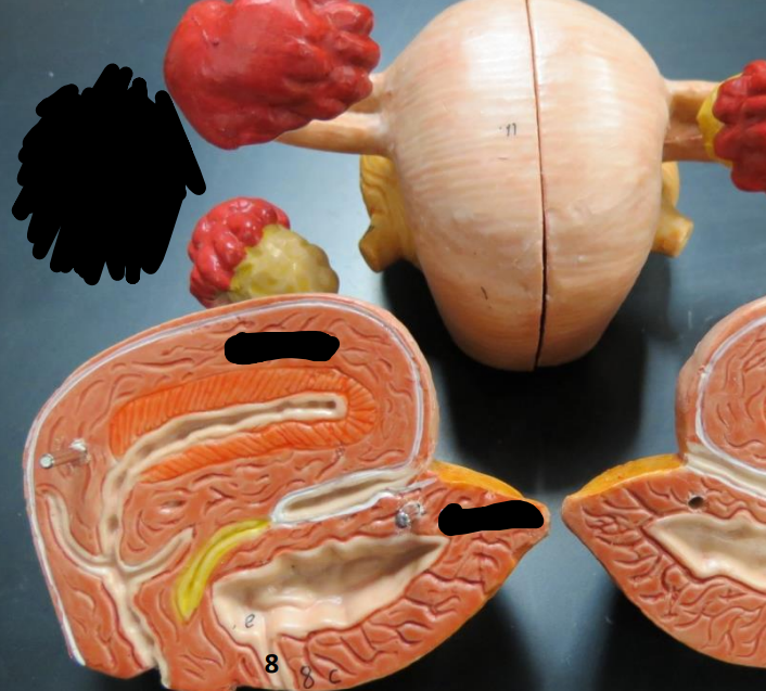
Label this image.
8/Top Black Blur: Urethra, Middle Black Blur: Uterus, Bottom Black Blur: Bladder
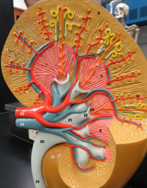
Label this image, 6-15.
6. Cortex, 7. Medulla, 8. Renal Pyramid, 10. Renal papilla, 12. Peritubular Capillaries, 13. Glomeruli, 14. Proximal tubule, 15. Ascending Limb of Nephron loop
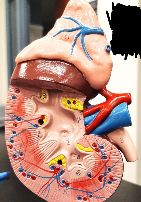
Label this image, 1-11 (no 10).
1. Renal Artery, 2. Renal Vein, 3. Ureter, 4. Renal Pelvis, 5. Renal Cortex, 6. Renal Medulla, 7. Renal Pyramid, 8. Minor Calyx, 9. Major Calyx, 11. Adrenal Gland
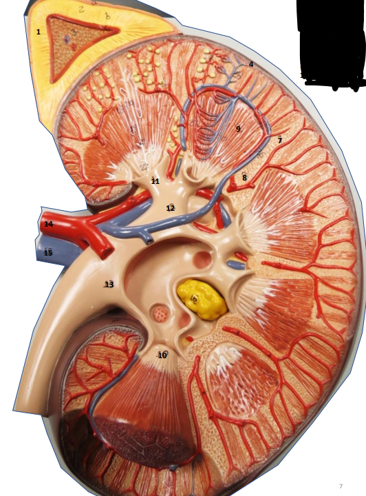
Label this image.
1. Adrenal Gland, 4. Kidney, 7. Renal Cortex, 8. Renal Columns, 9. Renal Pyramids, 10. Renal papillae, 11. Minor Calyx, 12. Major Calyx, 13. Renal Pelvis, 14. Renal Artery, 15. Renal Vein
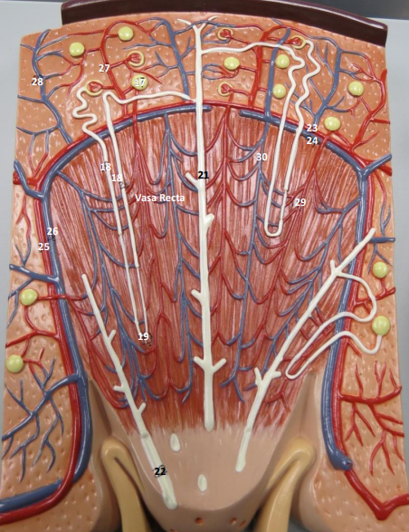
What is this model?
A nephron; the functional unit of the kidney (responsible for filtering blood and making urine).
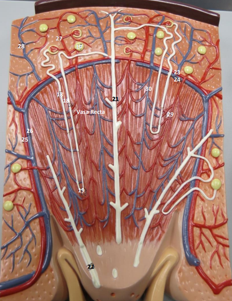
Label this image, 17-22.
17. Renal Corpuscle, 18. Descending Limb of Nephron Loop, 19. Nephron Loop (of Henle), 20. Ascending Limb of Nephron Loop, 21. Collecting Duct, 22. Papillary Duct
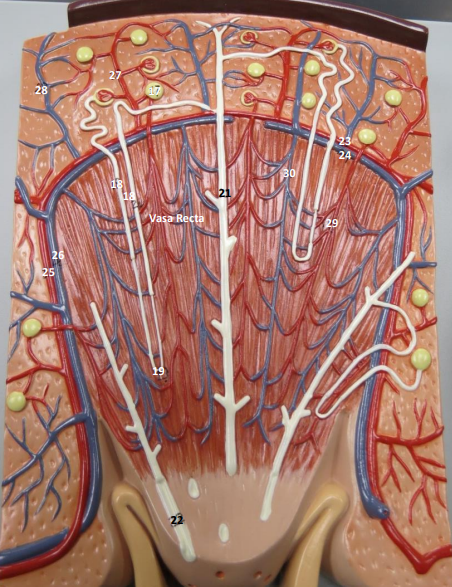
Label this image, 23-30.
23. Arcuate artery, 24. Arcuate Vein, 25. Interlobar artery, 26. Interlobar vein, 27. Interlobular artery, 28. Interlobular vein, 29&30. Vasa Recta capillaries (found in juxtamedullary nephrons only)
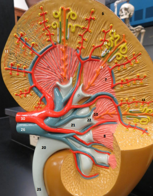
Label this image, 16, 17, 18, 20, 21, 22, 25, 26, 30.
16. Descending Limb of Nephron loop, 17. Distal tubule, 18. Collecting tubule, 20. Renal Pelvis, 21. Major calyx, 22. Minor calyx, 25. Ureter, 26. Renal vein, 30. Renal artery
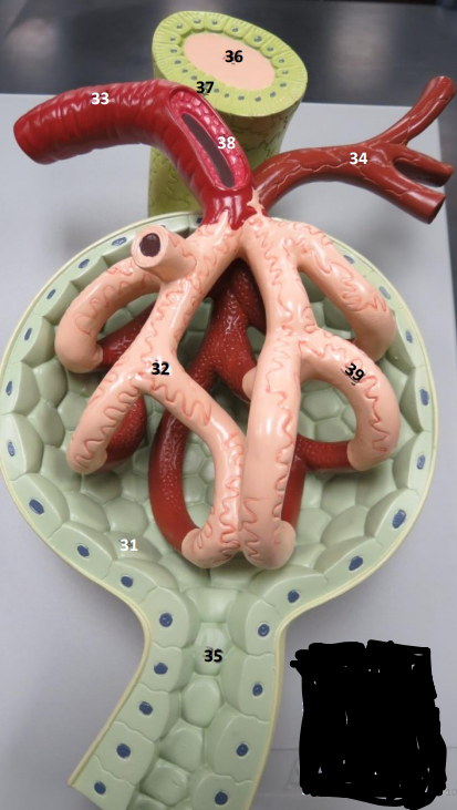
Label this image.
31. Renal Corpuscle, 32. Glomerulus, 33. Afferent Arteriole, 34. Efferent Arteriole, 35. Proximal Tubule, 36. Distal Tubule, 37. Macula Densa, 38. Juxtaglomerular Cell, 39. Podocyte
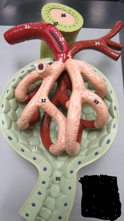
What is this?
A renal corpuscle.
What is the function of the macula densa?
A chemical sensor; monitors fluid flow and NaCl to regulate filtration and maintain blood pressue. Stimulates juxtaglomerular cells to release renin.
What is a juxtaglomerular cell? What does it do?
A mechanical sensor; acts as a blood pressure sensor along with baroreceptors who detect stretch.
What is the juxtaglomerular apparatus?
Macula densa and juxtaglomerular cells.
What does the adrenal cortex secrete? Is it the outer or inner part?
Secretes aldosterone, cortisol, and testosterone (androgen); outer part.
What does the adrenal medulla secrete? Is it the outer or inner part?
Secretes norepinephrine and epinephrine; inner part.
Where can the trigone be found?
In both males and females, but cannot be seen on the female models.
What are wrinkles also known as?
Rugae (in both bladder and stomach).
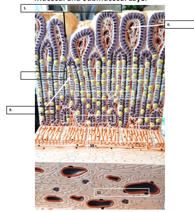
Label this image, 6-11.
6. capillary, 8. chief cell, 9. parietal cell, 10. muscularis mucosae, 11. submucosa