AQA Psychology A Level - Biopsychology
1/117
There's no tags or description
Looks like no tags are added yet.
Name | Mastery | Learn | Test | Matching | Spaced | Call with Kai |
|---|
No analytics yet
Send a link to your students to track their progress
118 Terms
Topic 1 - The Nervous System
…
Which two biological systems involved in adapting to change?
Nervous system and endocrine system
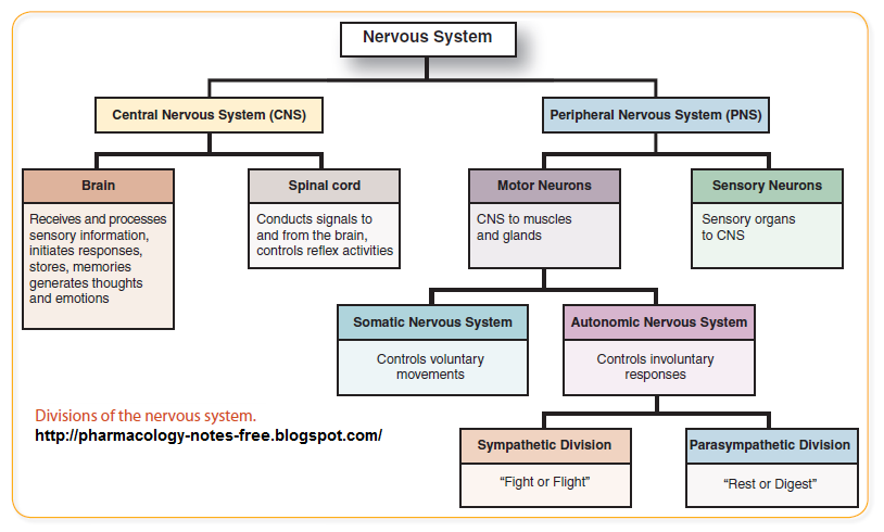
Name all the divisions of the nervous system
Names all the four lobes of the brain and state their location
1. What two things make up the central nervous system (CNS)?
2. What are the two main functions of the CNS?
1. brain and spinal cord
2. control of behaviour (e.g. higher mental processing, decision-making) and regulation of body’s physiological processes
What are the features of the spinal cord? (3)
- collection of nerve cells attached to brain
- runs length of spinal column
- attached to specific muscles and glands
What are the two main function of the spinal cord? (2)
(still considered CNS function)
to relay information between brain and rest of body (e.g. sensory receptors in eyes, skin, ears, etc.)
to perform simple reflex actions that don’t require brain involvement (e.g. pulling hand away from hot objects)
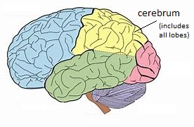
What four areas is the brain divided into?
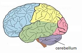
cerebrum
cerebellum
diencephalon
brain stem
Describe the structure of the cerebrum (3)
function (2)
(still considered function of CNS)
Cerebrum
largest part of brain
divided into four different lobes (frontal, temporal, parietal, occipital)
split into two cerebral hemispheres (connected by corpus callosum)
Function:
each lobe has different function e.g. frontal controls thought and speech production, occipital processes visual images
each hemisphere specialised for specific behaviour
Describe the structure of the cerebellum (1)
function (1)
(still considered function if CNS)
Cerebellum
beneath back of cerebrum
Function:
controls motor skills and balance to coordinate muscles for movement (e.g. precise movements in mouth for speech)
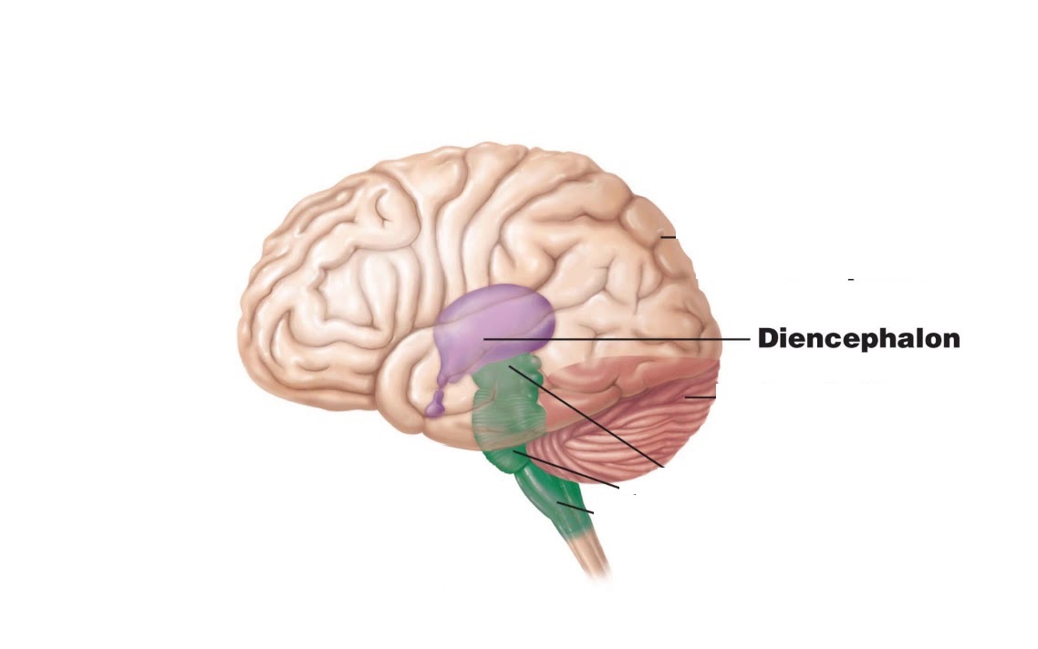
Describe the structure of the diencephalon (2)
function (2)
(still considered function of CNS)
Diencephalon
beneath cerebrum and above brainstem
contains the thalamus and hypothalamus
Function:
thalamus - relay station for nerve impulses coming from senses to be directed to appropriate part of brain for processing
hypothalamus - regulates body temp., hunger, thirst, etc. and links endocrine and nervous system to control hormone release from pituitary gland
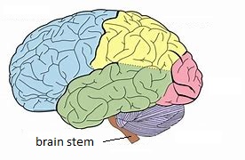
Describe the structure of the brain stem (2)
function (2)
(still considered function of CNS)
Brain stem
beneath brain
bridge between brain and spinal cord
Function:
regulates automatic functions e.g. breathing, heartbeat, swallowing
allows passing of impulses between brain and spinal cord via sensory and motor neurons
1. What makes up the peripheral nervous system (PNS)?
2. What is the main function of the PNS?
3. What are the two main divisions of the PNS?
1. all nerves outside of CNS (sensory and motor neurons)
2. relays nerve impulses between CNS and rest of body
3. somatic nervous system (SNS) and autonomic nervous system (ANS)
Describe the structure of the somatic nervous system (SNS) (2)
function (2)
Structure:
made up of cranial and spinal nerves
contains sensory and motor neurons
Function:
controls voluntary movements and reflex actions
sensory neurons relay info from body → CNS, while motor neurons relay info from CNS → body/effectors
Describe the structure of the autonomic nervous system (ANS) (1)
function (1)
Structure:
divided into two parts
Function:
controls involuntary actions e.g. breathing, heartbeat, digestion (so they can be completed efficiently without having to think about them)
Describe the function of the sympathetic nervous system (SNS) (4)
Function:
responds to emergencies - ‘fight or flight’
uses neurotransmitter noradrenaline (has stimulating effects)
increases heart rate, blood pressure, causes hair to stand on end
slows down less important bodily functions when under threat e.g. digestion and urination (so energy use can be redirected for more important processes)
Describe the function of the parasympathetic nervous system (PNS) (4)
Function:
relaxes after emergencies - ‘rest and digest’
uses neurotransmitter acetylcholine (has inhibiting effects)
decreases heart rate, blood pressure, less sweating
begins less important bodily functions again e.g. digestion and urination (energy no longer needed for threat)
What similarity do the SNS and PNS share?
Both tend to regulate the same organs/glands, but just have opposite effects.
Topic 2 - Neurons and Synaptic Transmission
…
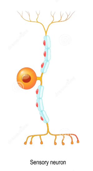
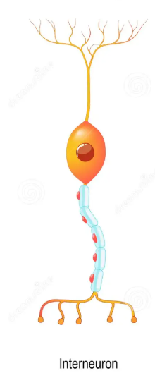
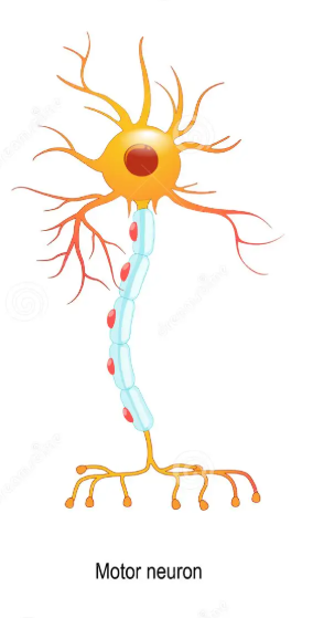
Name the three types of neurons
sensory, relay (intermediate), motor
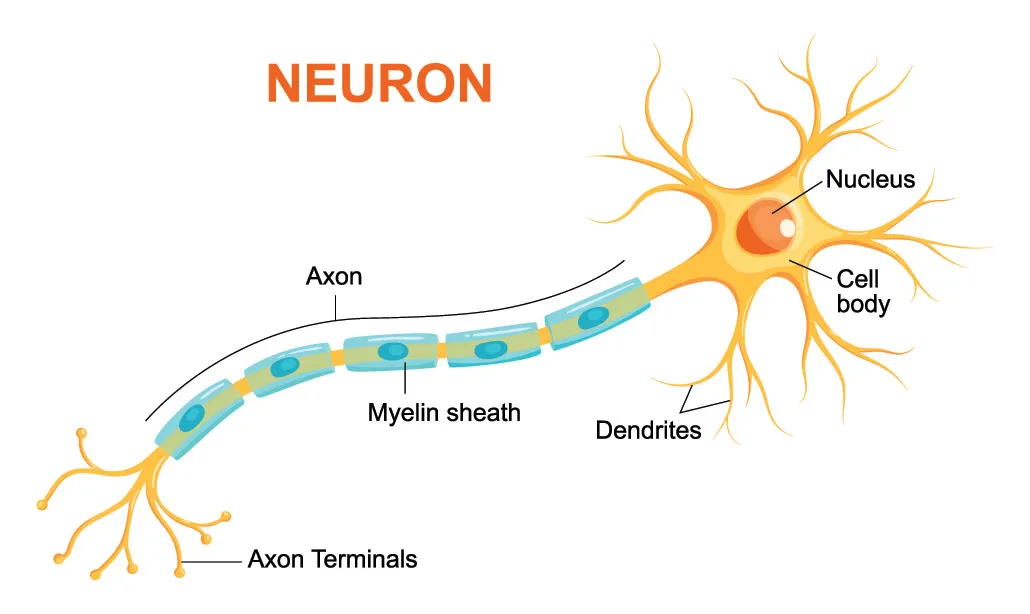
Name the typical parts of a neuron (4)
dendrites
cell body
axon
myelin sheath
Describe the function of each part of a neuron
dendrites (2)
cell body (2)
axon (2)
myelin sheath (2)
dendrites - on one end of neuron, receive signals from other neurons or sensory receptors
cell body - control centre of neuron, contains nucleus and other organelles
axon - long thin fibre where impulse is transmitted along (away from cell body towards axon terminal)
myelin sheath - insulating layer of Schwann cells, speeds up transmission along axon

Describe the function of sensory neurons (4)
where do impulses travel from and to
where is info converted to impulses
where is info translated
do all impulses go to brain
carry nerve impulses from sensory receptors to brain and spinal cord (receptor → CNS)
convert stimulus info from sensory receptors on one end (e.g. in skin, eyes, ears) into neural impulses
these then translated into sensations in brain (e.g. pain, heat, visual input)
some impulses only go to spinal cord for quick action (reflex)

Describe function of relay/intermediate neurons (2)
where do they carry impulses from and to
their abundance and where are they found
carry nerve impulses from sensory neurons to motor neurons within the brain and spinal cord (sensory neurons → CNS → motor neurons)
are the most abundant neurons and mostly found in brain and spinal cord (CNS)

Describe the function of motor neurons (3)
where do they carry impulses from and to
what impulse is carried to motor neuron and what are effectors
example of motor neuron action (+ what does strength of contraction depend on)
carry nerve impulses from brain and spinal cord to effector (CNS → effector)
reaction to stimulus made in CNS and travels as impulse to muscles or glands (effectors) to be carried out
example is muscle contraction, where neurotransmitters released for contraction to take place (strength of contraction depends on rate of axon firing)

Name the different parts of a synapse (8)
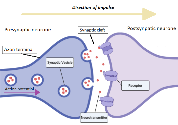
Explain the process of synaptic transmission (5)
action potential reaches presynaptic neuron
triggers neurotransmitter chemicals release from synaptic vesicles (exocytosis) in presynaptic neuron, into synaptic cleft
neurotransmitters diffuse across synapse/cleft
bind to receptor sites on postsynaptic neuron
neurotransmitters will either increase rate of firing (excitatory response - depolarisation) or decrease rate of firing (inhibitory response - hyperpolarisation) of postsynaptic neuron/membrane
Explain why impulses can only travel in one direction at a synapse (3)
synaptic vesicles containing neurotransmitters are only present and released from presynaptic neuron/membrane
receptors for neurotransmitters only present on postsynaptic neuron/membrane
diffusion of neurotransmitters only goes from high to low concentration, so only travels from presynaptic to postsynaptic neuron/membrane
Name two examples of excitatory neurotransmitters and two inhibitory neurotransmitters
Excitatory
noradrenaline
acetylcholine
Inhibitory
serotonin
GABA
How do excitatory and inhibitory neurotransmitters affect stimulation of the postsynpatic neurone/membrane?
mention binding to receptors, type of electrical change caused, post-synaptic-potential, and rate of firing
> Excitatory neurotransmitters bind to receptors on postsynaptic membrane, cause electrical change (depolarisation) in membrane, results in excitatory-post-synaptic-potential (EPSP) = postsynaptic neuron more likely to fire
> Inhibitory neurotransmitters bind to receptors on postsynaptic membrane, cause electrical change (hyperpolarisation) in membrane, results in inhibitory-post-synaptic-potential (IPSP) = postsynaptic neuron less likely to fire
Describe what is meant by summation (4)
when does summation occur
how is it calculated
what happens when excitatory input higher than inhibitory (does action potential occur, does cell fire, and effect on brain activity)
what happens when inhibitory input higher than excitatory (does action potential occur, does cell fire, and effect on brain activity)
when nerve cell/neuron receives both EPSPs and IPSPs at the same time
likelihood of cell firing determined by adding up excitatory and inhibitory input and finding out net result
if excitatory input higher than inhibitory, then cancels out inhibition and action potential occurs (cell more likely to fire) = increased brain activity
if inhibitory input higher than excitatory, then cancels out excitation and action potential doesn’t occur (cell less likely to fire) = reduced brain activity
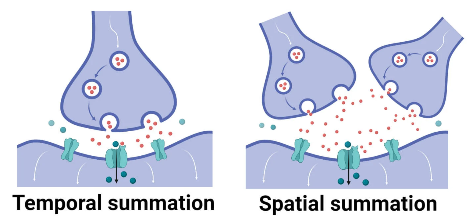
Describe the two types of summation (2)
what are they used for? (1)
Spatial summation - multiple impulses arrive simultaneously at several different presynaptic neurons to combine their impulse/neurotransmitters towards the same postsynaptic neuron
Temporal summation - rapid firing of multiple impulses on one presynaptic neuron over a period of time so outcome combines onto one postsynaptic neuron
> used to increase effect of either excitatory or inhibitory input (e.g. if single impulse is insufficient for excitatory or inhibitory response)
Topic 3 - The Endocrine System
…
State the function of endocrine glands (1)
define the term ‘hormones’ (1)
Endocrine glands → produce and secrete hormones
chemical messengers that circulate/travel in the bloodstream towards target sites to regulate bodily functions
What system is the endocrine system regulated by?
briefly explain how this works (3) - mention difference in effect between positive vs negative feedback
Feedback system (e.g. positive and negative feedback):
signal about body’s state sent from hypothalamus to pituitary gland
causes pituitary gland to secrete certain hormones into blood stream in reaction to bodily changes
if negative feedback, change is counteracted to return body to stable state, if positive feedback, change is further stimulated/amplified
Give the examples of major glands in the endocrine system (8)
- pituitary gland
- hypothalamus
- adrenal gland
- thyroid gland
- pancreas
- pineal gland
- testes (in males)
- ovaries (in females)
Name the hormone secreted for each major gland and outline their functions
pituitary gland
hypothalamus
adrenal gland
thyroid gland
pancreas
pineal gland
testes
ovaries
pituitary gland = (no specific hormone) → known as ‘master gland’, causes other glands to secrete hormones for effect or directly produces effects
hypothalamus = CRH (corticotrophin releasing hormone) → maintains homeostasis, sends signals to pituitary about body’s state
adrenal gland = adrenaline and cortisol → controls sympathetic division in fight or flight response, increases heart rate, blood pressure, sweat, etc.
thyroid gland = thyroxine → regulates metabolism and affects growth
pancreas = insulin and glucagon → regulates blood glucose levels (e.g. in diabetes)
pineal gland = melatonin → regulates circadian rhythms, day-night sleep schedule
testes = testosterone → stimulates development in secondary sexual characteristics in puberty in males
ovaries = oestrogen → stimulates development in secondary sexual characteristics in puberty in females
Describe how hormones work on target cells (2)
why is timing of hormone release important (1)
target cells have receptors that are particular to certain hormones
when enough receptor sites stimulated by hormones, results in physiological reaction in target cell
> timing of hormone release important for normal functioning e.g. too much or too little hormones at wrong time can result in dysfunction of bodily systems
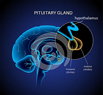
Describe the structure of the pituitary gland (2)
outline the function of the hormones released by the two parts of the pituitary gland (2)
Structure:
located just under hypothalamus
has two main parts - anterior (front) and posterior (back) pituitary (release different hormones that target different cells)
> anterior pituitary → releases ACTH as response to stress which stimulates adrenal glands to produce cortisol, releases LH and FSH for reproductive functioning and sexual characteristics
> posterior pituitary → releases oxytocin for contraction of uterus during childbirth
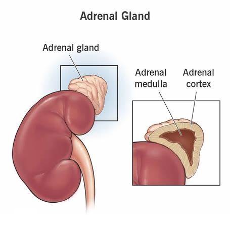
Structure of the adrenal glands (2)
function of each part, including hormone they release
cortex (3)
medulla (1)
Structure:
two adrenal glands sit on top of kidneys
each made up of two parts - outer region is adrenal cortex, inner region is adrenal medulla (release different hormones for different functions)
Function:
adrenal cortex → releases cortisol
regulates important body functions e.g. cardiovascular system
increased during stress
low levels results in low b.p., poor immune function, and inability to deal with stress
adrenal medulla → releases adrenaline and noradrenaline
prepare body for fight or flight e.g. increase heart rate, blood pressure and flow to muscles, conversion of glycogen to glucose for energy
Topic 4 - Fight or Fight Response
…
How are the amygdala and the hypothalamus involved in fight or flight? (4)
when individual faced with threat, amygdala in brain is mobilised
amygdala associates sensory signals (sight, hearing, smell) with emotions like fear or anger for fight or flight
amygdala sends distress signal to hypothalamus
hypothalamus communicates with rest of body via sympathetic nervous system (SNS)
What are the two systems for responding to stressors?
what stressors do each system respond to, give examples
SMA(P) system → response to short-term/acute (immediate) stressors e.g. being attacked
HPA system/axis → response to long-term/chronic (ongoing) stressors e.g. stressful job
Describe the SMA(P) system’s response to acute stressors (4)
S - sympathetic nervous system (SNS) triggered to prepare body for fight or flight, sends signal to adrenal glands via CNS
M - adrenal medulla releases adrenaline and smaller amounts of noradrenaline into bloodstream to circulate in body
A - adrenaline causes physiological changes for fight or flight e.g. increased heartbeat and blood pressure so blood reaches muscles, heart, and other vital organs, increased breathing rate to take in more oxygen, conversion of glycogen to glucose for energy
(P) - parasympathetic nervous system (PNS) dampens stress response when threat passed so body returned to normal state e.g. decreases heartbeat and blood pressure, begins digestion again
Describe the HPA axis/system’s response chronic stressors (3)
how are cortisol levels regulated? (1)
H - hypothalamus releases CRH (hormone) into bloodstream in response to ongoing stressor
P - pituitary gland receives CRH and is stimulated to produce and release ACTH (hormone) into bloodstream towards adrenal glands
A - adrenal cortex stimulated by ACTH to release various stress-related hormones (e.g. cortisol) for fight or flight response, some effects positive (e.g. quick burst of energy, low pain sensitivity) and some negative (e.g. impaired cognitive ability, lower immune system)
→ hypothalamus and pituitary gland have special receptors to monitor circulating cortisol levels, so feedback can be used to reduce CRH and ACTH levels if cortisol rises too high
1. PEE chain - weakness
Point
Evidence
Explain
Hint: Gender differences
P - gender differences in reaction to stress
E - research suggests females' behavioural response to stress focuses on 'tend and befriend' rather than 'fight or flight', which involves being nurturing and protective over their young and forming alliances with other women, due to evolutionary role of primary caregiver
E - shows stress response maybe not generalisable, more complex
2. PEE chain - weakness
Point
Evidence
Explain
Hint: Response not adaptive
P - fight-or-flight response has negative consequences
E - physiological responses in fight or flight are adaptive for stressors requiring physical/energetic activity, but these are not common nowadays, so can cause damage especially when repeatedly activated e.g. increased b.p. damaging blood vessels = heart disease
E - shows response is not adaptive to modern life, so not best way to cope with stress
3. PEE chain - weakness
Point
Evidence
Explain
Hint: Freeze response
P - ‘fight or flight’ response is too simplified
E - researchers argue first reaction to threat is to avoid confrontation/freeze rather than to flight or flee, as this makes animals/humans more hyper-vigilant and alert to danger to focus on info about best way to respond
E - shows this explanation is too simple and actual response to threats are more complex
Topic 5 - Localisation of Function
…
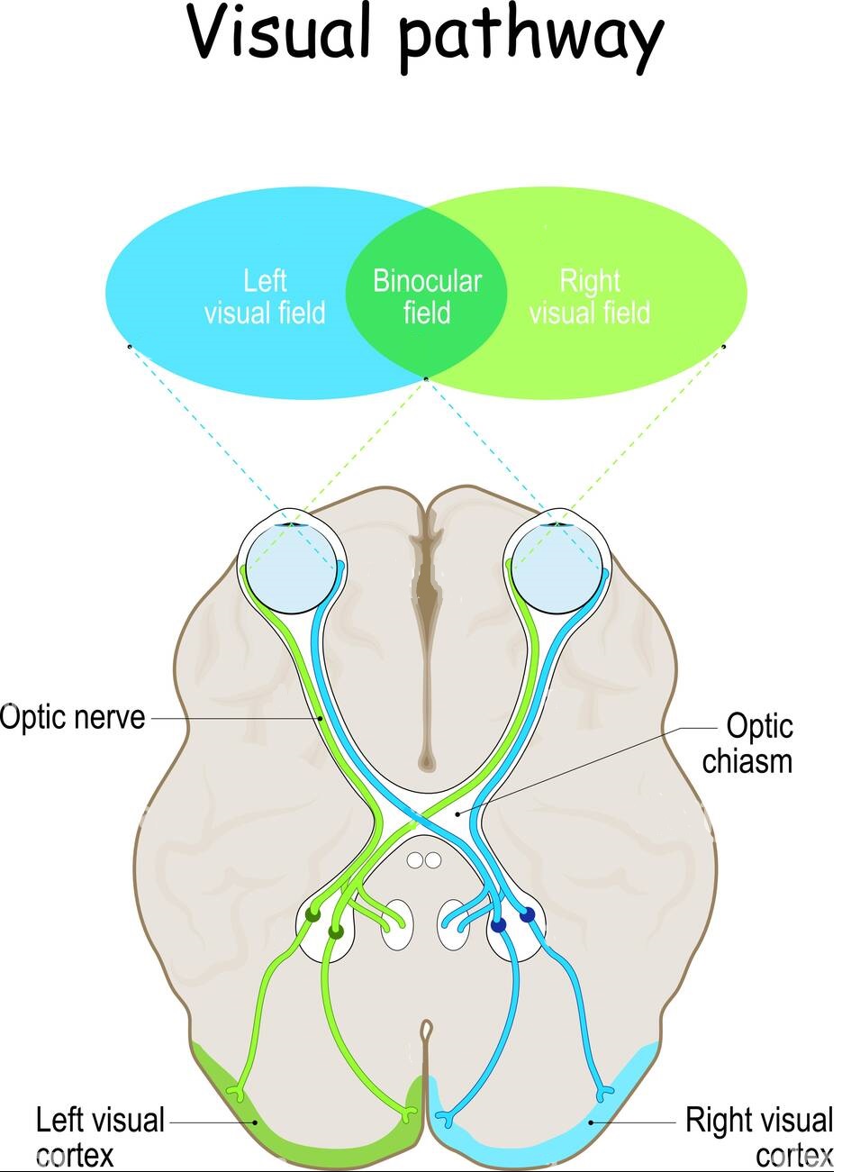
Where is the visual centre mainly located? (1)
describe the steps of visual processing (5)
Visual centre located in visual cortex, in occipital lobe (at back, in both hemispheres)
Visual processing:
light enters eye and hits photoreceptors (rods and cones) in retina at back of eye
nerve impulses from retina transmitted via optic nerve to brain
majority of impulses go to thalamus, which relays info to visual cortex
right hemisphere of visual cortex receives input from left-hand visual field, while left hemisphere receives input from right-hand visual field
different areas of visual cortex process info like colour, shape, movement
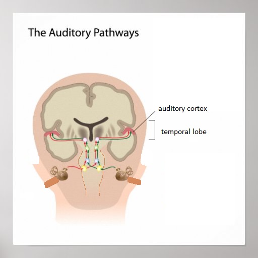
Where is the auditory centre located? (1)
describe the steps of auditory processing (5)
Auditory centre located in auditory cortex, in temporal lobe (on both hemispheres)
Auditory processing:
sound enters ear and reaches cochlea (inner ear)
sound waves converted to nerve impulses by cochlea
impulses transmitted via auditory nerve to brain
brain stem first does basic decoding of sound e.g. duration, intensity
thalamus further processes sound and relays info to auditory cortex, where any last processing occurs and response made
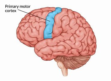
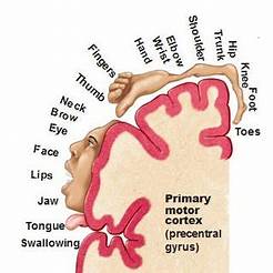
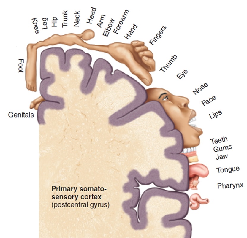
Where is the motor cortex located? (1)
describe how the motor cortex works (3)
Motor cortex located along precentral gyrus, in frontal lobe (in both hemispheres)
Motor processing:
each hemisphere with motor cortex controls opposite side of body’s muscles (e.g. right motor cortex controls left side of body)
different regions of motor cortex controls different parts of body
regions arranged logically next to each other in motor cortex e.g. region controlling foot next to region controlling leg, etc.
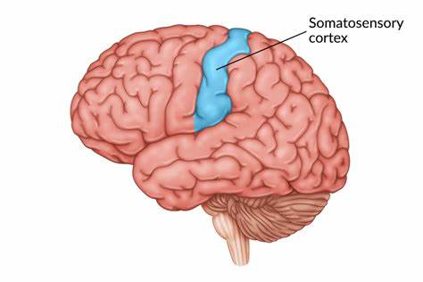
Where is the somatosensory cortex located? (1)
describe how the somatosensory cortex works (4)
Somatosensory cortex located along postcentral gyrus, in parietal lobe (in both hemispheres)
Somatosensory processing:
each hemisphere with somatosensory cortex receives opposite side of body’s sensory info (e.g. right somatosensory cortex receives sensory info from left side of body)
detects sensory events arising from different areas of body
sensory info from skin allows somatosensory cortex to produce sensations of touch, pressure, pain, temperature
this sensation localised to specific body region
Name the two language centres in the brain
Broca’s area
Wernicke’s area
Where is Broca’s area located? (1)
what did Broca study - who was his main patient and what issues did he have? (1)
what is Broca’s area responsible for? (1)
Broca’s area located in posterior portion of frontal lobe (in LEFT hemisphere)
Broca studied patients with lesions/damage in this area of brain e.g. patient ‘Tan’ - could only express syllable ‘Tan’, had unusual disorder so could understand spoken language but couldn’t speak or express thoughts in writing
Broca’s area believed to be responsible for speech production
Where is Wernicke’s area located? (1)
what could patients do with damage to this area? (1)
what is Wernicke’s area responsible for? (1)
Wernicke’s area located posterior portion of temporal lobe (in LEFT hemisphere)
patients with lesions/damage in this area of brain could speak but unable to understand language
Wernicke’s area believed to be responsible for understanding language
What did Wernicke propose about language? (1)
where are Broca’s and Wernicke’s area located in relation to motor and sensory regions? (2)
how do Wernicke’s and Broca’s area work together to understand and produce language? (3)
Wernicke proposed language involves separate motor and sensory regions:
Broca’s area located near motor region that controls mouth, tongue, vocal cords
Wernicke’s area located near sensory region that receives visual and auditory input
→ Visual and auditory input transferred to Wernicke’s area to be recognised as language and associated with meaning
→ Motor regions near Broca’s area then produce language to be spoken or written
→ Neural loop between Wernicke’s and Broca’s area communicate between the two
= language understood and spoken
1. PEE chain - weakness
Point
Evidence
Explain
Hint: Recovering after brain injury
P - equipotentiality theory challenges localisation theory
E - equipotentiality theory suggests basic motor and sensory functions are localised but higher mental functions aren’t, intact areas of cortex can take over responsibility for some cognitive functions after an injury to the normally responsible area
E - shows that localisation may not be practical to explain recovery after brain damage
2. PEE chain - weakness
Point
Evidence
Explain
Hint: Connection between regions more important
P - communication maybe more important than localisation
E - research suggests areas of the brain are very interdependent on each other and require interaction in order to work, complex behaviours such as language, reading, and movement are used gradually as stimulus moves through brain to produce response
E - shows that damage to localised areas may actually be damage to connections between areas, as they resemble each other
3. PEE chain - strength
Point
Evidence
Explain
Hint: Research from aphasia
(+ what is a general weakness for these studies?)
P - research support from aphasia studies
E - discovery of Broca’s and Wernicke’s area come from studies of aphasia (inability/impairment in understanding or producing speech due to brain damage), expressive aphasia showed brain damage to Broca’s area impaired production of language, receptive aphasia showed brain damage to Wernicke’s area impaired understanding language
E - shows that if damage to these areas result in impairment, then these areas must be involved in language functioning to begin with
(+ weakness = only studies brain damaged people, so maybe can’t generalise)
Topic 6 - Lateralisation and Split Brain
…
Define the term ‘lateralisation’
give an example of how the brain hemispheres differ
Lateralisation = fact that two halves/hemispheres of brain are not alike (e.g. in functional specialisations)
left hemisphere is dominant for speech and language, while right hemisphere dominant for visual-motor tasks
How are the two hemispheres connected? (1)
what does this allow for (1)
how were the differences in the hemispheres investigated (1)
Hemispheres connected by corpus callosum (connecting bundle of nerve fibres) - allows for communication of info between the two halves
investigated when surgery treatment for severe epilepsy had corpus callosum nerve bundle cut so that electrical activity causing seizures wouldn’t cross between hemispheres = ‘split brain’ patients
What did Sperry and Gazzaniga study? (1)
how did they do this (3)
findings (3)
Sperry and Gazzaniga = first to study capabilities of split-brain patients by testing hemispheric lateralisation:
info from left visual field goes to right hemisphere, while info from right visual field goes to left hemisphere
patients corpus callosum cut, so info presented to one hemisphere can’t travel to other hemisphere (only processed in hemisphere that received it)
patient fixates focus on dot in the centre of a screen while a picture/info (e.g. cat or dog image) shown to either left or right visual field
Findings:
if dog image shown to right visual field = patient could say ‘dog’
if cat image shown to left visual field = patient said they saw nothing
why? → picture in left visual field is processed by right hemisphere, however the language centres are only in the left hemisphere and info can’t travel to left hemisphere, so patient can’t verbally respond about seeing anything
1. PEE chain - strength
Point
Evidence
Explain
Hint: Research support for multitasking
P - advantages of hemispheric lateralisation
E - generally assumed brain lateralisation increases neural processing capacity as using only one hemisphere for particular task (e.g. language) leaves other one free to engage in another function e.g. brain lateralisation in chicken has been found to allow findings food and being vigilant of predators to occur simultaneously
E - there may be some research evidence regarding the practicality of lateralisation in multitasking
2. PEE chain - weakness
Point
Evidence
Explain
Hint: Case study J.W.
P - language may not be restricted to left hemisphere
E - recent studies disprove earlier beliefs that left hemisphere involved in language only, case study patient J.W. had damaged left hemisphere but developed ability to speak when info presented to left OR right hemisphere
E - shows the theory lacks temporal validity or reliability
3. PEE chain - weakness
Point
Evidence
Explain
Hint: Limited split-brain patients
P - split-brain research no longer useful/feasible
E - split brain procedure rarely carried out nowadays so there is lack of people in sufficient numbers for useful research, may be that the limited patients use had other confounding variables that affected them
E - shows the research is not valid or reliable and maybe not generalisable
Topic 7 - Plasticity and Functional Recovery of the Brain
…
Define the term ‘plasticity’
give three ways plasticity can occur
- The ability of the brain to change and adapt
through life experience, video games, and meditation
How can life experiences cause plasticity? (1)
how has brain plasticity been found for learning new skills? (1)
gaining new experiences allows nerve pathways frequently used to develop stronger connections, while neurons rarely used die = adapting to changing environment
increased age also kills nerve cells, though this can be reversed by learning new skill - research found 60 year olds taught new skill (juggling) increased grey matter in visual cortex, but when practising stopped changes were reversed = brain plasticity
How can video games cause plasticity?
what research showed this? (2)
research found ppts who played Super Mario for 30 mins every day for 2 months had significantly increased grey matter in various brain areas (e.g. cortex, cerebellum, hippocampus) than control group
showed new synaptic connections in brain areas involving spatial navigation, strategic planning, working memory and motor performance
How can meditation cause plasticity?
what research showed this? (2)
Tibetan monks experienced in meditation had superior gamma activity over students with no experience
these showed short and long-term/permanent changes in inner workings of brain
How was functional recovery after trauma first discovered? (2)
what two ways can recovery occur? (2)
stroke victims were able to recover some functioning
found neurons next to damaged areas could take over functioning, with two ways recovery can happen:
1. Neuronal unmasking
2. Stem cells
Describe neuronal unmasking role in functional recovery (3)
when brain area is damaged, this increases activity/input in surrounding areas
this causes dormant synapses (that have lost function/ineffective) to awaken
this opens connections to not normally activated areas of brain to develop new structures
Describe stem cells role in functional recovery (3)
stem cells are unspecialised, so can develop into nerve cells
could be implanted into brain to replace dead cells or to secrete growth hormones to rescue injured cells
could be transplanted in a nearby undamaged brain area to create new networks with damaged brain area
1. PEE chain - strength (for plasticity)
Point
Evidence
Explain
Hint: Human and animal studies
P - research support from animals and human studies
E - rats raised in an enriched environment developed more neurons in hippocampus than controls raised in lab cages, MRI scan showed more grey matter in hippocampi and larger posterior hippocampi (used for spatial navigation) of London taxi drivers who had more experience
E - shows reliable findings than can be generalised
2. PEE chain - strength (for functional recovery)
Point
Evidence
Explain
Hint: Animal (rat) study support
P - research support from animal studies
E - rats with traumatic brain injury injected with stem cells or solution with no stem cells, stem cell rats showed clear development of neurons in injured area but not control group
E - shows practicality of such research may improve brain damaged patients
3. PEE chain - weakness (for functional recovery)
Point
Evidence
Explain
Hint: Age differences
P - age differences in functional recovery
E - commonly accepted view that functional plasticity reduces with age and recovery much easier in children with traumatic brain injury, adults require much more practice to produce changes
E - shows age still plays role, so treatment maybe not generalisable/too simple
Topic 8 - Ways of studying the brain
…
Name the four main ways of studying the brain
Post-mortem examinations
fMRI
EEG
ERP’s
(2, 3, and 4 = scanning techniques)
Describe how post-mortem examinations can be used to study the brain (2)
give three examples of this in research
brains of those displaying unusual behaviour when alive are studied
when person dies, brain can be examined for abnormalities to explain behaviour
→ study of Tan discovered Broca’s area
→ HM case study led to hippocampus being linked to memory
→ depression been linked to reduced glial cells in frontal cortex
1. PEE chain - strengths and weaknesses (post-mortem examinations)
Point
Evidence
Explain
Hint: Deeper regions seen vs confounding variables
Strength = can examine deeper structural regions of brain than scanning techniques e.g. really helped our understanding of schizophrenia brains structural abnormalities
Weakness = confounding variables may affect post-mortem brain (e.g. length of time dead, cause of death, drug treatments, age etc.), also retrospective and offers no help to patient
Describe how fMRI’s can be used to study the brain (2)
measures changes in brain activity when person performs a task
increased blood flow and demand for oxygen in certain brain areas (lights up on scan) when doing tasks shows these areas involved in particular mental activity
2. PEE chain - strengths and weaknesses (fMRI)
Point
Evidence
Explain
Hint: Non-invasive and objective vs non-direct measuring and localisation
Strength = non-invasive with no radiation and offers more objective data than self-reports
Weakness = measure of blood flow not direct neural activity in certain brain areas, and it overlooks networked nature of brain (focuses on localisation rather than communication between areas)
Describe how EEG’s can be used to study the brain (2)
measure electrical activity of the brain cells by placing electrodes on scalp, which is mapped on a graph over time period
basic EEG patterns of alpha, beta, delta and theta waves used e.g. in sleep studies - each wave occurs during different stages of sleep
3. PEE chain - strength and weakness (EEG)
Point
Evidence
Explain
Hint: Real time changes vs no exact source
Strength = shows activity in real time so is good for diagnosing changes in brain activity e.g. Alzheimer's (slow activity) and epilepsy (spikes of activity)
Weakness = electrical activity can be detected by several neighbouring electrodes so EEG signal doesn’t give exact source of activity in brain
Describe how ERP’s (Event Related Potentials) can be used to study the brain (3)
small voltage changes in brain activity, triggered by stimulus or event
patients shown stimuli repeatedly to rule out neuronal noise and see a consistent pattern
then EEG divided into two categories: waves in first 100 milliseconds = sensory response (shows initial visual response), next 100 = cognitive response (shows information processing)
4. PEE chain - strength and weakness (ERP)
Point
Evidence
Explain
Hint: Continuous stimuli response seen vs need lots of trials
Strength = gives consistent measure of processing in response to certain stimuli and changes in stimuli, and can measure response to stimuli in absence of behavioural response
Weakness = need a large number of trials for meaningful data as ERP’s small and difficult to distinguish from other electrical brain activity, and deep brain activity cannot be recorded
Topic 9 - Circadian Rhythms
…
Name the master circadian pacemaker in humans (1)
what is it and where is it located (2)
what is its main function (1) - what areas does it reset (2) and what does this cause (1)
what is its primary input (1) - how does this work (2)
what is the main circadian rhythm that uses this process (1)
SCN (suprachiasmatic nucleus)
cluster of nerve cells in hypothalamus
acts as ‘master clock’ - constantly resets body clock and brain regions controlling sleep to be in synchrony with outside world via photoentrainment
light is primary input for photoentrainment - photoreceptors in eyes detect light levels and send to SCN (via optic nerve) to coordinate it’s built in circadian rhythm
= sleep wake cycle
Sleep wake cycle
how does circadian rhythm affect sleep wake cycle (1)
when are the strongest sleep drives (1)
why is this (1) - when is this more intense (1)
what other control is sleep wake cycle under and how (1)
circadian rhythm dips and rises in response to external light and darkness so we feel need to sleep and wake up
between 2-4am and 1-3pm
this is when our circadian rhythm dips in the day, so sleepiness felt - more intense if person is sleep deprived/insufficient sleep
homeostatic control also regulate sleep wake cycle - as energy used up in body, homeostasis increases drive for sleep throughout day
Name two other body functions that follow circadian rhythms
Core body temp. regulation
Hormone production
How is core body temperature regulated by circadian rhythm? (4)
when is it highest and lowest
what does temp. drop cause
what does temp. rise cause
why are we sleepy in afternoon
lowest around 4:30am and highest about 6pm
sleep occurs when core body temp. begins to drop
alertness occurs when core body temp. rises (e.g. during last hours of sleep)
also small drop in body temp. between 2-4pm (so people feel sleepy in afternoon)
How is hormone production regulated by circadian rhythm? (4)
when does melatonin production peak
what does this cause
when does melatonin production drop
what does this cause
melatonin production and release from pineal gland peaks in hours of darkness
this activates receptors in brain and encourages feelings of sleep
melatonin production and release drops in hours of light
this causes person to wake
1. PEE chain - strength
Point
Evidence
Explain
Hint: Cortisol levels in Antarctic
P - research support for importance of light
E - studies in Antarctic found cortisol levels had changed from being highest when ppts awake to being highest during noon, after having spent 3 months in continuous darkness of extreme Antarctic’s winter daylight pattern
E - shows extreme differences in light had affected body’s circadian rhythm and so affected hormone production
2. PEE chain - weakness
Point
Evidence
Explain
Hint: Morning vs evening people rhythm peaks
P - individual differences in circadian rhythms for sleep wake cycle
E - research finds circadian rhythms can vary between morning people (prefer to rise early and go to bed early - rhythm peaks 6am -10pm) and evening people (prefer to wake and go to bed later - rhythm peaks 10am - 1am)
E - shows patterns of circadian rhythms can’t be generalised
3. PEE chain - weakness
Point
Evidence
Explain
Hint: Role of artificial light in studies
(+ what is the strength of this finding)
P - issues with research methodology
E - early research of circadian rhythms (e.g Siffre’s free running circadian rhythm cave study - lived underground and found body clock ran slower/stretched more than 24hrs) involves ppts isolated from external cues affecting rhythm e.g. clocks and daylight, but not artificial light, research finds even dim artificial light alters circadian rhythms (stretched to 28hrs)
E - shows study’s may not be valid (+ light is still important factor = strength)
Topic 10 - Ultradian and Infradian Rhythms
…
Define an ultradian rhythm (1)
give an example (1)
how are sleep stages measured/recorded (1)
Ultradian Rhythm = when rhythm lasts less than 24 hours/occurs more than once every 24 hours
Example: stages of sleep cycle - alternating patterns of REM and NREM last 90-100 mins each during sleep stages
→ Stages measured using EEG (records electrical activity of brain to create pattern)
Briefly describe the 5 stages of sleep
mention brain activity, sleep depth, waves shown, muscle activity, and REM
light sleep but brain still quite active, some muscle activity
breathing and heart rate pattern slows, occasional sleep
deep sleep begins, brain shows delta waves
very deep sleep, rhythmic breathing, limited muscle activity, brain shows theta waves
REM - brainwaves speed up (resembles active person) while muscles still relaxed, dreaming occurs